

Influence of particle size and shape on their margination and wall-adhesion: implications in drug delivery vehicle design across nano-to-micro scale
- PMID: 30080212
- PMCID: PMC6247903
- DOI: 10.1039/c8nr04042g
Influence of particle size and shape on their margination and wall-adhesion: implications in drug delivery vehicle design across nano-to-micro scale
Abstract
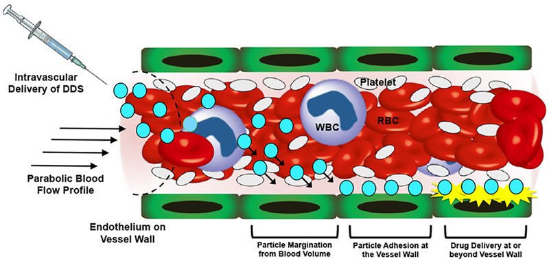
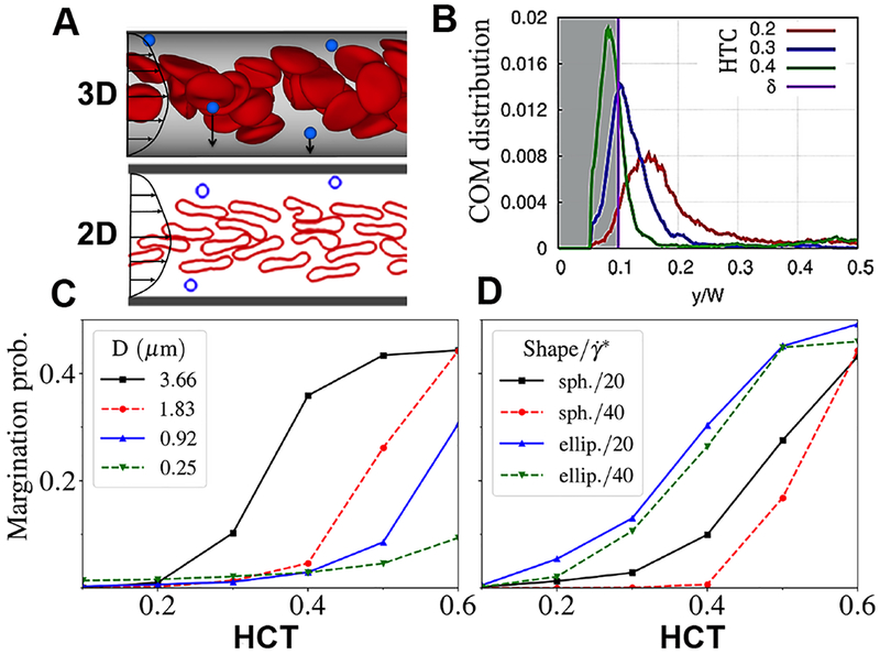
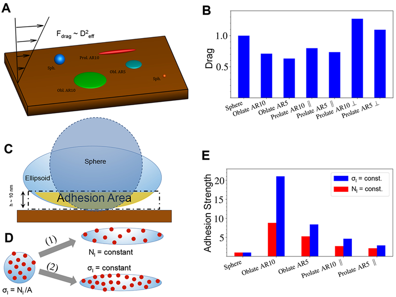
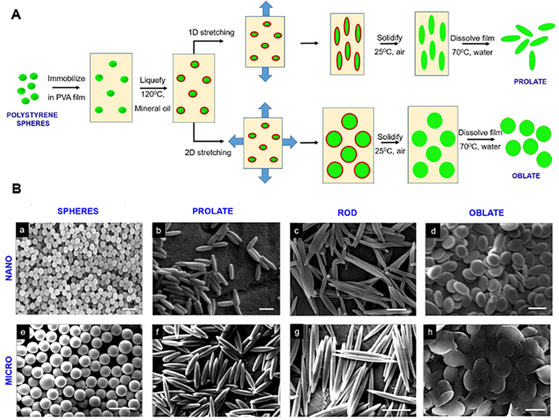
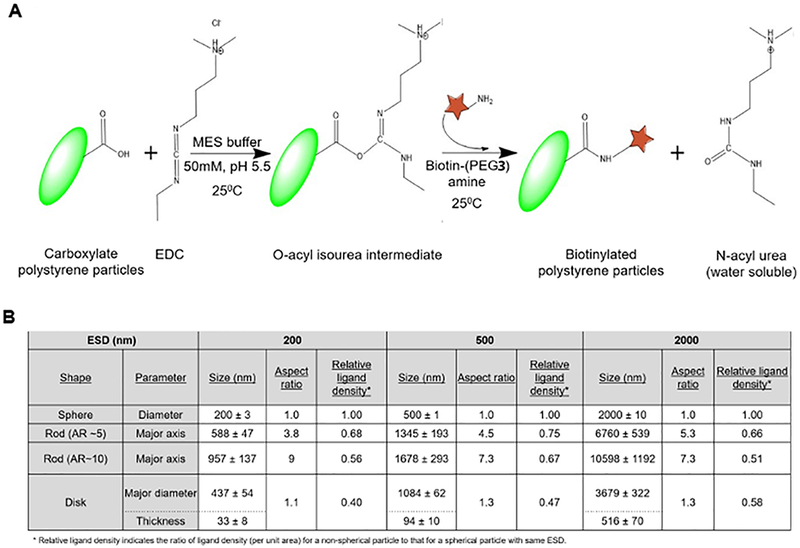
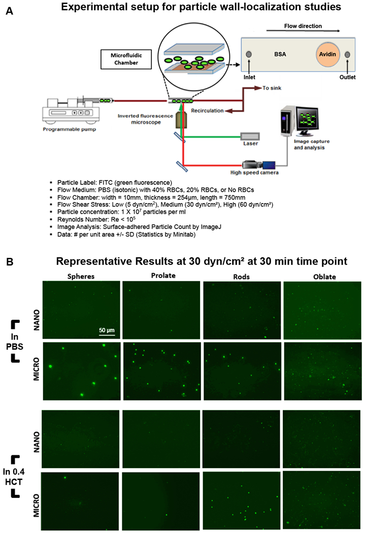
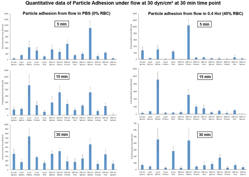
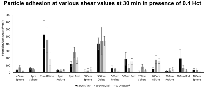
Intravascular drug delivery technologies majorly utilize spherical nanoparticles as carrier vehicles. Their targets are often at the blood vessel wall or in the tissue beyond the wall, such that vehicle localization towards the wall (margination) becomes a pre-requisite for their function. To this end, some studies have indicated that under flow environment, micro-particles have a higher propensity than nano-particles to marginate to the wall. Also, non-spherical particles theoretically have a higher area of surface-adhesive interactions than spherical particles. However, detailed systematic studies that integrate various particle size and shape parameters across nano-to-micro scale to explore their wall-localization behavior in RBC-rich blood flow, have not been reported. We address this gap by carrying out computational and experimental studies utilizing particles of four distinct shapes (spherical, oblate, prolate, rod) spanning nano- to-micro scale sizes. Computational studies were performed using the Large-scale Atomic/Molecular Massively Parallel Simulator (LAMMPS) package, with Dissipative Particle Dynamics (DPD). For experimental studies, model particles were made from neutrally buoyant fluorescent polystyrene spheres, that were thermo-stretched into non-spherical shapes and all particles were surface-coated with biotin. Using microfluidic setup, the biotin-coated particles were flowed over avidin-coated surfaces in absence versus presence of RBCs, and particle adhesion and retention at the surface was assessed by inverted fluorescence microscopy. Our computational and experimental studies provide a simultaneous analysis of different particle sizes and shapes for their retention in blood flow and indicate that in presence of RBCs, micro-scale non-spherical particles undergo enhanced 'margination + adhesion' compared to nano-scale spherical particles, resulting in their higher binding. These results provide important insight regarding improved design of vascularly targeted drug delivery systems.
Conflict of interest statement
Conflicts of Interest
There are no conflicts of interest to declare by any of the authors.
Figures
Similar articles
-
Margination of micro- and nano-particles in blood flow and its effect on drug delivery.Sci Rep. 2014 May 2;4:4871. doi: 10.1038/srep04871. Sci Rep. 2014. PMID: 24786000 Free PMC article.
-
Microparticle shape effects on margination, near-wall dynamics and adhesion in a three-dimensional simulation of red blood cell suspension.Soft Matter. 2015 Mar 21;11(11):2097-109. doi: 10.1039/c4sm02686a. Soft Matter. 2015. PMID: 25601616
-
The margination propensity of ellipsoidal micro/nanoparticles to the endothelium in human blood flow.Biomaterials. 2013 Jul;34(23):5863-71. doi: 10.1016/j.biomaterials.2013.04.011. Epub 2013 May 2. Biomaterials. 2013. PMID: 23642534
-
Particle margination and its implications on intravenous anticancer drug delivery.AAPS PharmSciTech. 2014 Jun;15(3):762-71. doi: 10.1208/s12249-014-0099-6. Epub 2014 Apr 2. AAPS PharmSciTech. 2014. PMID: 24687242 Free PMC article. Review.
-
The effects of particle size, shape, density and flow characteristics on particle margination to vascular walls in cardiovascular diseases.Expert Opin Drug Deliv. 2018 Jan;15(1):33-45. doi: 10.1080/17425247.2017.1316262. Epub 2017 Apr 13. Expert Opin Drug Deliv. 2018. PMID: 28388248 Review.
Cited by
-
Biodegradable polyester-based nano drug delivery system in cancer chemotherapy: a review of recent progress (2021-2023).Front Bioeng Biotechnol. 2023 Nov 1;11:1295323. doi: 10.3389/fbioe.2023.1295323. eCollection 2023. Front Bioeng Biotechnol. 2023. PMID: 38026861 Free PMC article. Review.
-
Pulmonary endothelium-targeted nanoassembly of indomethacin and superoxide dismutase relieves lung inflammation.Acta Pharm Sin B. 2023 Nov;13(11):4607-4620. doi: 10.1016/j.apsb.2023.05.024. Epub 2023 May 26. Acta Pharm Sin B. 2023. PMID: 37969734 Free PMC article.
-
Recent Review on Biological Barriers and Host-Material Interfaces in Precision Drug Delivery: Advancement in Biomaterial Engineering for Better Treatment Therapies.Pharmaceutics. 2024 Aug 16;16(8):1076. doi: 10.3390/pharmaceutics16081076. Pharmaceutics. 2024. PMID: 39204421 Free PMC article. Review.
-
Synthesis of Novel Conjugated Linoleic Acid (CLA)-Coated Superparamagnetic Iron Oxide Nanoparticles (SPIONs) for the Delivery of Paclitaxel with Enhanced In Vitro Anti-Proliferative Activity on A549 Lung Cancer Cells.Pharmaceutics. 2022 Apr 11;14(4):829. doi: 10.3390/pharmaceutics14040829. Pharmaceutics. 2022. PMID: 35456663 Free PMC article.
-
Alleviating experimental pulmonary hypertension via co-delivering FoxO1 stimulus and apoptosis activator to hyperproliferating pulmonary arteries.Acta Pharm Sin B. 2023 Jun;13(6):2369-2382. doi: 10.1016/j.apsb.2022.12.002. Epub 2022 Dec 8. Acta Pharm Sin B. 2023. PMID: 37425053 Free PMC article.
References
-
- Maeda H and Matsumara Y, Tumoritropic and lymphotropic principles of macromolecular drugs, Crit. Rev. Ther. Drug Carrier Syst, 1989, 6, 193–210. - PubMed
-
- Wilczewska AZ, Niemirowicz K, Markiewicz KH and Car H, Nanoparticles as drug delivery systems, Pharmacol. Rep, 2012, 64, 1020–1037. - PubMed
-
- Brannon-Peppas L and Blanchette JO, JNanoparticle and targeted systems for cancer therapy, Adv. Drug. Del. Rev, 2012, 64, 206–212. - PubMed
MeSH terms
Grants and funding
LinkOut - more resources
Full Text Sources
Other Literature Sources
Miscellaneous