. 2018 Aug 6;13(8):e0201880.
doi: 10.1371/journal.pone.0201880. eCollection 2018.

Interaction between the cellular E3 ubiquitin ligase SIAH-1 and the viral immediate-early protein ICP0 enables efficient replication of Herpes Simplex Virus type 2 in vivo

Affiliations

Interaction between the cellular E3 ubiquitin ligase SIAH-1 and the viral immediate-early protein ICP0 enables efficient replication of Herpes Simplex Virus type 2 in vivo

Julia S Czechowicz et al. PLoS One. .

Abstract

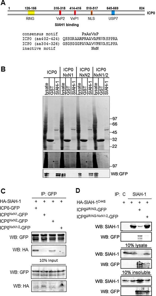
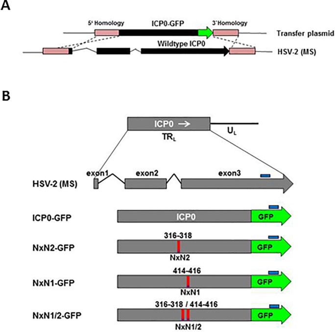
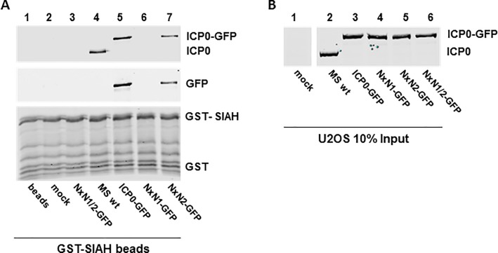
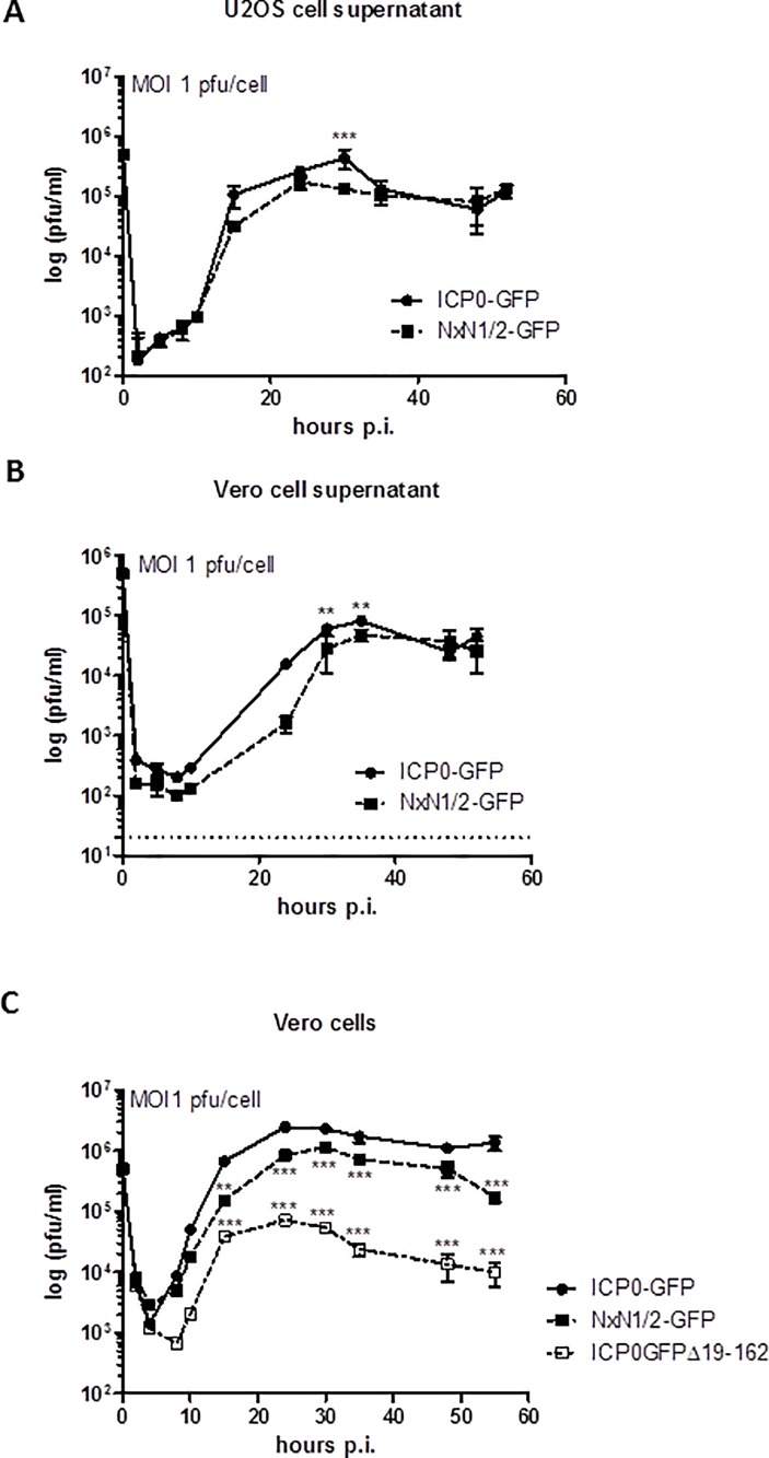
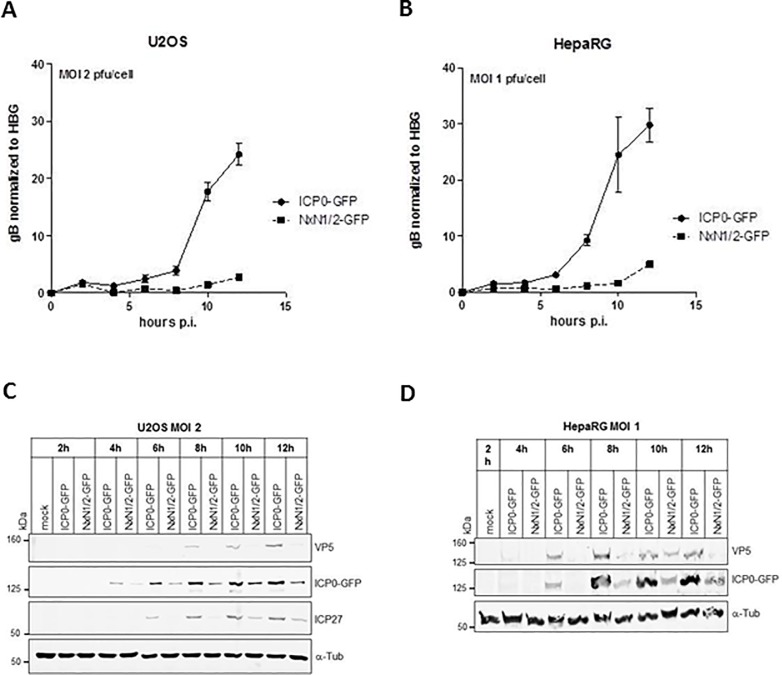
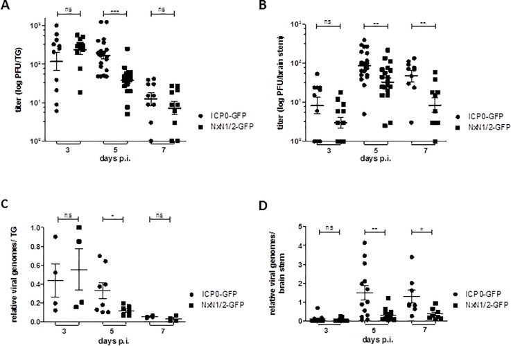
Herpes Simplex Virus type 2 (HSV-2) is a neurotropic human pathogen. Upon de novo infection, the viral infected cell protein 0 (ICP0) is immediately expressed and interacts with various cellular components during the viral replication cycle. ICP0 is a multifunctional regulatory protein that has been shown to be important for both efficient viral replication and virus reactivation from latency. In particular, as previously demonstrated in transfected tissue culture models, ICP0 interacts with the cellular E3 ubiquitin ligase SIAH-1, which targets ICP0 for proteasomal degradation. However, the consequence of this virus-host interaction during the establishment of HSV-2 infection in vivo has not yet been elucidated. Here we confirmed that ICP0 of HSV-2 interacts with SIAH-1 via two conserved PxAxVxP amino acid binding motifs. We also demonstrate in vitro that a SIAH-1 binding-deficient HSV-2 strain, constructed by homologous recombination technology, exhibits an attenuated growth curve and impaired DNA and protein synthesis. This attenuated phenotype was also confirmed in an in vivo ocular infection mouse model. Specifically, viral load of the SIAH-1 binding-deficient HSV-2 mutant was significantly reduced in the trigeminal ganglia and brain stem at day 5 and 7 post infection. Our findings indicate that the interplay between ICP0 and SIAH-1 is important for efficient HSV-2 replication in vivo, thereby affecting viral dissemination kinetics in newly infected organisms, and possibly revealing novel targets for antiviral therapy.


Conflict of interest statement

The commercial affiliation of an author of this study [MME] (Biomedizinische Forschungsgesellschaft mbH, Vienna, Austria) does not alter our adherence to all PLOS ONE policies on sharing data and materials.

Figures

Fig 1
Fig 1. ICP0 interacts with SIAH-1 via two minimal interaction motifs.
(A) Schematic representation of HSV-2 ICP0 indicating the position of the RING domain (yellow), the USP7 interaction domain (blue), the nuclear localization signal (brown) and the two SIAH interaction motifs VxP1 and VxP2 (red). The primary amino acid sequence surrounding the predicted SIAH binding motifs is depicted together with the consensus motif and the position of the inactivating NxN mutation. (B) HEK293T cells were transfected with plasmids encoding GFP-tagged ICP0 and its mutants and the cell lysates were incubated with GST or GST-SIAH-1-loaded glutathione sepharose beads. The upper SDS-PAGE gel shows a Coomassie staining of the respective input control (lysate) and the eluates from the GST or GST-SIAH-1 beads. Below, a contrast enhanced section of the gel with putative ICP0 bands indicated by asterisks. ICP0 was detected by Western blotting using an antibody against the C-terminal GFP tag. Size markers in kDa. (C) HEK293T cells were transfected with plasmids encoding GFP-tagged ICP0 and its mutants and HA-tagged SIAH-1. The ICP0-GFP proteins were immunoprecipitated from the lysates, ICP0 and SIAH-1 were detected by SDS-PAGE and Western blotting using antibodies against the GFP and HA-tags. (D) HEK293T cells were transfected with plasmids encoding the inactive mutant HA-SIAH-1C44S and GFP-ICP0ΔRING or GFP-ICP0ΔRING/NxN1/2 as indicated. Immunoprecipitation from the cell lysates was performed using control mouse IgG or anti-SIAH-1. ICP0 and SIAH-1 were detected by SDS-PAGE and Western blotting using antibodies against SIAH-1 or the GFP-tag. The lower panel shows the analysis of the RIPA buffer-insoluble pellet after cell lysis.
Fig 2
Fig 2. Construction of HSV-2 mutant viruses with mutated SIAH binding motifs in ICP0.
(A) Depiction of homologous recombination. The native ICP0 sequence in HSV-2 strain MS was exchanged by a sequence encoding ICP0-eGFP fusion protein. (B) Maps of the various ICP0 constructs. The wild-type gene is located in the HSV-2 TRL region. The various ICP0-GFP DNA sequences were accordingly adjusted to reduce their GC content. The NxN1 and NxN2 mutations in the SIAH-1 binding domains are marked in red. Sequences recognized by Southern blot probes are marked in blue.
Fig 3
Fig 3. Virally expressed ICP0NxN1/2 does not bind to SIAH-1.
(A) U2OS cells were infected for 48 h with the indicated mutants and HSV-2 strain MS (MS wt) at an MOI 0.01 pfu/cell. Cell lysates were incubated with GST-SIAH-1-loaded glutathione sepharose beads. Eluates were analyzed by SDS-PAGE and Western blotting using antibodies directed against HSV-2 ICP0 and GFP. (B) Input controls (10%) of the GST-pulldown were analyzed as before using ICP0-specific antibody.
Fig 4
Fig 4. The NxN1/2 mutation in ICP0 of HSV-2 results in delayed viral growth.
(A) U2OS cells were infected in duplicates with an MOI of 1 pfu/cell of HSV-2-ICP0-GFP (ICP0-GFP) and HSV-2-ICP0NxN1/2-GFP (NxN1/2-GFP). At the indicated time points supernatants were harvested and titrated on Vero cell monolayers. Mean ± SD of viral titers (n = 2 per data point) were plotted as a function of time. (B) Vero cells were infected in duplicates and evaluated as above. (C) Vero cells were infected in duplicates with HSV-2-ICP0-GFP, HSV-2-ICP0NxN1/2-GFP and HSV-2-GFP-ICP0Δ19–162 (i.e. ICP0ΔRING mutant) at an MOI of 1 pfu/cell. At the indicated time points supernatants were harvested, titrated and evaluated as above. Titer differences were analyzed using two-way ANOVA, with ** p<0.01 and *** p<0.001. The detection limits are shown by a dashed line.
Fig 5
Fig 5. NxN1/2 mutation in ICP0 results in delayed and reduced vital DNA and protein synthesis.
(A) U2OS cells, and (B) HepaRG cells were infected with HSV-2-ICP0-GFP (ICP0-GFP) or HSV-2-ICP0NxN1/2-GFP (NxN1/2-GFP) at an MOI of 2 or 1 pfu/cell, respectively. Infected cells were harvested at the indicated time points post infection (p.i.) for isolation of genomic and viral DNA. Viral DNA copies were determined by quantitative real-time PCR using oligonucleotide-primers and probes directed against the UL27 gene encoding glycoprotein gB. Copy numbers were normalized to the sequence of the human single-copy beta-globin gene (HBG). (C) U2OS cells, and (D) HepaRG cells were infected with HSV-2-ICP0-GFP or HSV-2-ICP0NxN1/2-GFP at an MOI of 2 for up to 24 hours, or 1 pfu/cell for 12 hours, respectively. At the indicated time points, cells were harvested. Whole cell lysates were analyzed by SDS-PAGE and Western blot against the indicated proteins. Signals were quantified in S2 Fig.
Fig 6
Fig 6. HSV-2 replication in the cornea of C57Bl/6J mice.
(A) Schematic representation of the murine ocular HSV-2 infection model. Both corneas of anesthetized mice were scarified. Virus was placed on the eyes using 105 pfu/eye. Ocular swabs were taken at the indicated time points post infection (p.i.). Animals were sacrificed at day 3, 5 and 7 post infection, and trigeminal ganglia (TG) and the brain stem were isolated for further analyses. (B) Ocular swabs from HSV-2-ICP0-GFP (ICP0-GFP) or HSV-2-ICP0NxN1/2-GFP (NxN1/2-GFP) infected animals taken at the indicated time points p.i. were analyzed for viral titers. For every experiment a group of n = 10 mice were infected and experiments were repeated four times. Each symbol represents the mean ± SEM of the titers, determined on Vero cell monolayers. The limit of detection is indicated by a dashed line. Statistical differences were calculated using two-way ANOVA with *** p< 0.001.
Fig 7
Fig 7. The NxN1/2 mutation in ICP0 leads to impaired replication in the TG and brain stem of C57Bl/6J mice.
At day 3, 5 and 7 post infection (p.i.), (A) the trigeminal ganglia (TG), and (B) brain stem was extracted from HSV-2-ICP0-GFP and HSV-2-ICP0NxN1/2-GFP infected mice, homogenized and viral titers were determined as in Fig 6. Symbols represent individual animals; floating bars represent the mean ± SEM. Titer differences were determined by the Mann-Whitney test. (C) Half of the homogenized TG tissue from panel A, and (D) half of the tissue from panel B was lysed and total DNA was isolated to determine viral genome copy numbers. Oligonucleotide-primers specific for the HSV-2 UL27 gene were used and copy numbers were normalized to murine β-actin sequences. Each symbol represents the relative genome number of an individual animal, relative to the genome copy number of a single HSV-2-ICP0-GFP infected mouse, which was set as 100%. Floating bars represent the mean ± SEM. Statistical differences were determined as above.

Similar articles

Cited by

References

    1. Roizman B, Knipe DM (2001) Herpes Simplex viruses and their replication In: Knipe DM, Howley P, Griffin DE, Lamb RA, Martin MA et al., editors. Fields Virology. Philadelphia: Lippincott Williams & Wilkins; pp. 2399–2459.
    1. Roizman B, Whitley RJ (2013) An inquiry into the molecular basis of HSV latency and reactivation. Annu Rev Microbiol 67: 355–374. 10.1146/annurev-micro-092412-155654 - DOI - PubMed
    1. Thellman NM, Triezenberg SJ (2017) Herpes Simplex Virus Establishment, Maintenance, and Reactivation: In Vitro Modeling of Latency. Pathogens 6 pathogens6030028 [pii]; 10.3390/pathogens6030028 - DOI - PMC - PubMed
    1. Smith G (2012) Herpesvirus transport to the nervous system and back again. Annu Rev Microbiol 66: 153–176. 10.1146/annurev-micro-092611-150051 - DOI - PMC - PubMed
    1. Sandri-Goldin RM (2007) Initiation of transcription and RNA synthesis, processing and transport in HSV and VZV infected cells. NBK47363 [bookaccession]. - PubMed

Publication types

MeSH terms