. 2018 Aug 21;115(34):E7932-E7941.
doi: 10.1073/pnas.1802177115. Epub 2018 Aug 6.

Structure of the EmrE multidrug transporter and its use for inhibitor peptide design

Affiliations

Structure of the EmrE multidrug transporter and its use for inhibitor peptide design

Victor Ovchinnikov et al. Proc Natl Acad Sci U S A. .

Abstract

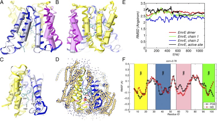
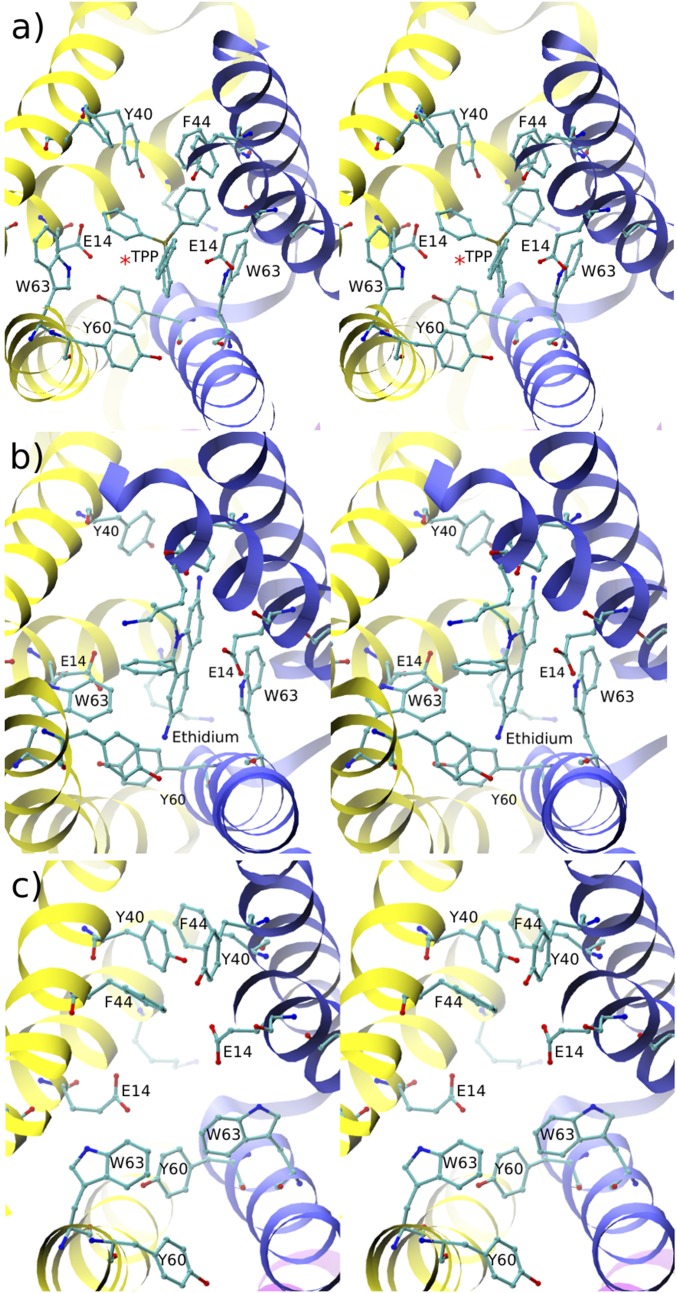
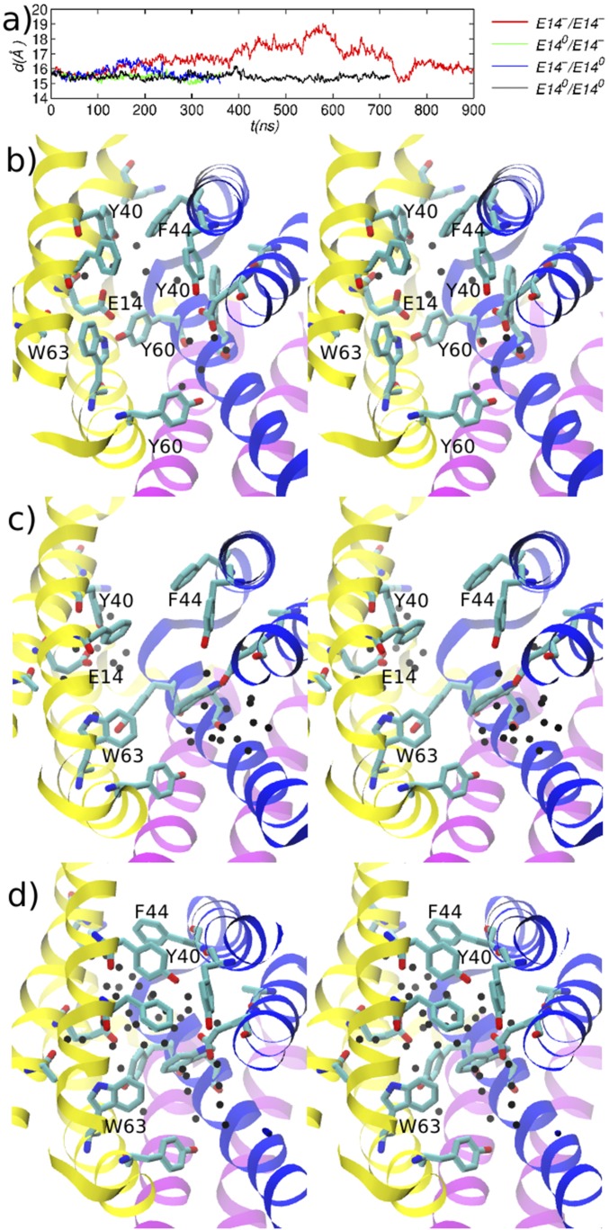
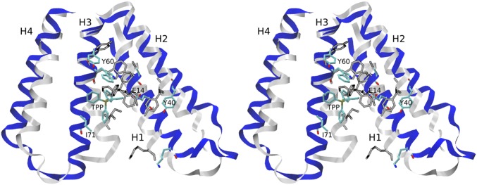
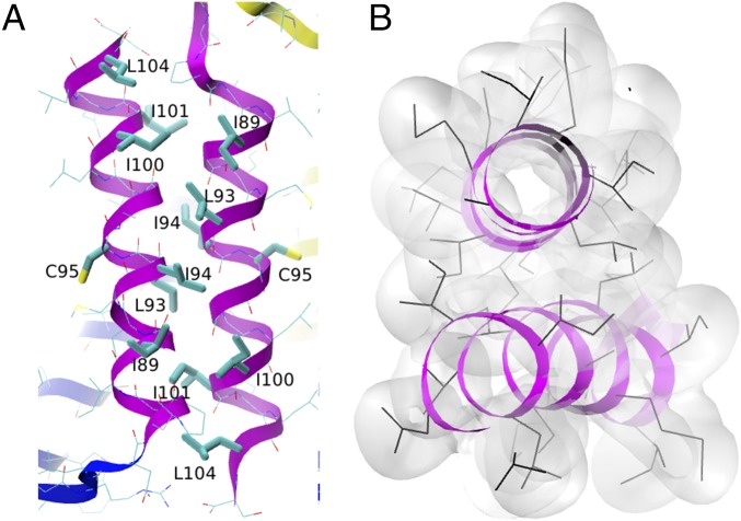
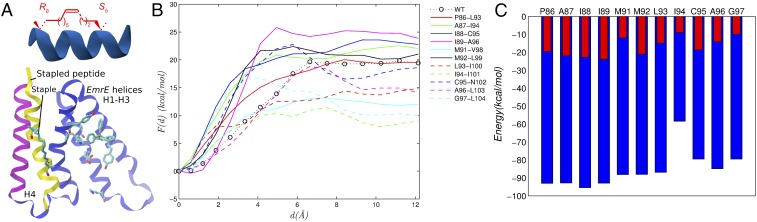
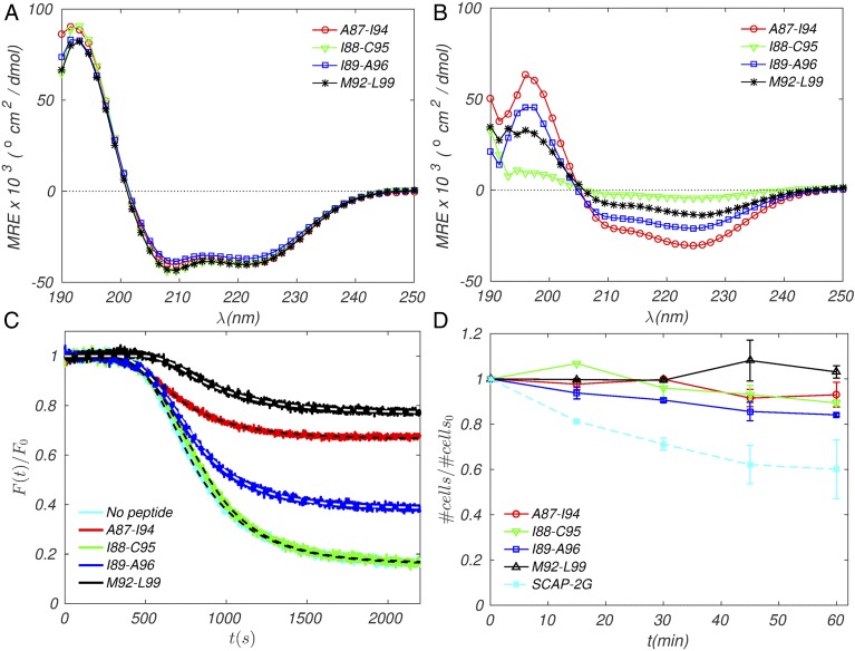
Small multidrug resistance (SMR) pumps represent a minimal paradigm of proton-coupled membrane transport in bacteria, yet no high-resolution structure of an SMR protein is available. Here, atomic-resolution structures of the Escherichia coli efflux-multidrug resistance E (EmrE) multidrug transporter in ligand-bound form are refined using microsecond molecular dynamics simulations biased using low-resolution data from X-ray crystallography. The structures are compatible with existing mutagenesis data as well as NMR and biochemical experiments, including pKas of the catalytic glutamate residues and the dissociation constant ([Formula: see text]) of the tetraphenylphosphonium+ cation. The refined structures show the arrangement of residue side chains in the EmrE active site occupied by two different ligands and in the absence of a ligand, illustrating how EmrE can adopt structurally diverse active site configurations. The structures also show a stable, well-packed binding interface between the helices H4 of the two monomers, which is believed to be crucial for EmrE dimerization. Guided by the atomic details of this interface, we design proteolysis-resistant stapled peptides that bind to helix H4 of an EmrE monomer. The peptides are expected to interfere with the dimerization and thereby inhibit drug transport. Optimal positions of the peptide staple were determined using free-energy simulations of peptide binding to monomeric EmrE Three of the four top-scoring peptides selected for experimental testing resulted in significant inhibition of proton-driven ethidium efflux in live cells without nonspecific toxicity. The approach described here is expected to be of general use for the design of peptide therapeutics.

Keywords: drug resistance; membrane proteins; molecular dynamics; stapled peptides; structure refinement.


Figures

Fig. 1.
Fig. 1.
Refined structure of EmrE. (A and B) Side views showing monomer chains 1 (yellow) and 2 (blue), with helices H4 of both monomers drawn in purple. (C) Side view showing the dimer interface and TPP inside the binding pocket; the orientation in C is obtained from those (A and B) by 90o and –90o rotations, respectively, about the vertical axis using the right-hand rule. The refined structure in ribbon representation is superimposed on the Cα-only PDB structure, drawn as connected cylinders. (D) Side view showing the solvation environment of the dimer, with the lipids with any atom within 2.7 Å of the protein backbone outlined in dark blue and water oxygens within 5 Å of a protein atom drawn as small green spheres. (E) RMSD between the evolving simulation structure and the initial minimized structure. Only the helix backbones were used for the calculation. (F) RMSFs computed from MD with those obtained from B-factors in the PDB; for each residue, the RMSF shown represents an average over the coordinates of the residue heavy atoms; a 3-point smoothing filter was applied to the simulation and PDB data.
Fig. 2.
Fig. 2.
Stereoviews of the active site conformations of EmrE. (A) With ligand TPP. (B) With ligand ethidium. (C) Ligand-free. EmrE monomers 1 and 2 are shown in yellow and in blue, respectively. The active site is visualized through the open side of EmrE; the closed side is thus farther from the reader in the direction perpendicular to the page. In this, as in all stereo figures that follow, side-by-side wall-eyed arrangement is used. In A, the red asterisk indicates the average position of a water molecule that mediates the interaction between E14[1] and TPP.
Fig. 3.
Fig. 3.
Evolution of EmrE dimer after removal of TPP. (A) Distance between the centers-of-mass (COMs) of helices H1–H3 of EmrE monomers 1 and 2; the legend entries indicate the protonation states of E14 residues in monomer 1/monomer 2, respectively. B–D correspond to the structure of the doubly deprotonated EmrE near the beginning, middle, and end of the 900 ns MD trajectory, respectively. Black spheres represent water oxygens within 5 Å of an E14 residue. The closed side of EmrE is at the top and away from the reader in the direction perpendicular to the page.
Fig. 4.
Fig. 4.
Monomeric EmrE. Shown is a stereoview of equilibrated EmrE monomer bound to TPP (colors) overlaid on the conformation of the monomer in the dimer (gray scale). Before deletion of monomer 1, the closed side of the EmrE dimer was at the top.
Fig. 5.
Fig. 5.
Helices H4. (A) Side view and (B) top view show the close packing of hydrophobic side chains. In A, side chains of hydrophobic residues that are in contact with the opposite H4 helix (and residue C95, which faces the lipid) are drawn in thick lines; the remaining residues are drawn in thin lines. In B, side chains are drawn as black lines, and side chain atoms are also drawn as a transparent surface formed by the union of spheres with the corresponding vdW radii. Hydrogen atoms are not shown.
Fig. 6.
Fig. 6.
Design of stapled peptides. (A) Illustration of a hydrocarbon staple (Top); a stable EmrE-stapled peptide complex stapled at positions M92–L99 (Bottom). (B) Profiles of the free energy as a function of monomer–peptide displacement. (C) Monomer–peptide interaction of vdW energy (blue) computed using the Generalized Born membrane model (37) and monomer–peptide interaction of free energy (red); the correlation coefficient between the interaction energy and the free energy is 0.74.
Fig. 7.
Fig. 7.
Experimental tests of inhibitors. (A and B) CD spectra of hydrocarbon-stapled EmrE TM4 peptides in bacterial membrane mimetics: (A) 20 μM peptides in buffer (10 mM Tris HCl, 10 mM NaCl, pH 7.4) and 140 mM SDS; (B) 20 μM peptides in 2.5 mM POPC:POPG (3:1 M ratio). Spectra shown represent the average of three independent samples. (C) Inhibition of ethidium efflux by stapled peptides. E. coli cells were grown in minimal media and incubated with the ionophore CCCP, EtBr, and either DMSO or peptide (4 μM) in DMSO. Cells were placed in fresh media (lacking CCCP) to observe fluorescence decay of ethidium as it is pumped from the cells. Fluorescence intensity is normalized to the initial value. Spectra shown represent the average of three independent experiments; dashed lines represent computed fits to the exponential decay model with initial plateau (see Materials and Methods). (D) Cell toxicity assay. E. coli cells were grown in minimal media in the presence or absence of peptide (4 μM). OD600 was measured over 1 h in 15-min intervals. All time points are normalized to the starting time point and cell growth in the absence of peptide. S-CAP-2G is included as a positive control for cell toxicity [sequence: KKKKKK-AGFAAWAAFGA-NH2; hydrocarbon-stapled positions indicated by A; the shorter i,i + 4 hydrocarbon staple was used (41)]. Each curve represents the average of two independent experiments. Error is indicated as SEM.

References

    1. Nikaido H. Multidrug resistance in bacteria. Annu Rev Biochem. 2009;78:119–146. - PMC - PubMed
    1. Paulsen I, Brown M, Skurray R. Proton-dependent multidrug efflux systems. Microbiol Rev. 1996;60:575–608. - PMC - PubMed
    1. Bay D, Rommens K, Turner R. Small multidrug resistance proteins: A multidrug transporter family that continues to grow. Biochim Biophys Acta. 2008;1778:1814–1838. - PubMed
    1. Bay DC, Turner RJ. Diversity and evolution of the small multidrug resistance protein family. BMC Evol Biol. 2009;9:140. - PMC - PubMed
    1. Tal N, Schuldiner S. A coordinated network of transporters with overlapping specificities provides a robust survival strategy. Proc Natl Acad Sci USA. 2009;106:9051–9056. - PMC - PubMed

Publication types

MeSH terms

LinkOut - more resources