. 2018 Aug 21;115(34):E7960-E7969.
doi: 10.1073/pnas.1807811115. Epub 2018 Aug 6.

MICU1 imparts the mitochondrial uniporter with the ability to discriminate between Ca2+ and Mn2+

Affiliations

MICU1 imparts the mitochondrial uniporter with the ability to discriminate between Ca2+ and Mn2+

Kimberli J Kamer et al. Proc Natl Acad Sci U S A. .

Abstract

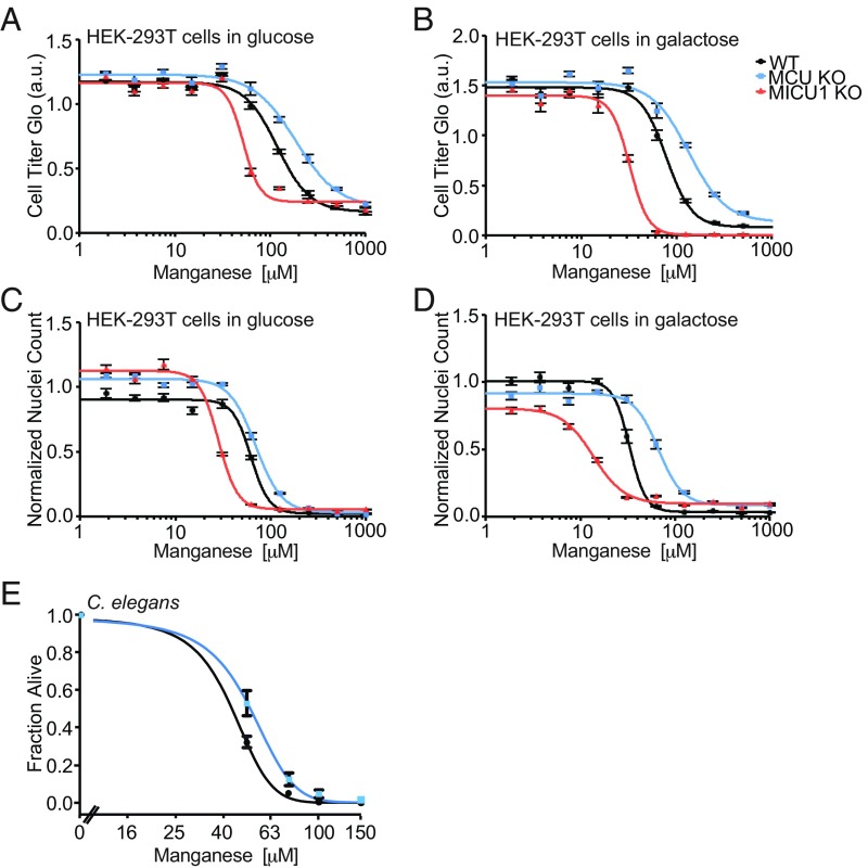
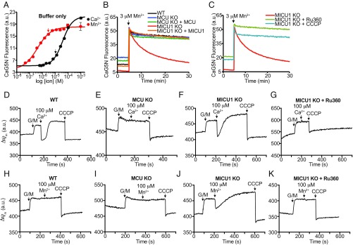
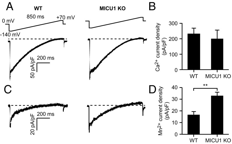
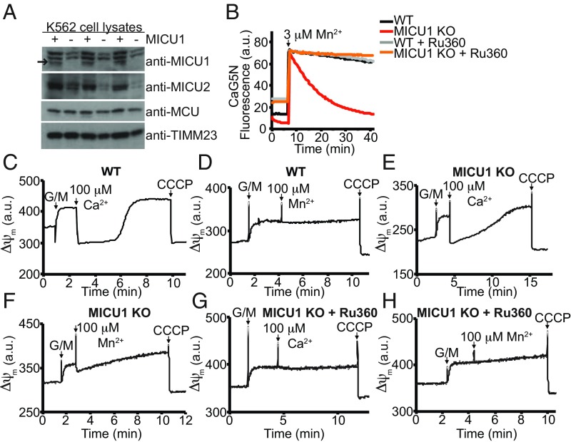
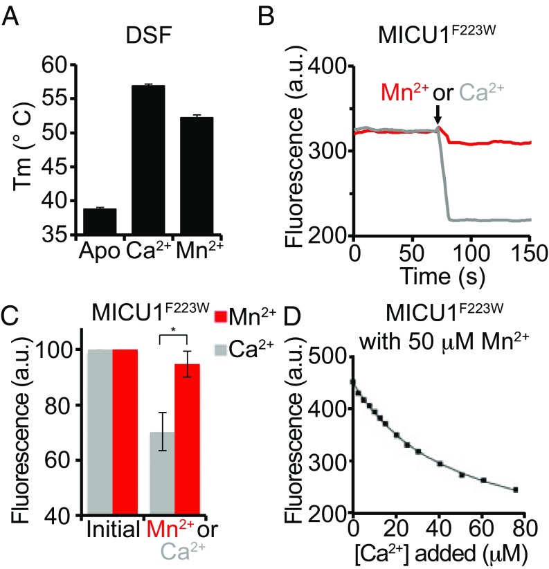
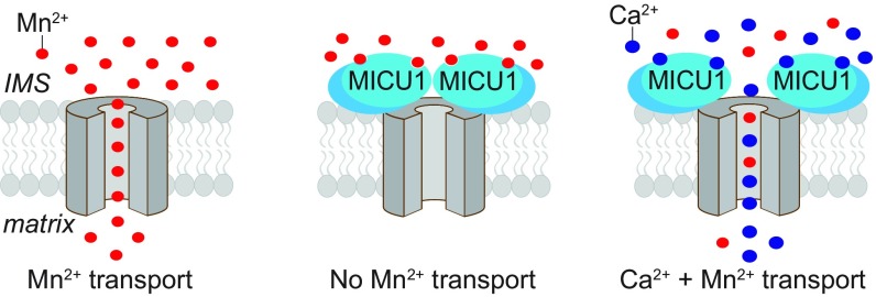
The mitochondrial uniporter is a Ca2+-activated Ca2+ channel complex that displays exceptionally high conductance and selectivity. Here, we report cellular metal toxicity screens highlighting the uniporter's role in Mn2+ toxicity. Cells lacking the pore-forming uniporter subunit, MCU, are more resistant to Mn2+ toxicity, while cells lacking the Ca2+-sensing inhibitory subunit, MICU1, are more sensitive than the wild type. Consistent with these findings, Caenorhabditis elegans lacking the uniporter's pore have increased resistance to Mn2+ toxicity. The chemical-genetic interaction between uniporter machinery and Mn2+ toxicity prompted us to hypothesize that Mn2+ can indeed be transported by the uniporter's pore, but this transport is prevented by MICU1. To this end, we demonstrate that, in the absence of MICU1, both Mn2+ and Ca2+ can pass through the uniporter, as evidenced by mitochondrial Mn2+ uptake assays, mitochondrial membrane potential measurements, and mitoplast electrophysiology. We show that Mn2+ does not elicit the conformational change in MICU1 that is physiologically elicited by Ca2+, preventing Mn2+ from inducing the pore opening. Our work showcases a mechanism by which a channel's auxiliary subunit can contribute to its apparent selectivity and, furthermore, may have implications for understanding how manganese contributes to neurodegenerative disease.

Keywords: EF hand; calcium; manganese; neurodegeneration; selectivity.


Conflict of interest statement

The authors declare no conflict of interest.

Figures

Fig. 1.
Fig. 1.
MICU1 KO HEK-293T cells are sensitized to Mn2+, whereas MCU KO cells and mcu-1 KO C. elegans are less sensitive than WT. (A) HEK-293T cells (WT, MCU KO, and MICU1 KO) were treated with a range of concentrations of MnCl2 in media containing high glucose. Cell viability was measured using CellTiter-Glo. (B) HEK-293T cells (WT, MCU KO, and MICU1 KO) were treated with a range of concentrations of MnCl2 in media lacking glucose, containing galactose. Cell viability was measured using CellTiter-Glo. (C) HEK-293T cells (WT, MCU KO, and MICU1 KO) were treated with a range of concentrations of MnCl2 in media containing high glucose. Cell viability was measured by counting nuclei. (D) HEK-293T cells (WT, MCU KO, and MICU1 KO) were treated with a range of concentrations of MnCl2 in media lacking glucose, containing galactose. Cell viability was measured by counting nuclei. (E) C. elegans young adults were treated with a range of concentrations of manganese, and the fraction living after 24 h is plotted. Data shown represent the average ± SEM of four (E) or six (AD) technical replicates.
Fig. 2.
Fig. 2.
Loss of MICU1 promotes MCU-mediated Mn2+ uptake into mitochondria in HEK-293T cells. (A) Titration curves for Calcium Green-5N (CaG5N) fluorescence with varying concentrations of Ca2+ or Mn2+. Fluorescence was measured using a PerkinElmer Envision fluorimeter. Arrows indicate 3 μM Mn2+ (red) and 12 μM Ca2+ (black) for reference. Titrations were performed using the same buffer as in subsequent ion clearance experiments, with no cells. (B and C) Cells were permeabilized with digitonin in the presence of the CaG5N fluorescent dye and given a pulse of 3 μM Mn2+. CaG5N fluorescence was monitored using a PerkinElmer LS55 fluorimeter and is reporting on relative extramitochondrial [Mn2+]. Where indicated, 1 μM CCCP was added to uncouple mitochondria or 1 μM Ru360 was added to inhibit the uniporter and were present throughout the trace. (DK) Mitochondrial membrane potential was monitored with TMRM, and the signal is inverted (such that higher signal corresponds to increased membrane potential). G/M indicates the addition of 5 mM glutamate and malate to energize the mitochondria. Pulses of Ca2+ or Mn2+ indicated are 100 μM, and CCCP indicates the addition of 1 μM CCCP to uncouple mitochondria. Where indicated, 1 μM Ru360 was present throughout the trace. Representative traces of three biological replicates are shown for each experiment.
Fig. 3.
Fig. 3.
MICU1 KO HEK-293T mitoplasts have increased Mn2+ current. (A and C) Exemplar traces of Ca2+ (A) and Mn2+ (C) currents obtained during voltage ramps from −140 to +80 mV, for WT (Left) or MICU1 KO (Right) cells. Voltage ramp protocol is shown above. Currents have been normalized for mitoplast capacitance to control for mitoplasts of different sizes. (B and D) Summary data for current density at −140 mV (n = 6–7). **P < 0.01.
Fig. 4.
Fig. 4.
Loss of MICU1 promotes MCU-mediated Mn2+ uptake into mitochondria in K562 cells. (A) Immunoblot of WT or MICU1 KO K562 whole-cell lysates is shown. MICU1 antibody shows some nonspecific bands, but the MICU1 band is indicated by the arrow. Immunoblots show three replicates, which were performed on the same gel, but prepared from independently prepared cell lysates. (B) Cells were permeabilized with digitonin in the presence of the CaG5N fluorescent dye and given a pulse of 3 μM Mn2+. CaG5N fluorescence was measured using a PerkinElmer LS55 fluorimeter and is reporting on extramitochondrial Mn2+ levels. Where indicated, 1 μM Ru360 was added to inhibit the uniporter. (CH) Mitochondrial membrane potential was monitored with TMRM, and the signal is inverted (such that higher signal corresponds to increased membrane potential). G/M indicates the addition of 5 mM glutamate and malate to energize the mitochondria. Pulses of Ca2+ or Mn2+ indicated are 100 μM, and CCCP indicates the addition of 1 μM CCCP to uncouple mitochondria. Where indicated, 1 μM Ru360 was present throughout the trace. Representative traces of three biological replicates are shown.
Fig. 5.
Fig. 5.
MICU1 binds Mn2+ but does not undergo the Ca2+-specific conformational change. (A) Differential scanning fluorimetry (DSF) of MICU1 in the presence and absence of 10 mM Ca2+ or Mn2+. Data shown represent the average ± SD of five independent biological replicates. (B and C) Intrinsic tryptophan fluorescence of purified MICU1F223W is monitored (λexc = 290 ± 5 nm; λem = 350 ± 2.5 nm) before and after the addition of 1 mM Mn2+ or Ca2+. Real-time monitoring is shown (B). The average ± SD of three independent biological replicates is shown for the initial, baseline fluorescence, and the fluorescence after addition of Mn2+ or Ca2+ (C). The large difference between the fluorescence after addition of Ca2+ vs. Mn2+ is statistically significant (P = 0.004) and indicates inability of Mn2+ to induce a conformational transition in MICU1. (D) A Ca2+/Mn2+ competition experiment was performed with MICU1F223W by titrating Ca2+ in the presence of 50 μM Mn2+ and monitoring the conformational change (as a proxy for Ca2+ binding) using intrinsic tryptophan fluorescence (λexc = 290 ± 5 nm; λem = 350 ± 2.5 nm). Fitting the data to a quadratic equation (Materials and Methods) yields KCa/KMn = 1.1.
Fig. 6.
Fig. 6.
MICU1 augments the overall ion selectivity of the uniporter. Schematic representation of the response of the uniporter pore (in gray) to Mn2+ under three conditions: Left, in the absence of MICU1; Middle, in the presence of MICU1; and Right, in the presence of both MICU1 and Ca2+. In the absence of MICU1, the uniporter pore can transport Mn2+ (with or without the presence of Ca2+). When MICU1 is present, Mn2+ binds to MICU1 but does not elicit a conformational transition, so the uniporter does not transport Mn2+. When MICU1 is present along with Ca2+, Ca2+ binds to MICU1’s EF hands, eliciting a conformational transition in MICU1, which allows both Ca2+ and Mn2+ passage through the pore. Note that it is not known how Ca2+ binding to MICU1 leads to uniporter ion transport, nor is the stoichiometry of uniporter components known. MICU1 is depicted as a heterodimer with MICU2 (in darker blue).

References

    1. Kirichok Y, Krapivinsky G, Clapham DE. The mitochondrial calcium uniporter is a highly selective ion channel. Nature. 2004;427:360–364. - PubMed
    1. Sather WA, McCleskey EW. Permeation and selectivity in calcium channels. Annu Rev Physiol. 2003;65:133–159. - PubMed
    1. Oxenoid K, et al. Architecture of the mitochondrial calcium uniporter. Nature. 2016;533:269–273. - PMC - PubMed
    1. Cao C, Wang S, Cui T, Su XC, Chou JJ. Ion and inhibitor binding of the double-ring ion selectivity filter of the mitochondrial calcium uniporter. Proc Natl Acad Sci USA. 2017;114:E2846–E2851. - PMC - PubMed
    1. Maynard LS, Cotzias GC. The partition of manganese among organs and intracellular organelles of the rat. J Biol Chem. 1955;214:489–495. - PubMed

Publication types

MeSH terms