Review
. 2018 Sep 24;200(20):e00249-18.
doi: 10.1128/JB.00249-18. Print 2018 Oct 15.

Relationship between the Viable but Nonculturable State and Antibiotic Persister Cells

Affiliations
Review

Relationship between the Viable but Nonculturable State and Antibiotic Persister Cells

Mesrop Ayrapetyan et al. J Bacteriol. .

Abstract

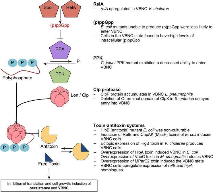
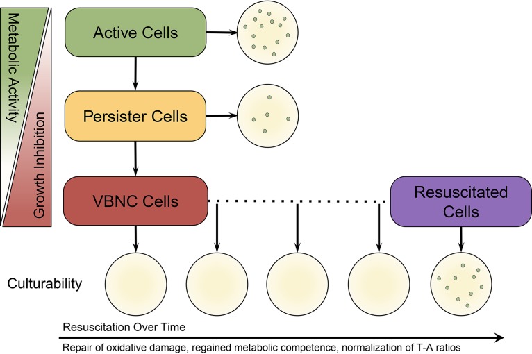
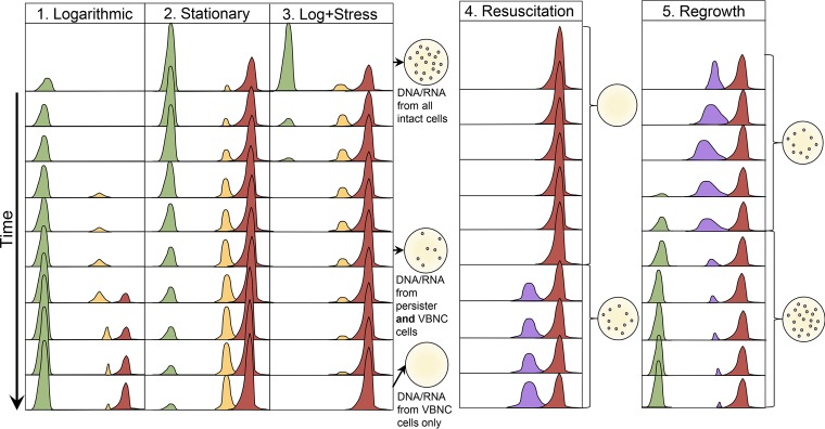
Bacteria have evolved numerous means of survival in adverse environments with dormancy, as represented by "persistence" and the "viable but nonculturable" (VBNC) state, now recognized to be common modes for such survival. VBNC cells have been defined as cells which, induced by some stress, become nonculturable on media that would normally support their growth but which can be demonstrated by various methods to be alive and capable of returning to a metabolically active and culturable state. Persister cells have been described as a population of cells which, while not being antibiotic resistant, are antibiotic tolerant. This drug-tolerant phenotype is thought to be a result of stress-induced and stochastic physiological changes as opposed to mutational events leading to true resistance. In this review, we describe these two dormancy strategies, characterize the molecular underpinnings of each state, and highlight the similarities and differences between them. We believe these survival modes represent a continuum between actively growing and dead cells, with VBNC cells being in a deeper state of dormancy than persister cells.

Keywords: VBNC; antibiotic resistance; antibiotic tolerance; dormancy; drug discovery; persistence; persister; viable but nonculturable.


Figures

FIG 1
FIG 1
Experimental dormancy dynamics of antibiotic persistence and the VBNC state. Persister cells are isolated by exposing a growing culture of cells to a lethal dose of antibiotics. The cells that remain culturable after treatment are called persisters. This typically produces the classic biphasic killing curve, with the slope of the initial phase (green dotted line) generated by the death of sensitive cells and the slope of the second phase (yellow dotted line) generated by the surviving persisters. Similarly, VBNC cells are isolated by applying a stress (e.g., cold temperature) or an antibiotic to a growing culture. Upon exposure to this stress, cells lose culturability in a varied amount of time (depending on the stress used) (blue line); however, a large portion of the population remains viable but nonculturable (red dotted line), as is determined by a variety of assays that test for gene expression, membrane stability, and metabolic activity. When the inducing stress is removed and adequate conditions are met (vertical dotted gray line), cells begin to alter their physiology toward resuscitation. After a lag period (dependent on the stress and bacterial species), cells regain the ability to grow on nutrient media.
FIG 2
FIG 2
The breadth of the VBNC state. This phylogenetic diagram was created using PhyloT (http://phylot.biobyte.de), using NCBI taxonomy. The VBNC state has been discovered in many bacteria but also in archaea and in fungi. Whether the described phenomenon is truly the same across these three domains of life has not yet been answered; however, this certainly upholds the idea that dormancy is a fundamental aspect of life on earth. Organisms written in bold have previously been shown to resuscitate from the VBNC state, while all others have either been detected in a nonculturable state or were induced but unable to resuscitate under the conditions provided. Coryn., Corynebacterium.
FIG 3
FIG 3
Mechanism of persister formation with supporting evidence of its role in the VBNC state. In this model, the activation of RelA and SpoT due to amino acid starvation leads to the intracellular accumulation of the alarmone (p)ppGpp. This leads to the inhibition of exopolyphosphatase (PPX), an enzyme that degrades polyphosphates, causing their accumulation due to continued activity of polyphosphate kinase. Lon protease is activated by polyphosphate and begins to selectively degrade proteins, including antitoxins. This disturbs toxin-antitoxin ratios and allows free toxins to inhibit cell growth, leading to persistence. It is important to note that even if this exact mechanism is not at play, each component has independently been shown to have a significant role in AP and VBNC. Modified from reference . PPK, polyphosphate kinase.
FIG 4
FIG 4
The dormancy continuum hypothesis. Stochasticity and environmental cues lead to the degradation of proteins (including antitoxins), reduction in metabolic activity, and inhibition of growth. In the very beginning stages of dormancy, antibiotic-tolerant persister cells are produced. When these persister cells are transferred to laboratory media, their metabolic competence allows them to resuscitate and grow on media. However, if the stressful conditions continue, these persister cells go deeper into dormancy and lose their culturability (i.e., become VBNC) as their metabolism further declines, protein damage continues, and they become growth arrested. When VBNC cells are transferred to media, they do not produce colonies. However, if the inducing stress is removed and adequate time and conditions are provided for VBNC, cells will resuscitate and regain their ability to grow on media. Resuscitation is the molecular process by which cells repair oxidative damage, regain metabolic competence, and normalize their toxin-antitoxin ratios. After resuscitation has occurred, cells are once again able to produce colonies. Attempts at culturing these cells any time prior to the completion of the resuscitation process will result in no growth.
FIG 5
FIG 5
Model of population dynamics as part of the dormancy continuum. Each column numbered 1 to 5 represents a different phase or treatment. Actively growing cells are represented by the green curve, persister cells are represented by the yellow curve, VBNC cells are represented by the red curve, and resuscitated cells are represented by the purple curve. Each set of curves represents a snapshot on an arbitrary time scale that proceeds downwards. 1) In logarithmic phase, persisters start to form and transition to the VBNC state. Persisters are in a transitory phase and exist at a low number, while VBNC cells represent deep dormancy and accumulate over time. 2) As stationary phase progresses, the number of transitioning persisters increase and reach a steady state with the VBNC population, while actively growing cells decline in number. 3) When logarithmic-phase cells encounter a stressful condition, some actively growing cells rapidly enter the persister state, while many susceptible cells die. Over time, with continued stress, these persisters will transition to the VBNC state. The drawings of petri plates to the right of the curves represent the culture status of the population. It is important to note that there are scenarios (pictured) when only persisters are culturable but DNA/RNA studies would be affected by genomic material from both persisters and VBNC cells. 4) Resuscitation is a variable process depending on the inducing stress and the species being studied but will eventually lead to regained culturability in a large portion of the remaining population. Even after resuscitation, some VBNC cells remain. Resuscitated cells may be in a unique physiological state and are likely antibiotic tolerant, as they are not actively dividing. 5) Regrowth occurs when resuscitated cells that have regained their ability to grow transition back to a rapidly growing phenotype when nutrients are supplied.

References

    1. Bergkessel M, Basta DW, Newman DK. 2016. The physiology of growth arrest: uniting molecular and environmental microbiology. Nat Rev Microbiol 14:549–562. doi:10.1038/nrmicro.2016.107. - DOI - PMC - PubMed
    1. Xu HS, Roberts N, Singleton FL, Attwell RW, Grimes DJ, Colwell RR. 1982. Survival and viability of nonculturable Escherichia coli and Vibrio cholerae in the estuarine and marine environment. Microb Ecol 8:313–323. doi:10.1007/BF02010671. - DOI - PubMed
    1. Kamruzzaman M, Udden SM, Cameron DE, Calderwood SB, Nair GB, Mekalanos JJ, Faruque SM. 2010. Quorum-regulated biofilms enhance the development of conditionally viable, environmental Vibrio cholerae. Proc Natl Acad Sci U S A 107:1588–1593. doi:10.1073/pnas.0913404107. - DOI - PMC - PubMed
    1. Manina G, McKinney JD. 2013. A single-cell perspective on non-growing but metabolically active (NGMA) bacteria. Curr Top Microbiol Immunol 374:135–161. doi:10.1007/82_2013_333. - DOI - PubMed
    1. Nelson EJ, Chowdhury A, Flynn J, Schild S, Bourassa L, Shao Y, LaRocque RC, Calderwood SB, Qadri F, Camilli A. 2008. Transmission of Vibrio cholerae is antagonized by lytic phage and entry into the aquatic environment. PLoS Pathog 4:e1000187. doi:10.1371/journal.ppat.1000187. - DOI - PMC - PubMed

MeSH terms

Substances

LinkOut - more resources