. 2018 Nov 16;11(11):dmm035873.
doi: 10.1242/dmm.035873.

Drosophila melanogaster as a function-based high-throughput screening model for antinephrolithiasis agents in kidney stone patients

Affiliations

Drosophila melanogaster as a function-based high-throughput screening model for antinephrolithiasis agents in kidney stone patients

Sohrab N Ali et al. Dis Model Mech. .

Abstract

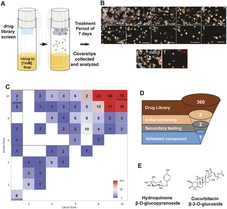
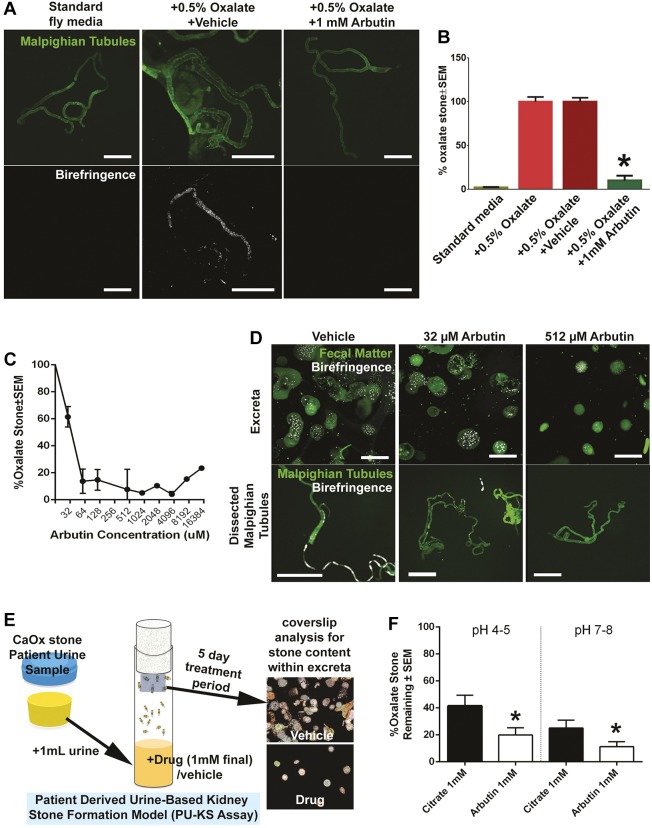
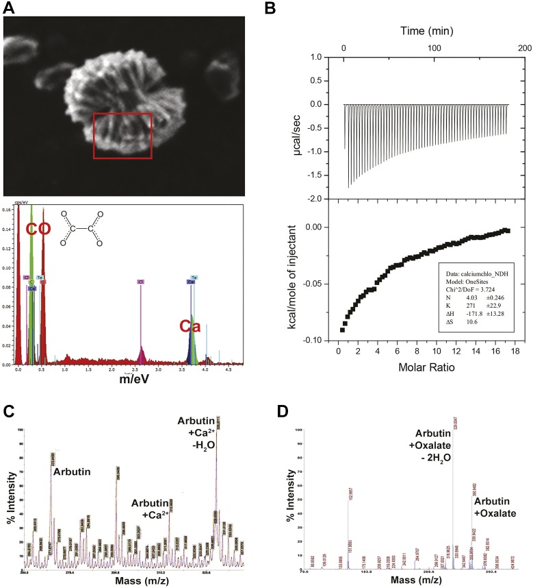
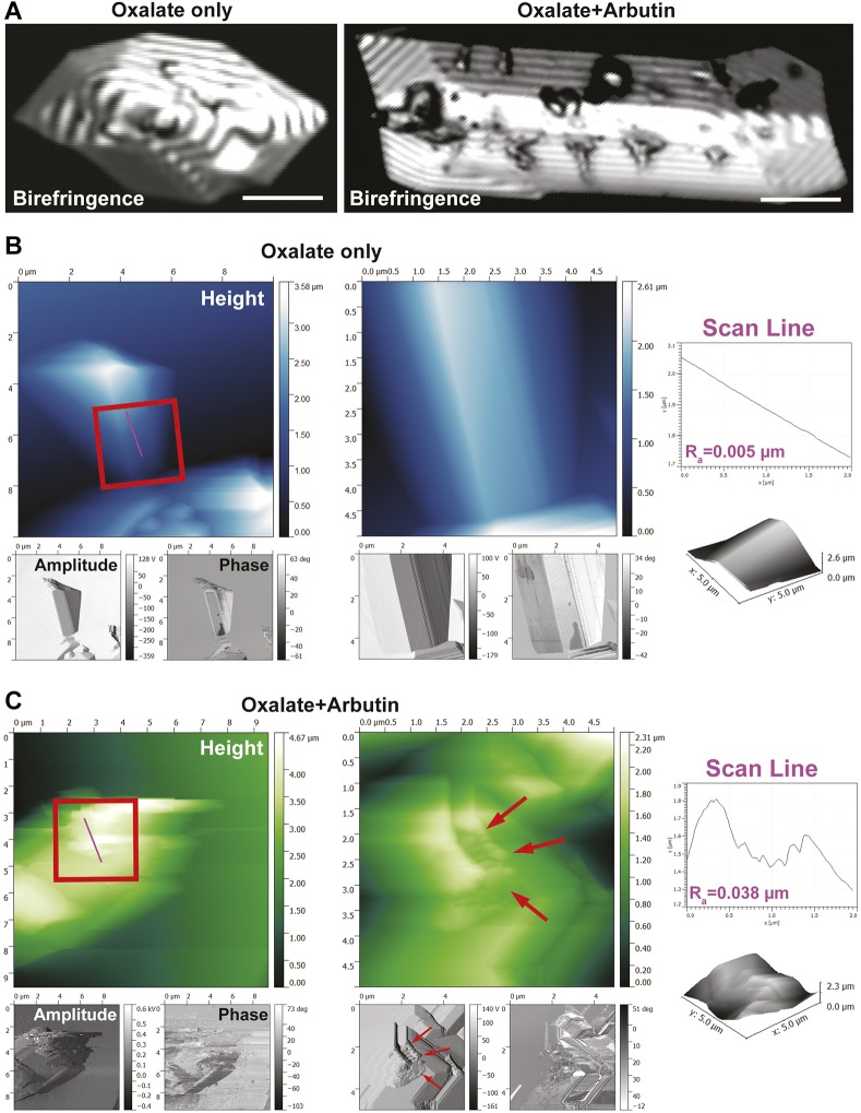
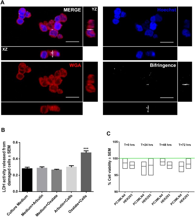
Kidney stone disease involves the aggregation of stone-forming salts consequent to solute supersaturation in urine. The development of novel therapeutic agents for this predominantly metabolic and biochemical disorder have been hampered by the lack of a practical pre-clinical model amenable to drug screening. Here, Drosophila melanogaster, an emerging model for kidney stone disease research, was adapted as a high-throughput functional drug screening platform independent of the multifactorial nature of mammalian nephrolithiasis. Through functional screening, the therapeutic potential of a novel compound commonly known as arbutin that specifically binds to oxalate, a key component of kidney calculi, was identified. Through isothermal titration calorimetry, high-performance liquid chromatography and atomic force microscopy, arbutin was determined to interact with calcium and oxalate in both free and bound states, disrupting crystal lattice structure, growth and crystallization. When used to treat patient urine samples, arbutin significantly abrogated calculus formation in vivo and outperformed potassium citrate in low pH urine conditions, owing to its oxalate-centric mode of action. The discovery of this novel antilithogenic compound via D. melanogaster, independent of a mammalian model, brings greater recognition to this platform, for which metabolic features are primary outcomes, underscoring the power of D. melanogaster as a high-throughput drug screening platform in similar disorders. This is the first description of the use of D. melanogaster as the model system for a high-throughput chemical library screen. This article has an associated First Person interview with the first authors of the paper.

Keywords: Calcium oxalate; Drosophila melanogaster; Fluorescent bisphosphonate; Intravital imaging; Nephrolithiasis.


Conflict of interest statement

Competing interestsThe authors declare no competing or financial interests.

Figures

Fig. 1.
Fig. 1.
Calcium oxalate calculi formation in D. melanogaster. (A) Confocal and birefringence images of pulverized human calcium oxalate crystals (upper row) and synthetic hydroxyapatite particles (lower row) stained with alendronate-FITC probe. Scale bars: 1 mm. (B) Schematic of D. melanogaster model of calcium oxalate calculi formation within Malpighian tubules (MTs). (C) Intravital imaging of birefringence signal representing oxalate-based calculi within MTs of D. melanogaster larvae. Intravital imaging of RFP-expressing diet-induced calcium-oxalate-stone-carrying D. melanogaster larvae. Arrow indicates an RFP+ MT with birefringent signal (C, bottom). Scale bars: 1 mm. (D) Dissected MTs reveal the presence of alendronate-FITC-positive deposits (arrows) within RFP+ MTs, confirming the presence of oxalate-based calculi. Scale bars: 25 µm. (E) Dissected MTs as imaged by brightfield or scanning electron microscopy (SEM). Dashed arrow represents fly Malpighian tubule and gut complex. Solid arrow shows an MT calculus. (F) SEM/energy-dispersive X-ray (EDX) analysis of calcium oxalate monohydrate calculi extracted from MTs. Scale bar: 3 µm. (G) SEM/EDX analysis of calcium oxalate dehydrate calculi extracted from MTs. Scale bar: 3 µm.
Fig. 2.
Fig. 2.
Calculi present in fecal excreta of Drosophila as a drug discovery platform. (A) Increasing amounts of sodium oxalate in fly medium induced kidney stone formation, present within MTs as evidenced by birefringence signal and alendronate-FITC signal. (B) D. melanogaster survival on sodium oxalate-treated fly medium over a 60-day period. (C) D. melanogaster deposit calculi-rich fecal excreta on the fly tube wall (green, autofluorescence of fecal matter) and coverslip attached to sponge lid. Flies grown in sodium oxalate-rich fly medium produce fecal excrement-containing birefringent bodies representing calculi (white, birefringence signal).
Fig. 3.
Fig. 3.
Drug screening results using fecal excreta and calculi present within fecal excreta from D. melanogaster colonies. (A) Schematic of in vivo drug library screens for antilithogenic compounds based on the calculi-fecal excreta coverslip assay. (B) Representative images of calculi or excreta density based on an arbitrary scale from 10 (highest/most dense) to 1 (lowest). A DMSO vehicle control with a score of 10 for calculi is shown. A representative ‘hit’ with a score of 1 for calculi is shown. Scale bars: 4 mm. (C) Drug screening results from a chemical library representing 360 naturally occurring compounds. ‘Hits’ were defined as coverslips that yielded a ‘1’ score for calculi deposition with no toxic impact on D. melanogaster viability (5-10 in terms of fecal excreta score, green gate). (D,E) A second focused library screen with the eight hits (D) yielded a final list of two active compounds (E). After tertiary analysis of the two active compounds, one was validated, arbutin.
Fig. 4.
Fig. 4.
Arbutin and its antilithogenic effects on oxalate-based calculi. (A) Polarized microscopy of MTs treated with arbutin compared with standard medium and oxalate-supplemented fly medium. (B) Percentage of fecal excreta area that contains birefringence signal/calculi when flies are grown in various fly medium treatments, including arbutin. *P<0.01 with two-way ANOVA, Scheffe α correction. (C) Dose-dependent inhibition of oxalate-based fecal excreta deposited by Drosophila with various concentrations of arbutin. (D) Microscopy of coverslips deposited with calculi-rich fecal excreta and dissected MTs when fly medium is supplemented with 0, 32 and 512 µM arbutin. (E) Schematic of patient urine-based kidney stone in vivo formation assay. Patient urine samples are added to fly media with and no other supplementation. Coverslips containing calculi-rich fecal excreta are analyzed via microscopy. Vehicle image for coverslip analysis is DMSO sample reproduced from Fig. 3B; drug image is from the image labeled 2 in Fig. 3B. (F) The patient urine-based kidney stone in vivo formation assay was used to compare potassium citrate with arbutin at various pHs. Coverslips containing calculi-containing fecal excreta were analyzed by birefringence microscopy. *P<0.05, two-way ANOVA, Scheffe α correction.
Fig. 5.
Fig. 5.
Arbutin and its interactions with calcium and oxalate. (A) SEM image of arbutin complexed with calcium. EDX spectra reveal four calcium ions for every molecule of oxalate. (B) Isothermal titration calorimetric analysis of arbutin and calcium chloride, revealing a molar ratio of four calcium ions for each arbutin molecule. (C) Matrix-assisted laser desorption/ionization (MALDI) spectrum of calcium and arbutin complexes formed in solution. (D) MALDI spectrum of arbutin and oxalate complexes formed in solution.
Fig. 6.
Fig. 6.
Calcium oxalate and arbutin crystal structure interaction analysis. (A) Confocal birefringence images of pure oxalate crystals prior to arbutin exposure (left), and following exposure to arbutin (right). Scale bars: 10 µm. (B) Atomic force microscopy (AFM) image of pure oxalate crystals. Insets in the middle panel provide higher magnification views of the crystal surface. Scan line analysis of the height channel within the inset reveals a smooth topography. (C) Oxalate crystals exposed to arbutin reveal a highly active surface topography decorated with arbutin drug molecules (red arrows). Insets in the left panel reveal a rough surface topography, as shown by scan line analysis of the height channel.
Fig. 7.
Fig. 7.
Cytotoxicity of arbutin and oxalate on human kidney epithelial cells. (A) Confocal fluorescence images of HEK293 cells stained with CellTracker Red to label the cell surface and Hoechst to label the nuclei. HEK293 cells were treated with 20 µM sodium oxalate for 30 min. Birefringence signal was observed within HEK293 cells (white signal). Scale bars: 25 µm. (B) Activity of LDH released from HEK293 cells after 30 min incubation with oxalate in the presence or absence of arbutin. (C) Cell viability in arbutin-treated HEK293 and PC3MLN4 cells over a 3-day period. Green line represents control normalized to 100% at that same timepoint. ***P<0.05, two-way ANOVA, Scheffe α correction.

References

    1. Balcke P., Schmidt P., Zazgornik J., Kopsa H. and Minar E. (1983). Pyridoxine therapy in patients with renal calcium oxalate calculi. Proc. Eur. Dial. Transplant Assoc. 20, 417-421. - PubMed
    1. Chi T., Kim M. S., Lang S., Bose N., Kahn A., Flechner L., Blaschko S. D., Zee T., Muteliefu G., Bond N. et al. (2015). A Drosophila model identifies a critical role for zinc in mineralization for kidney stone disease. PLoS ONE 10, e0124150 10.1371/journal.pone.0124150 - DOI - PMC - PubMed
    1. Cognigni P., Bailey A. P. and Miguel-Aliaga I. (2011). Enteric neurons and systemic signals couple nutritional and reproductive status with intestinal homeostasis. Cell Metab. 13, 92-104. 10.1016/j.cmet.2010.12.010 - DOI - PMC - PubMed
    1. Cole L. E., Vargo-Gogola T. and Roeder R. K. (2014). Bisphosphonate-Functionalized Gold Nanoparticles for Contrast-Enhanced X-Ray Detection of Breast Microcalcifications. Biomaterials 35, 2312 -2321. 10.1016/j.biomaterials.2013.11.077 - DOI - PMC - PubMed
    1. Deisinger P. J., Hill T. S. and English J. C. (1996). Human exposure to naturally occurring hydroquinone. J. Toxicol. Environ. Health 47, 31-46. 10.1080/009841096161915 - DOI - PubMed

Publication types