. 2018 Aug 6;8(1):11718.
doi: 10.1038/s41598-018-30226-x.

Mechanical ventilation and Streptococcus pneumoniae pneumonia alter mitochondrial homeostasis

Affiliations

Mechanical ventilation and Streptococcus pneumoniae pneumonia alter mitochondrial homeostasis

Mathieu Blot et al. Sci Rep. .

Abstract

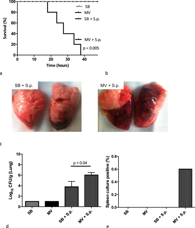
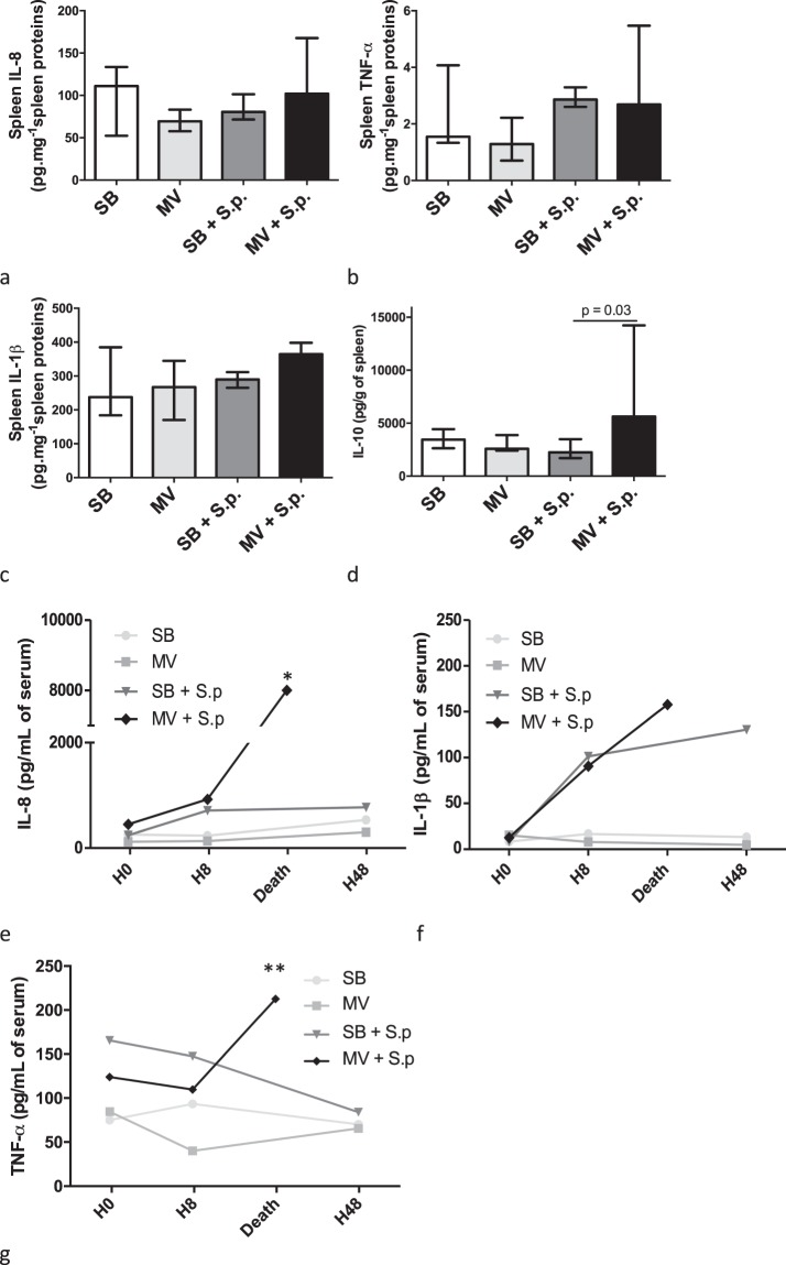
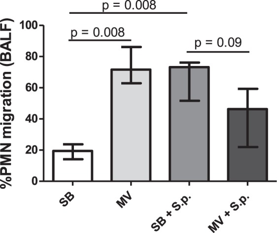
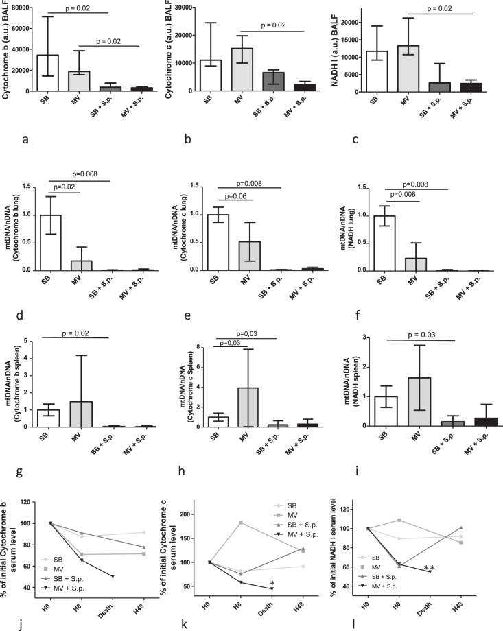
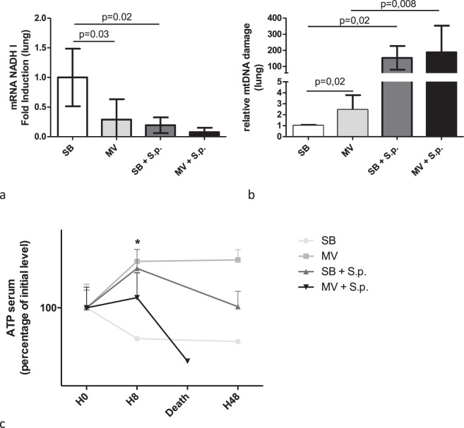
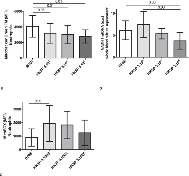
Required mechanical ventilation (MV) may contribute to bacterial dissemination in patients with Streptococcus pneumoniae pneumonia. Significant variations in plasma mitochondrial DNA (mtDNA) have been reported in sepsis according to the outcome. The impact of lung stretch during MV was addressed in a model of pneumonia. Healthy or S. pneumoniae infected rabbits were submitted to MV or kept spontaneously breathing (SB). Bacterial burden, cytokines release, mitochondrial DNA levels, integrity and transcription were assessed along with 48-hour mortality. Compared with infected SB rabbits, MV rabbits developed more severe pneumonia with greater concentrations of bacteria in the lungs, higher rates of systemic dissemination, higher levels of circulating inflammatory mediators and decreased survival. Pulmonary mtDNA levels were significantly lower in infected animals as compared to non-infected ones, whenever they were SB or MV. After a significant early drop, circulating mtDNA levels returned to baseline values in the infected SB rabbits, but remained low until death in the MV ones. Whole blood ex-vivo stimulation with Streptococcus pneumoniae resulted in a reduction of polymorphonuclear leukocytes mitochondrial density and plasma mtDNA concentrations. Thus, persistent mitochondrial depletion and dysfunction in the infected animals submitted to MV could account for their less efficient immune response against S. pneumoniae.


Conflict of interest statement

The authors declare no competing interests.

Figures

Figure 1
Figure 1
Main features of Streptococcus pneumoniae pneumonia in either spontaneously breathing or mechanically ventilated rabbits. (a) Time-dependent probability of survival (log rank test, p = 0.005); (b) Lung injury assessment according to macroscopic score calculation; (c) Lung pictures after pneumonia in either SB (left) or MV (right) animals; (d) pulmonary bacterial concentrations (Log10 CFU per gram of lung); (e) Pulmonary-to-systemic bacterial translocation expressed as the spleen positive culture rate 48 hours after bacteria instillation (or at the time of death if earlier). CFU: colony forming unit; MV: mechanical ventilation, SB: spontaneously breathing, S.p.: Streptococcus pneumoniae, IQR: interquartile range.
Figure 2
Figure 2
Inflammatory cytokine concentrations and gene expression in the lung tissue of spontaneously breathing or mechanically ventilated rabbits with or without Streptococcus pneumoniae pneumonia. Median (IQR) inflammatory cytokine concentrations (IL-8 [a], IL-1β [b] and TNF-α [c]) in lung homogenates obtained 48 hours (or at the time of death if earlier) after tracheal instillation of saline (controls) or 5.108 CFU of Streptococcus pneumoniae in either spontaneously breathing or mechanically ventilated rabbits. Relative mRNA copies number of IL-8, IL-1β and TNF-α genes within the lung were measured by the reverse transcriptase-polymerase chain reaction. All values are shown as the fold increase compared with uninfected spontaneous breathing rabbits - value set to 1. The Kruskall Wallis test and the Mann-Whitney U test were used successively for all intergroup comparisons and followed by post hoc corrections for multiple comparisons using the Bonferroni method. CFU: colony forming unit; SB: spontaneously breathing, MV: mechanical ventilation, S.p.: Streptococcus pneumoniae, IL: interleukin, TNF: tumor necrosis factor, mRNA: messenger ribonucleic acid, BALF: broncho-alveolar lavage fluid; IQR: interquartile range.
Figure 3
Figure 3
Inflammatory cytokine concentrations and gene expression in the systemic compartment of spontaneously breathing or mechanically ventilated rabbits with or without Streptococcus pneumoniae pneumonia. Median (IQR) inflammatory cytokine concentrations (IL-8 [A], IL-1β [B] and TNF-α [C]) in spleen homogenates, 48 hours (or at the time of death if earlier) after tracheal instillation of saline (controls) or 5.108 CFU of Streptococcus pneumoniae in spontaneously breathing or mechanically ventilated rabbits. Relative mRNA copies numbers of IL-8, IL-1β and TNF-α genes within the spleen were measured by the reverse transcriptase-polymerase chain reaction. All values are shown as the fold increase compared with uninfected spontaneous breathing rabbits - value set to 1. Median plasma concentrations of inflammatory cytokines (IL-8 [e], IL-1β [f] and TNF-α [g]) were measured by ELISA at baseline, 8 or 48 hours (or at the time of death if earlier) after challenge. The Kruskall Wallis test and the Mann-Whitney U test were used successively for all intergroup comparisons and followed by post hoc corrections for multiple comparisons using the Bonferroni method.* and ** denote p < 0.05 between infected SB and MV rabbits at H48 or time of death, respectively. CFU: colony forming unit, SB: spontaneously breathing, MV: mechanical ventilation, S.p.: Streptococcus pneumoniae, IL: interleukin, TNF: tumor necrosis factor, mRNA: messenger ribonucleic acid, BALF: broncho-alveolar lavage fluid; IQR: Interquartile Range.
Figure 4
Figure 4
Polymorphonuclear leukocytes chemotaxis induced by broncho-alveolar lavage fluid in spontaneously breathing or mechanically ventilated rabbits with or without Streptococcus pneumoniae pneumonia. PMN chemotaxis was measured using a Boyden chamber, 48 hours (or at the time of death if earlier) after tracheal instillation of saline (controls) or 5.108 CFU of Streptococcus pneumoniae in either spontaneously breathing or mechanically ventilated rabbits. Results are expressed as the mean (SD) % of PMNs from healthy volunteers attracted by rabbit BALF. The Kruskall Wallis test and the Mann-Whitney U test were used successively for all intergroup comparisons and followed by post hoc corrections for multiple comparisons using the Bonferroni method. SB: spontaneous breathing, MV: mechanical ventilation, BALF: broncho-alveolar lavage fluid, IL: interleukin, PMN: polymorphonuclear leukocytes, S.p.: Streptococcus pneumoniae, SD: standard deviation.
Figure 5
Figure 5
Mitochondrial DNA levels in broncho-alveolar lavage fluid, lung tissue, spleen and plasma in spontaneously breathing or mechanically ventilated rabbits with or without Streptococcus pneumoniae pneumonia. Median (IQR) mitochondrial DNA levels (Cytochrome b [a], Cytochrome c [b] and NADH I [c]) were measured in BALF, 48 hours (or at the time of death if earlier) after tracheal instillation of saline (controls) or 5.108 CFU of Streptococcus pneumoniae in spontaneously breathing rabbits or mechanically ventilated rabbits. Mitochondrial DNA levels were also measured in the lung (Cytochrome b [d], Cytochrome c [e] and NADH I [f]) and in the spleen (Cytochrome b [g], Cytochrome c [h] and NADH I [i]). Plasma concentrations (Cytochrome b [j], Cytochrome c [k] and NADH I [l]) were measured at baseline, 8 or 48 hours (or at the time of death if earlier) after saline or bacterial challenge. Results are expressed as percentage of baseline level. The Kruskall Wallis test and the Mann-Whitney U test were used successively for all intergroup comparisons and followed by post hoc corrections for multiple comparisons using the Bonferroni method. * denotes p = 0.05 and ** denotes p = 0.08 between infected SB and MV rabbits at H48 or time of death, respectively. SB: spontaneous breathing, MV: mechanical ventilation, BALF: broncho-alveolar lavage fluid, NADH: Nicotinamide adenine dinucleotide, PMN: polymorphonuclear leukocytes, S.p.: Streptococcus pneumonia, IQR: interquartile range.
Figure 6
Figure 6
Pulmonary mitochondrial gene transcription, ATP plasma level and deleted mitochondrial DNA proportion in spontaneously breathing or mechanically ventilated rabbits with or without Streptococcus pneumoniae pneumonia. Pulmonary mitochondrial NADH-I mRNA was measured by reverse transcriptase-PCR (RT-qPCR), and normalized to GAPDH mRNA, 48 hours (or at the time of death if earlier) after tracheal instillation of saline (controls) or 5.108 CFU of Streptococcus pneumoniae in spontaneously breathing or mechanically ventilated rabbits [a]. Pulmonary mtDNA damage was assessed by quantifying the 4977bp deletion in mtDNA by PCR, and normalized to the mitochondrial NADH I gene [b]. The level of plasma ATP was measured at baseline, 8 or 48 hours (or at the time of death if earlier) after challenge [c]. The Kruskall Wallis test and the Mann-Whitney U test were used successively for all intergroup comparisons and followed by post hoc corrections for multiple comparisons using the Bonferroni method.ATP: adenosine triphosphate; SB: spontaneous breathing, MV: mechanical ventilation, RNA: ribonucleic acid, GAPDH: glyceraldehyde-3-phosphate dehydrogenase, NADH: Nicotinamide adenine dinucleotide, PCR: polymerase chain reaction, S.p.: Streptococcus pneumoniae.
Figure 7
Figure 7
Mitochondrial homeostasis disturbance in whole blood stimulated ex vivo by Heat Killed Streptococcus pneumoniae (HKSP). (a) Neutrophil mitochondrial density assessed by flow cytometry with Mitotracker Green FM, and expressed as a median fluorescence intensity (MFI), in whole blood stimulated 16 hours with different concentrations of HKSP (5×10³, 5×10⁴, 5×10⁵ bacteria/mL) or unstimulated (RPMI). Experiments were performed eight times. Data represent mean ± SD, n=8 per condition. Normality distribution was verified with D’Agostino-Pearson normality test and t-test was used for all intergroup comparisons. A statistically significant correlation was demonstrated by the Spearmann test (r = −0.34; p = 0.03). (b) Mitochondrial DNA NADH I level in cell culture supernatant after whole blood stimulation by HKSP. t-test was used for all intergroup comparisons. A statistically significant correlation was demonstrated by the Spearmann test (r = −0.34; p = 0.03). (c) Mitochondrial ROS assessed by flow cytometry with MitoSOX expressed as a median fluorescence intensity (MFI) in the experimental conditions described above. Data represent mean ± SD, n=8 per condition. t-test was used for all intergroup comparisons.DNA: deoxyribonucleic acid, HKSP: Heat Killed Streptococcus pneumoniae, MFI: Median fluorescence intensity, NADH: nicotinamide adenine dinucleotide dehydrogenase, RPMI: Roswell Park Memorial Institute medium, SD: standard deviation.
Figure 8
Figure 8
Lung mitochondrial biogenesis evaluation in spontaneously breathing or mechanically ventilated rabbits with or without Streptococcus pneumoniae pneumonia. Relative mRNA copy numbers of mitochondrial biogenesis genes (PGC1-α and TFAM) within the lung were measured by the reverse transcriptase-polymerase chain reaction. All values are shown as the fold increase compared with uninfected spontaneous breathing rabbits - value set to 1. The Kruskall Wallis test and the Mann-Whitney U test were used successively for all intergroup comparisons and followed by post hoc corrections for multiple comparisons using the Bonferroni method.

Similar articles

Cited by

References

    1. Mandell LA, et al. Infectious Diseases Society of America/American Thoracic Society consensus guidelines on the management of community-acquired pneumonia in adults. Clin Infect Dis. 2007;44(Suppl 2):S27–72. doi: 10.1086/511159. - DOI - PMC - PubMed
    1. Mongardon N, et al. Epidemiology and outcome of severe pneumococcal pneumonia admitted to intensive care unit: a multicenter study. Crit Care. 2012;16:R155. doi: 10.1186/cc11471. - DOI - PMC - PubMed
    1. Blot M, et al. A leukocyte score to improve clinical outcome predictions in bacteremic pneumococcal pneumonia in adults. Open Forum Infect Dis. 2014;1:ofu075. doi: 10.1093/ofid/ofu075. - DOI - PMC - PubMed
    1. Slutsky AS, Ranieri VM. Ventilator-induced lung injury. N Engl J Med. 2013;369:2126–2136. doi: 10.1056/NEJMra1208707. - DOI - PubMed
    1. Pugin J, Ricou B, Steinberg KP, Suter PM, Martin TR. Proinflammatory activity in bronchoalveolar lavage fluids from patients with ARDS, a prominent role for interleukin-1. Am J Respir Crit Care Med. 1996;153:1850–1856. doi: 10.1164/ajrccm.153.6.8665045. - DOI - PubMed

Publication types

MeSH terms