. 2019 Jan;38(1):33-46.
doi: 10.1038/s41388-018-0430-x. Epub 2018 Aug 6.

FGF18, a prominent player in FGF signaling, promotes gastric tumorigenesis through autocrine manner and is negatively regulated by miR-590-5p

Affiliations

FGF18, a prominent player in FGF signaling, promotes gastric tumorigenesis through autocrine manner and is negatively regulated by miR-590-5p

Jinglin Zhang et al. Oncogene. 2019 Jan.

Abstract

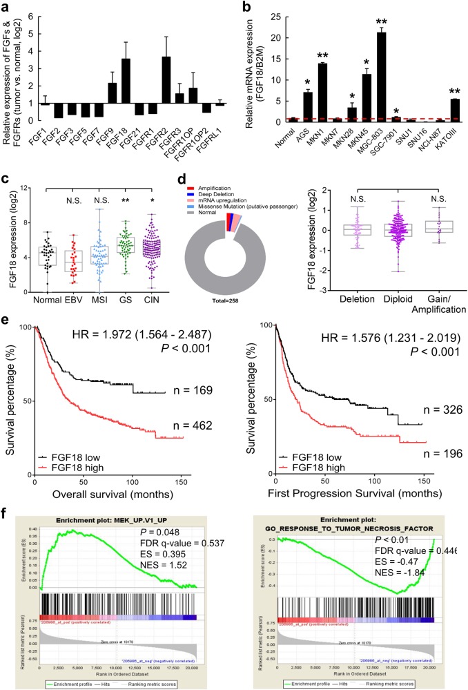
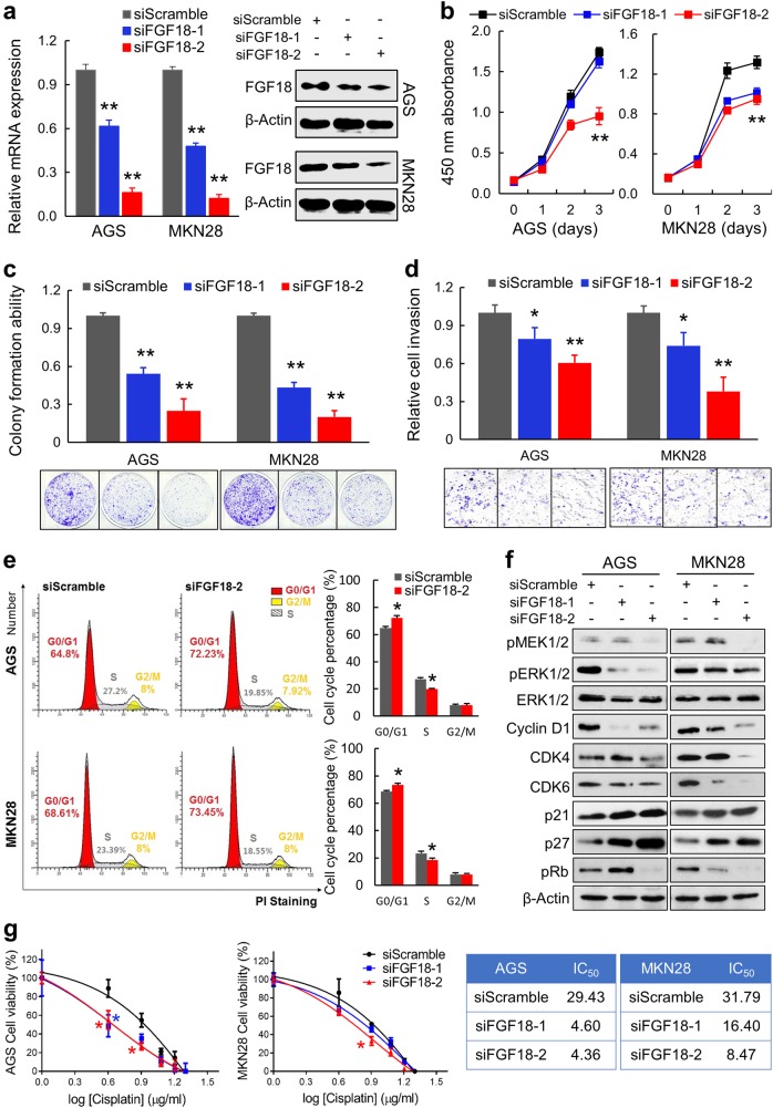
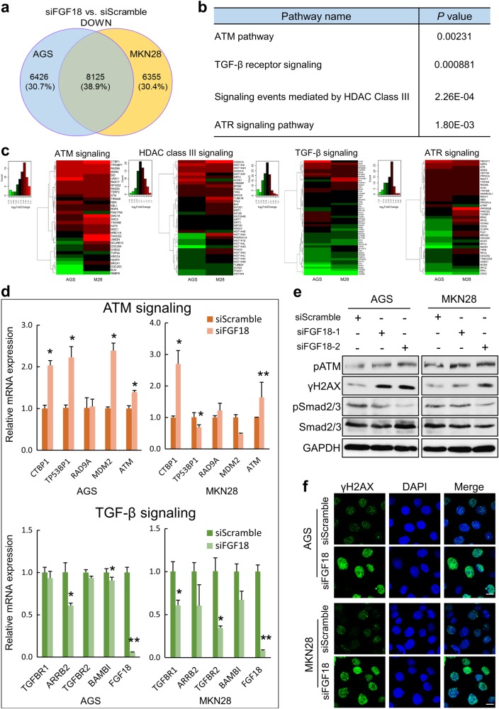
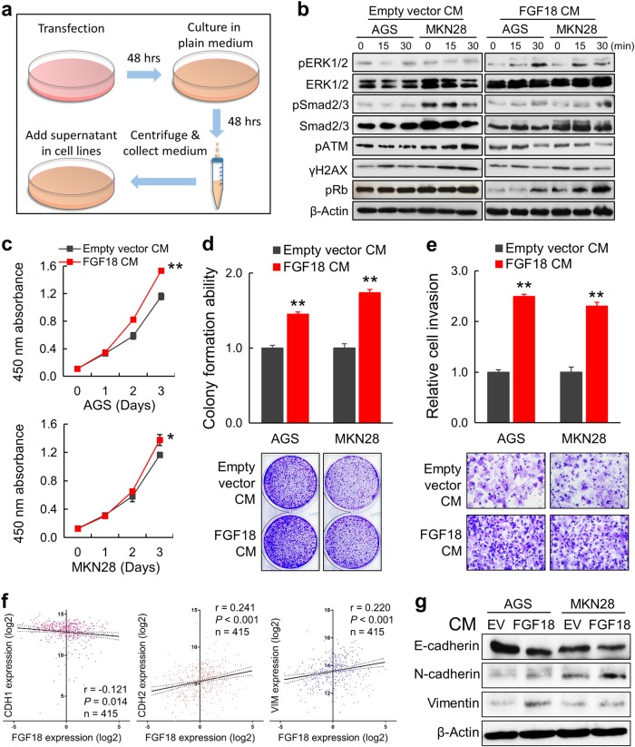
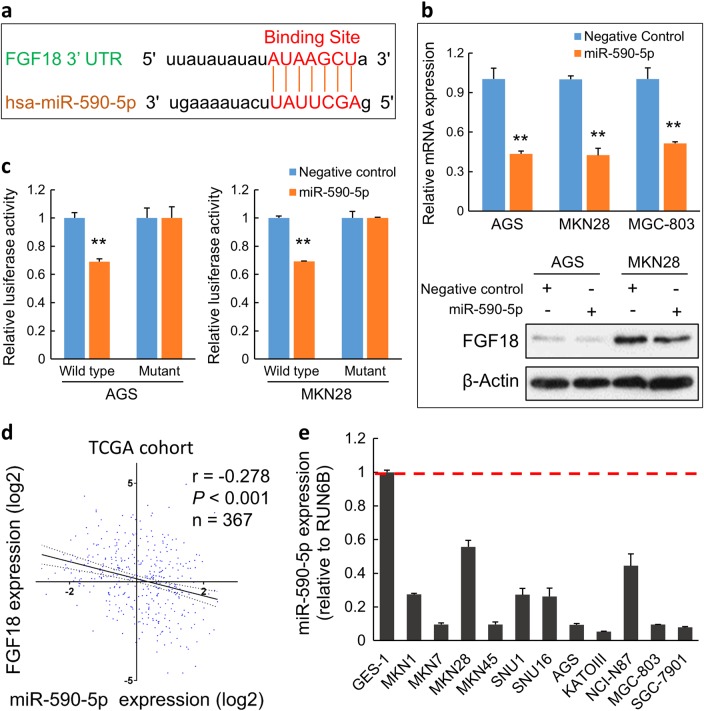
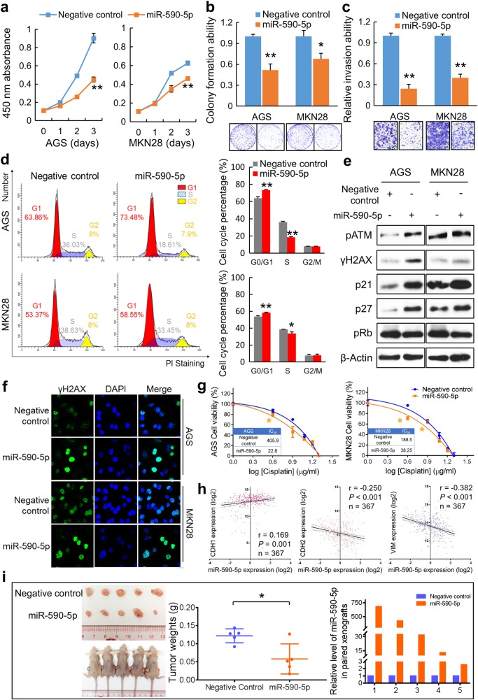
Fibroblast growth factors (FGFs) and their receptors are significant components during fundamental cellular processes. FGF18 plays a distinctive role in modulating the activity of both tumor cells and tumor microenvironment. This study aims to comprehensively investigate the expression and functional role of FGF18 in gastric cancer (GC) and elucidate its regulatory mechanisms. The upregulation of FGF18 was detected in seven out of eleven (63.6%) GC cell lines. In primary GC samples, FGF18 was overexpressed in genomically stable and chromosomal instability subtypes of GC and its overexpression was associated with poor survival. Knocking down FGF18 inhibited tumor formation abilities, induced G1 phase cell cycle arrest and enhanced anti-cancer drug sensitivity. Expression microarray profiling revealed that silencing of FGF18 activated ATM pathway but quenched TGF-β pathway. The key factors that altered in the related signaling were validated by western blot and immunofluorescence. Meanwhile, treating GC cells with human recombinant FGF18 or FGF18-conditioned medium accelerated tumor growth through activation of ERK-MAPK signaling. FGF18 was further confirmed to be a direct target of tumor suppressor, miR-590-5p. Their expressions showed a negative correlation in primary GC samples and more importantly, re-overexpression of FGF18 partly abolished the tumor-suppressive effect of miR-590-5p. Our study not only identified that FGF18 serves as a novel prognostic marker and a therapeutic target in GC but also enriched the knowledge of FGF-FGFR signaling during gastric tumorigenesis.


Conflict of interest statement

The authors declare that they have no conflict of interest.

Figures

Fig. 1
Fig. 1
FGF18 shows overabundance in GC. a FGF18 has the highest expression level in FGFs and FGFRs among GC cell lines. b FGF18 is overexpressed in seven out of eleven GC cell lines (*, P < 0.05; **, P < 0.001). Dash line indicated the normalized expression of FGF18 in GES-1. c Expression pattern of FGF18 based on molecular classification. EBV EBV-positive, MSI microsatellite unstable, GS genomically stable, CIN chromosomal instability (N.S. not significant; *, P < 0.05; **, P < 0.001). d Left panel: Different types of FGF18 genetic alterations in primary GC samples (n = 258). Right panel: the correlation of FGF18 mRNA expression with its copy number aberrations (N.S. not significant). e Upregulation of FGF18 indicated worse outcomes (P < 0.001) based on the Kaplan Meier plotter (www.kmplot.com) analysis. f Enrichment plots of gene expression signatures according to FGF18 expression levels in a breast cancer cohort (NCBI/GEO/GSE57303; left panel: MEK signaling, P = 0.048; right panel: tumor necrosis factor signaling, P< 0.01). The barcode plot indicated the position of the genes in each gene set; red and blue colors represented the high and low expression of FGF18, respectively
Fig. 2
Fig. 2
Silencing of FGF18 in GC cells displays anti-tumor function in vitro. a Transfection of siFGF18s significantly reduced both the mRNA and protein levels of FGF18 (**, P < 0.001). b Three-day cell proliferation assays presented that siFGF18-transfection significantly suppressed proliferation in GC cells (**, P < 0.001). The mean and SDs obtained from six wells were plotted. c Monolayer colony formation assays suggested that siFGF18-transfection inhibited anchorage-dependent colony formation ability (**, P < 0.001). Assays were performed in triplicate. Error bars represented SDs. d siFGF18-transfected cells showed retarded cell invasion (*, P < 0.05; **, P < 0.001). Vision fields were randomly picked for thrice, from which the SDs were achieved. e Cell cycle distribution examined by flow cytometry indicated G1 arrest in siFGF18-transfected cells. Experiments were performed in triplicate. Statistical analysis of cell cycle percentages was presented by histograms (*, P < 0.05). f Western blot analysis demonstrated the protein levels of cell cycle regulators, as well as the phosphorylated MEK1/2, ERK1/2 after FGF18 silencing. g Drug sensitivity was enhanced by treating cells with siFGF18s (*, P < 0.05). The cell viability upon different concentrations of Cisplatin was detected after 48 h by CCK8 cell proliferation assay. The mean and SDs were obtained from 6 wells. The largest mean was defined as 100% and the smallest mean was defined as 0%. IC50 values were calculated and listed in the right panel
Fig. 3
Fig. 3
FGF18 crosstalks with ATM and TGF-β pathways. a Selection of downregulated genes in both siFGF18-treated cell lines. b The genes downregulated in both cell lines with FGF18 knockdown significantly enriched in four signaling pathways. c The heat maps demonstrated the differentially expressed genes in these four signaling pathways respectively. d High-ranked upregulated genes in ATM signaling pathway and downregulated genes in TGF-β signaling pathway were validated by qRT-PCR (*, P < 0.05; **, P < 0.001). e Western blot analysis indicated that ATM and histone H2AX were activated, while phosphorylation of Smad2 and Smad3 was reduced due to FGF18 knockdown. f Immunofluorescent staining validated that γH2AX was significantly increased in cells with FGF18 knockdown
Fig. 4
Fig. 4
FGF18-conditioned medium (CM) enhances tumor growth of GC cells. a Schematic diagram for the CM preparation and cell treatment. b pERK1/2, pSMAD2/3, and pRb were activated by FGF18-CM, while ATM cascade was inactivated by FGF18-CM treatment. All changes were time-dependent. Cells under empty vector (EV)-CM treatment were applied as control. c Three-day cell proliferation assays indicated that FGF18-CM significantly increased proliferation of the GC cells (*, P < 0.05; **, P < 0.001). The mean and SDs obtained from six wells were plotted. d FGF18-CM enhanced the anchorage-dependent colony formation ability (**, P < 0.001). Assays were conducted in triplicate independently. Error bars represent SDs. e Treating with FGF18-CM accelerated cell invasion ability (**, P < 0.001). SDs were achieved from the cell number in each random vision field. f Correlation between FGF18 and related EMT markers based on the TCGA data. g Immunoblotting of EMT markers in the cells treated with CM for 48 h (empty vector and FGF18)
Fig. 5
Fig. 5
FGF18 is directly regulated by miR-590-5p in GC. a The putative binding site of miR-590-5p was located in the FGF18 3′ UTR according to miRNA database miRDB. b Both mRNA and protein expression of FGF18 were decreased after miR-590-5p overexpression in AGS and MKN28 cells (**, P < 0.001). c miR-590-5p suppressed the luciferase activity in the constructs containing the wild-type binding site in 3′ UTR of FGF18 (**, P < 0.001), but without effect in the constructs containing a corresponding mutated binding site. d The expression association between FGF18 mRNA and miR-590-5p in TCGA cohort. e The expression pattern of miR-590-5p in 11 GC cell lines and an immortalized gastric epithelium cell line GES-1. All GC cell lines showed a uniform decrease of miR-590-5p. Dash line indicated the normalized expression of miR-590-5p in GES-1
Fig. 6
Fig. 6
miR-590-5p functions as a tumor suppressor in GC. a Ectopic transfection of miR-590-5p significantly suppressed proliferation in GC cell lines (**, P< 0.001). The mean and SDs obtained from six wells were plotted. b miR-590-5p transfection significantly inhibited anchorage-dependent colony formation ability (**, P < 0.001). Experiments were performed in triplicate. Error bars represent SDs. c Cell invasion ability was suppressed by miR-590-5p significantly (*, P < 0.05; **, P < 0.001). SDs were achieved from visions randomly selected. d Cell cycle distribution was examined by flow cytometry, which suggested G1 phase arrest in miR-590-5p-transfected cells. Experiments were conducted in triplicate. Statistical analysis of cell cycle percentages were presented by histograms (*, P < 0.05; **, P < 0.001). e Western blot analysis demonstrated the increased level of p21, p27, and reduction of pRb. Key factors in ATM signaling were activated in the miR-590-5p transfectants. f Immunofluorescence showed that γH2AX was significantly increased in GC cells with ectopic miR-590-5p expression. g Drug sensitivity was enhanced by miR-590-5p (*, P < 0.05). The cell viability was detected with different concentrations of Cisplatin. The mean and SDs were obtained from six wells. The largest mean was defined as 100% and the smallest mean defined as 0%. IC50 values were calculated and listed in tables. h The expression correlation of miR-590-5p and related EMT markers in TCGA dataset. i The xenograft formation ability with stable miR-590-5p abundance was significantly inhibited compared with the negative control (*, P < 0.05). Black circles indicate the negative controls and red circles show the xenografts derived from miR-590-5p-transfected cells
Fig. 7
Fig. 7
Re-overexpression diminished the suppressive effects of miR-590-5p in GC. a FGF18 mRNA was restored in FGF18 re-overexpressed AGS and MKN28 cells (*, P < 0.05; **, P < 0.001). b FGF18 re-overexpression promoted growth in the miR-590-5p treated cells (**, Negative control + Empty vector vs. miR-590-5p + Empty vector, P < 0.001; ##, miR-590-5p + Empty vector vs. miR-590-5p + FGF18, P < 0.001). c Monolayer colony formation ability of AGS and MKN28 cells, which were transfected with miR-590-5p, were rescued by FGF18 re-overexpression (**, P < 0.001). d The invasive ability was significantly raised in FGF18 re-overexpressed cells compared with miR-590-5p treated cells (**, P < 0.001). SDs were achieved from triplicate experiments. e FGF18 re-overexpression in MGC-803 cells formed bigger xenografts compared with miR-590-5p transfection group (**, P < 0.001). f Schematic figure summarized all the study. In normal gastric epithelium cells, miR-590-5p suppresses FGF18 expression and blocks FGF18 signaling. The ATM signaling is normally activated, which in turn induces DNA damage repair, cell cycle arrest and apoptosis. In GC cells, silenced miR-590-5p fails to inhibit FGF18 expression. Thus the activated FGF18 axis triggers MEK-ERK and Smad2/3 signalings through FGFR, which promotes downstream expression and causes aberrant proliferation and invasion

Similar articles

Cited by

References

    1. Jemal A, Bray F, Center MM, Ferlay J, Ward E, Forman D. Global cancer statistics. CA Cancer J Clin. 2011;61:69–90. doi: 10.3322/caac.20107. - DOI - PubMed
    1. McLean MH, El-Omar EM. Genetics of gastric cancer. Nat Rev Gastroenterol Hepatol. 2014;11:664–74. doi: 10.1038/nrgastro.2014.143. - DOI - PubMed
    1. Network CGAR. Comprehensive molecular characterization of gastric adenocarcinoma. Nature. 2014;513:202–9. doi: 10.1038/nature13480. - DOI - PMC - PubMed
    1. Wiedemann M, Trueb B. Characterization of a novel protein (FGFRL1) from human cartilage related to FGF receptors. Genomics. 2000;69:275–9. doi: 10.1006/geno.2000.6332. - DOI - PubMed
    1. Belov AA, Mohammadi M. Molecular mechanisms of fibroblast growth factor signaling in physiology and pathology. Cold Spring Harb Perspect Biol. 2013;5:a015958.. doi: 10.1101/cshperspect.a015958. - DOI - PMC - PubMed

Publication types

MeSH terms