. 2018 Jul 23:62.
doi: 10.29219/fnr.v62.1439. eCollection 2018.

Glutamine supplementation improves intestinal cell proliferation and stem cell differentiation in weanling mice

Affiliations

Glutamine supplementation improves intestinal cell proliferation and stem cell differentiation in weanling mice

Siyuan Chen et al. Food Nutr Res. .

Abstract

Background: Intestinal stem cells can be differentiated into absorptive enterocytes and secretory cells, including Paneth cells, goblet cells, and enteroendocrine cells. Glutamine is a primary metabolic fuel of small intestinal enterocytes and is essential for the viability and growth of intestinal cells.

Objective: Whether glutamine supplementation affects the differentiation of intestinal stem cells is unknown.

Design: Three-week-old ICR (Institute of Cancer Research) male mice were divided randomly into two groups: 1) mice receiving a basal diet and normal drinking water and 2) mice receiving a basal diet and drinking water supplemented with glutamine. After 2 weeks, the mice were sacrificed to collect the ileum for analysis.

Results: The study found that glutamine supplementation in weanling mice decreases the crypt depth in the ileum, leading to higher ratio of villus to crypt in the ileum, but promotes cell proliferation of intestinal cells and mRNA expression of Lgr5 (leucine-rich repeat-containing g-protein coupled receptor5) in the ileum. Glutamine has no effect on the number of Paneth cells and goblet cells, and the expression of markers for absorptive enterocytes, Paneth cells, goblet cells, and enteroendocrine cells.

Conclusion: These findings reveal the beneficial effects of dietary glutamine supplementation to improve intestinal morphology in weanling mammals.

Keywords: Paneth cells; glutamine; goblet cells; intestinal stem cells; weaning.


Figures

Fig. 1
Fig. 1
The weight gain in weanling mice. The weight gain (a), food intake (b), and water intake (c) were monitored during the experiment. Weight gain data were analyzed by unpaired t-test, while the data about food intake and water intake were analyzed by Mann–Whitney test. The data are Mean ± SD with an n = 11 in control group and 12 in glutamine group.
Fig. 2
Fig. 2
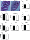
Glutamine improves the ratio of villus to crypt in the ileum of weanling mice. (a) Representative images of hematoxylin and eosin (H&E) staining in the ileum of weanling mice were shown (×200; n = 8). The villus length and crypt depth were measured as indicated in the image. (b–d) The statistical analysis of villus length (b), crypt depth (c), and the ratio of villus to crypt (d) from images shown at (a). The data related to villus length and crypt depth were analyzed by unpaired t-test, while the data related to the ratio of villus to crypt were analyzed by Mann–Whitney test. The data are Mean ± SD with an n = 8. (e–g) mRNA expressions of sucrase (e), enteroendocrine cells-expressed chromogranin A (CHGA) and peptide YY (PYY) (f), as well as Hes1 and Math1 (g) were analyzed in the ileum. N = 10, data were analyzed with unpaired t-test. The data are Mean±SEM.
Fig. 3
Fig. 3
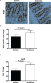
The intestinal cell proliferation after glutamine supplementation in weanling mice. (a) The representative images of immunohistochemistry (IHC) staining with Ki67 antibody in the ileum of weanling mice were shown (×100; n = 8). (b) The statistical analysis of Ki67-positive cells in each crypt from images shown on the (a). (c) The mRNA expression of Lgr5 in the ileum. The data were analyzed by unpaired t-test. The data are Mean ± SD (b) or SEM (c) with an n = 8 (a, b) or 10 (c).
Fig. 4
Fig. 4
Paneth cells in the ileum after glutamine supplementation in weanling mice. (a) The mRNA expression of lysozyme (Lyz) and angiogenin 4 (Ang4) in the ileum. The data were analyzed by unpaired t-test. The data are Mean ± SEM with an n = 10. (b) The number of Paneth cells from HE staining in the ileum. Left, the representative images of Paneth cells in the ileum of weanling mice were shown (×400; n = 8). Right, the statistical analysis of the number of Paneth cells in each crypt from images shown on the Left. The data were analyzed by Mann–Whitney test. The data are Mean ± SD with an n = 8. (c) The number of Paneth cells was analyzed with lysozyme antibody. Top, the representative images of IHC staining with lysozyme antibody in the ileum of weanling mice were shown (×200; n = 8). Bottom, the statistical analysis of lysozyme positive cells in each crypt from images shown on the Top. The data were analyzed by unpaired t-test. The data are Mean ± SD with an n = 8.
Fig. 5
Fig. 5
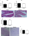
Goblet cells in the ileum after glutamine supplementation in weanling mice. (a) The mRNA expression of mucin2 (Muc2) and trefoil factor 3 (Tff3) in the ileum. The data were analyzed by unpaired t-test. The data are Mean ± SEM with an n = 10. (b, c) The number of goblet cells from HE staining in the ileum. (b) The representative images of goblet cells in the ileum of weanling mice were shown (×200; n = 8). (c) The statistical analysis of number of goblet cells in each villus from images shown on (b). The data were analyzed by Mann–Whitney test. The data are Mean ± SD with an n = 8. (d, e) The number of goblet cells was evaluated with Alcian blue staining. (d) The representative images of Alcian blue staining in the ileum of weanling mice were shown (×100; n = 8). (e) The statistical analysis of goblet cells in each villus from images shown on the (d). The data were analyzed using unpaired t-test. The data are Mean ± SD with an n = 8.

References

    1. Barker N, van Es JH, Kuipers J, Kujala P, van den Born M, Cozijnsen M, et al. . Identification of stem cells in small intestine and colon by marker gene Lgr5. Nature 2007; 449(7165): 1003–7. - PubMed
    1. de Lau W, Peng WC, Gros P, Clevers H. The R-spondin/Lgr5/Rnf43 module: regulator of Wnt signal strength. Genes Dev 2014; 28(4): 305–16. - PMC - PubMed
    1. Peng HS, Pooyaiah N, Forrester M, Cochran E, Wang Q. Ex vivo culture of primary intestinal stem cells in collagen gels and foams. ACS Biomater Sci Eng 2015; 1(1): 37–42. - PubMed
    1. Bloemendaal AL, Buchs NC, George BD, Guy RJ. Intestinal stem cells and intestinal homeostasis in health and in inflammation: a review. Surgery 2016; 159(5): 1237–48. - PubMed
    1. Tian H, Biehs B, Warming S, Leong KG, Rangell L, Klein OD, et al. . A reserve stem cell population in small intestine renders Lgr5-positive cells dispensable. Nature 2011; 478(7368): 255–9. - PMC - PubMed

LinkOut - more resources