. 2018 Jul 23:9:797.
doi: 10.3389/fphar.2018.00797. eCollection 2018.

Hepatoprotective Effect of Steroidal Glycosides From Dioscorea villosa on Hydrogen Peroxide-Induced Hepatotoxicity in HepG2 Cells

Affiliations

Hepatoprotective Effect of Steroidal Glycosides From Dioscorea villosa on Hydrogen Peroxide-Induced Hepatotoxicity in HepG2 Cells

Maqsood A Siddiqui et al. Front Pharmacol. .

Abstract

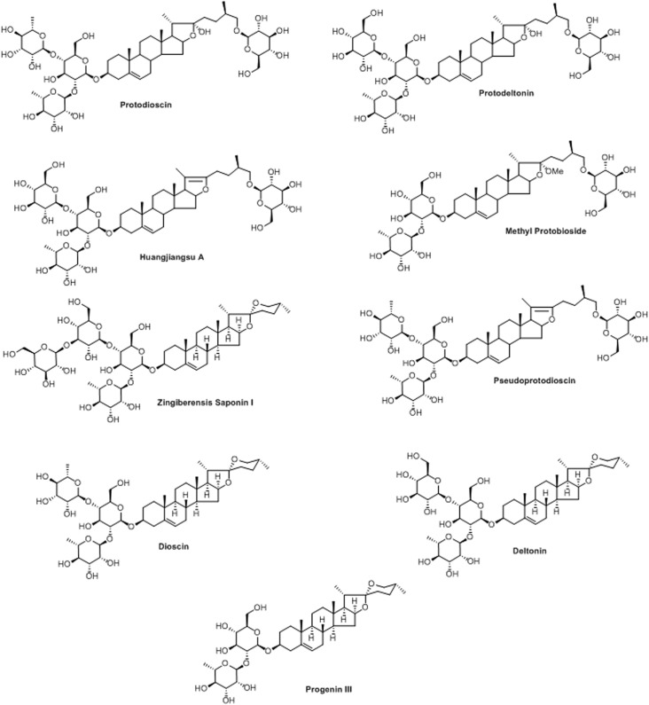
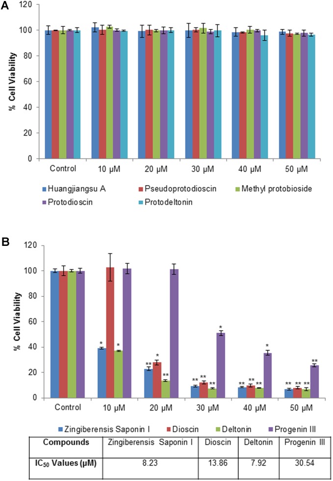
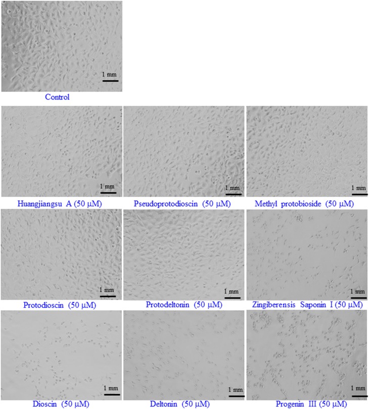
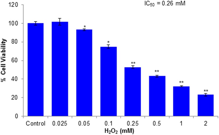
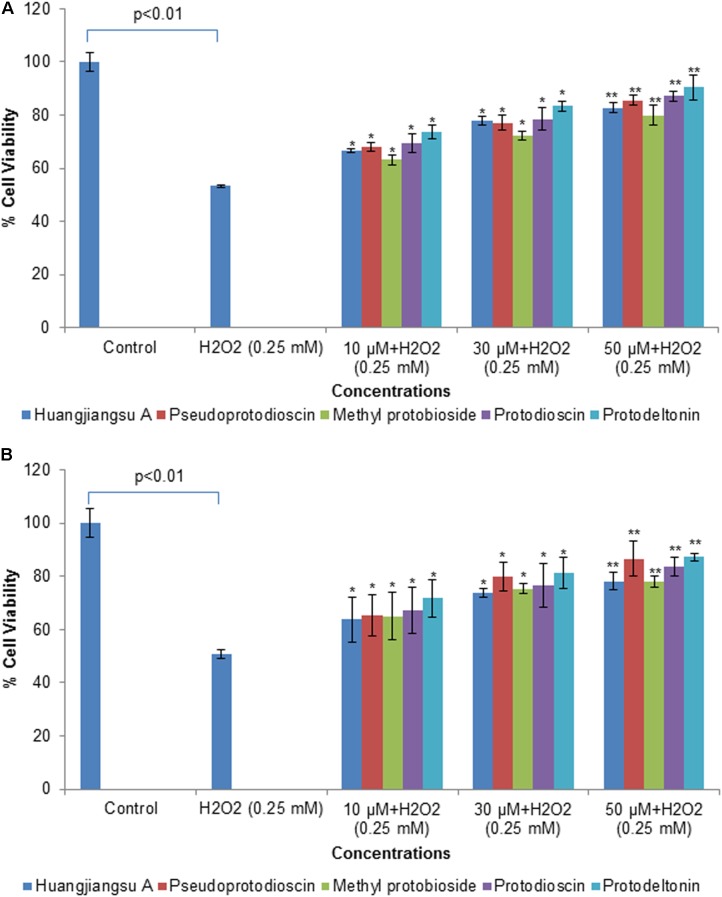
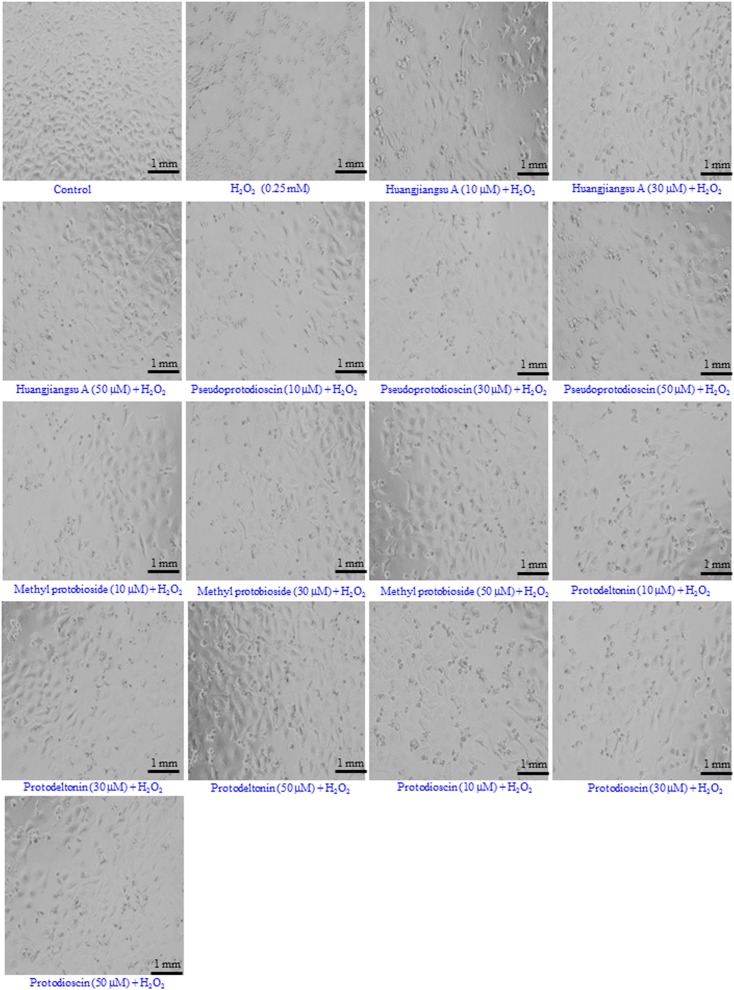
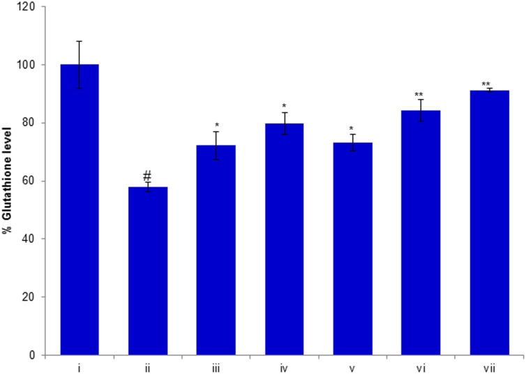
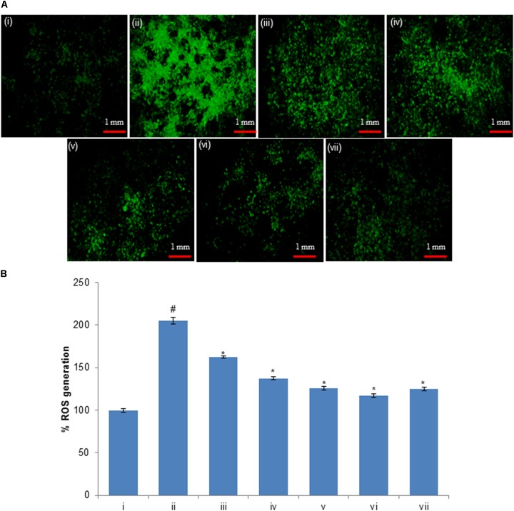
Dioscorea villosa, commonly known as "Wild Yam" and native to North America, is well documented for its pharmacological properties due to the presence of steroidal glycosides. However, the hepatoprotective potential of these compounds has not been studied so far. The present investigation was aimed to study the hepatoprotective effect of the steroidal glycosides from D. villosa against H2O2, a known hepatotoxin, in human liver cell line (HepG2). Cytotoxicity assessment was carried out in cells exposed to various concentrations (10-50 μM) of compounds for 24 h using MTT assay and morphological changes. All tested compounds were known and among them, spirostans (zingiberensis saponin I, dioscin, deltonin and progenin III) were found to be cytotoxic whereas, furostans (huangjiangsu A, pseudoprotodioscin, methyl protobioside, protodioscin, and protodeltonin) were non-cytotoxic. Further, HepG2 cells were pretreated with biologically safe concentrations (10, 30, and 50 μM) of non-cytotoxic compounds and then cytotoxic (0.25 mM) concentration of H2O2. After 24 h, cell viability was assessed by MTT and NRU assays, while morphological changes were observed under the microscope. The results showed that treatment of HepG2 cells with compounds prior to H2O2 exposure effectively increased cell viability in a concentration-dependent manner. Furthermore, huangjiangsu A, pseudoprotodioscin, methyl protobioside, protodioscin, and protodeltonin at 50 μM increased GSH level and decreased intracellular ROS generation against H2O2-induced damages. The results from this study revealed that compounds isolated from D. villosa have hepatoprotective potential against H2O2-induced cytotoxicity and ROS generation and could be promising as potential therapeutic agents for liver diseases.

Keywords: Dioscorea villosa; Dioscoreaceae; H2O2; ROS generation; cytotoxicity; steroidal glycosides.


Figures

FIGURE 1
FIGURE 1
Structures of isolated compounds from Dioscorea villosa.
FIGURE 2
FIGURE 2
Cytotoxicity assessment by MTT assay in HepG2 cells following the exposure of different concentrations of compounds isolated from Dioscorea villosa. All values are given as mean ± SD. (A) Non-cytotoxic compounds and (B) Cytotoxic compounds. p < 0.05, ∗∗p < 0.01 versus control.
FIGURE 3
FIGURE 3
Representative images of the morphological changes in HepG2 cells after the exposure of the compounds isolated from Dioscorea villosa. All the images were acquired using a phase contrast inverted microscope at 20× magnification. Each scale bar = 1 mm.
FIGURE 4
FIGURE 4
Cytotoxicity assessments by MTT assay in HepG2 cells following the exposure of various concentrations of H2O2 for 24 h. All values are given as mean ± SD. p < 0.05, ∗∗p < 0.01 versus control.
FIGURE 5
FIGURE 5
Assessment of protective potential of compounds on cell viability of HepG2 cells by (A) MTT assay and (B) NRU assay. Cells were exposed to 10, 30, and 50 μM of non-cytotoxic compounds for 24 h. Then the cells were exposed to H2O2 at 0.25 mM for 24 h. All values are given as mean ± SD. p < 0.05, ∗∗p < 0.01 versus H2O2.
FIGURE 6
FIGURE 6
Morphological changes observed in HepG2 cells. Cells were pre-exposed with non-cytotoxic compounds for 24 h and then H2O2 (0.25 mM) for 24 h. Images were acquired using a phase contrast inverted microscope at 20× magnification. Each scale bar = 1 mm.
FIGURE 7
FIGURE 7
Protective potential of compounds isolated from Dioscorea villosa on depletion in glutathione level. HepG2 cells were exposed to 50 μM of non-cytotoxic compounds for 24 h prior to the addition of H2O2 for 24 h. All values are represented as mean ± SD. (i) Control; (ii) H2O2 (0.25 mM); (iii) Huangjiangsu A (50 μM) + H2O2 (0.25 mM); (iv) Pseudoprotodioscin (50 μM) + H2O2 (0.25 mM); (v) Methyl protobioside (50 μM) + H2O2 (0.25 mM); (vi) Protodioscin (50 μM) + H2O2 (0.25 mM); and (vii) Protodeltonin (50 μM) + H2O2 (0.25 mM). #p < 0.01 versus control, p < 0.05 and ∗∗p < 0.01 versus H2O2 exposure.
FIGURE 8
FIGURE 8
(A) H2O2-induced ROS generation and ameliorative effect of pre-treatment of compounds in HepG2 cells and (B) percent change in ROS generation. ROS generation was evaluated using dichlorodihydrofluorescein diacetate (DCFH-DA) dye. (i) Untreated control; (ii) H2O2 (0.25 mM); (iii) Huangjiangsu A (50 μM) + H2O2 (0.25 mM); (iv) Pseudoprotodioscin (50 μM) + H2O2 (0.25 mM); (v) Methyl protobioside (50 μM) + H2O2 (0.25 mM); (vi) Protodioscin (50 μM) + H2O2 (0.25 mM); and (vii) Protodeltonin (50 μM) + H2O2 (0.25 mM). #p < 0.01 versus control, p < 0.05 versus H2O2 exposure. Each scale bar = 1 mm.

References

    1. Ali Z., Smillie T. J., Khan I. A. (2013a). 7-Oxodioscin, a new spirostan steroid glycoside from the rhizomes of Dioscorea nipponica. Nat. Prod. Commun. 8 319–321. - PubMed
    1. Ali Z., Smillie T. J., Khan I. A. (2013b). Cholestane steroid glycosides from the rhizomes of Dioscorea villosa (wild yam). Carbohydr. Res. 370 86–91. 10.1016/j.carres.2012.12.022 - DOI - PMC - PubMed
    1. Ali Z., Smillie T. J., Khan I. A. (2013c). Two spirostan steroid glycoside fatty esters from Dioscorea cayenensis. Nat. Prod. Commun. 8 323–326. - PMC - PubMed
    1. Al-Sheddi E. S., Farshori N. N., Al-Oqail M. M., Musarrat J., Al-Khedhairy A. A., Siddiqui M. A. (2016). Protective effect of Lepidium sativum seed extract against hydrogen peroxide-induced cytotoxicity and oxidative stress in human liver cells (HepG2). Pharm. Biol. 54 314–321. 10.3109/13880209.2015.1035795 - DOI - PubMed
    1. Aruoma O. I. (1998). Free radicals, oxidative stress, and antioxidants in human health and disease. J. Am. Oil Chem. Soc. 75 199–212. 10.1007/s11746-998-0032-9 - DOI - PMC - PubMed

LinkOut - more resources