. 2018 Jul 31:6:e5186.
doi: 10.7717/peerj.5186. eCollection 2018.

Identification of exosome-like nanoparticle-derived microRNAs from 11 edible fruits and vegetables

Affiliations

Identification of exosome-like nanoparticle-derived microRNAs from 11 edible fruits and vegetables

Juan Xiao et al. PeerJ. .

Abstract

Edible plant-derived exosome-like nanoparticles (EPDELNs) are novel naturally occurring plant ultrastructures that are structurally similar to exosomes. Many EPDELNs have anti-inflammatory properties. MicroRNAs (miRNAs) play a critical role in mediating physiological and pathological processes in animals and plants. Although miRNAs can be selectively encapsulated in extracellular vesicles, little is known about their expression and function in EPDELNs. In this study, we isolated nanovesicles from 11 edible fruits and vegetables and subjected the corresponding EPDELN small RNA libraries to Illumina sequencing. We identified a total of 418 miRNAs-32 to 127 per species-from the 11 EPDELN samples. Target prediction and functional analyses revealed that highly expressed miRNAs were closely associated with the inflammatory response and cancer-related pathways. The 418 miRNAs could be divided into three classes according to their EPDELN distributions: 26 "frequent" miRNAs (FMs), 39 "moderately present" miRNAs (MPMs), and 353 "rare" miRNAs (RMs). FMs were represented by fewer miRNA species than RMs but had a significantly higher cumulative expression level. Taken together, our in vitro results indicate that miRNAs in EPDELNs have the potential to regulate human mRNA.

Keywords: Cross-kingdom; Exosome-like nanoparticles; Illumina sequencing; miRNA expression profile; miRNAs expression profile.


Conflict of interest statement

The authors declare there are no competing interests.

Figures

Figure 1
Figure 1. EPDELN morphological ultrastructure and size distribution.
The morphological ultrastructure was visualized by AFM, and the size distribution of EPDELNs was analyzed by DLS in coconut (A), Hami melon (B), and tomato (C). Photographs by Juan Xiao.
Figure 2
Figure 2. Identification and expression profiles of EPDELN-related miRNAs.
(A) Number of miRNA species in different EPDELN samples. (B) Length distribution and frequency of filtered reads with matches to mature miRNAs in miRBase 21.0. (C–E) Normalized expression values and proportions (relative to all miRNAs of each EPDELN) of miRNAs in EPDELNs of coconut (C), orange (D), and tomato (E). An asterisk denotes that a miRNA with identical nomenclature is annotated in different species.
Figure 3
Figure 3. Distribution, classification, and functional analysis of miRNAs with high expression in EPDELNs.
(A–C) Expression distribution of miRNAs in coconut (A), orange (B), and tomato (C). The ordinate and abscissa correspond to the ranking of miRNA expression levels and the number of co-expressed species, respectively. The terms “frequent miRNAs” (FMs), “moderately present miRNAs” (MPMs), and “rare miRNAs” (RMs) are used to describe miRNAs present almost simultaneously in 8–11, 4–7, or 1–3 EPDELN samples, respectively. The solid line is used to demarcate the top 20 expressed miRNAs of each EPDELN sample. (D) Gene Ontology and KEGG pathway analyses of categories enriched in the specific target genes of miRNAs of each EPDELN sample. The size of each circle represents the number of genes, and the color signifies the p-value. (E) Diagram of putative MIR-168c binding sites in TSC22D3 aligned against wild-type (WT) or mutant (MUT) MIR-168c putative target sites in the luciferase reporter plasmid. Paired bases are indicated by a black vertical line, and a mismatch is indicated by two dots. (F) Luciferase activity in HeLa cells cotransfected with MIR-168c or scrambled control oligonucleotides and the reporter constructs from (E) (n = 3). Statistical significance was determined by the Student’s t-test (*, p < 0.05).
Figure 4
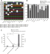
Figure 4. Characteristics of EPDELN-related miRNAs.
(A) Expression of 26 conserved FMs in 11 EPDELN-derived miRNA libraries. The white region indicates that expression of the designated miRNA was not detected in the given ELN sample. (B) Percentage breakdown of total normalized expression of the three specific classes of miRNAs in each EPDELN sample. (C) Relationship between number of miRNA species and the proportion of normalized expression. The difference in the proportion of normalized expression between FMs and RMs was highly significant (**) according to the t-test.

References

    1. Araceli C, Madelourdes PH, Maelena P, Joséa R, Carlosandrés G. Chemical studies of anthocyanins: a review. Food Chemistry. 2009;113:859–871. doi: 10.1016/j.foodchem.2008.09.001. - DOI
    1. Bang C, Thum T. Exosomes: new players in cell–cell communication. The International Journal of Biochemistry & Cell Biology. 2012;44:2060–2064. doi: 10.1016/j.biocel.2012.08.007. - DOI - PubMed
    1. Bartel DP. MicroRNAs: genomics, biogenesis, mechanism, and function. Cell. 2004;116:281–297. doi: 10.1016/S0092-8674(04)00045-5. - DOI - PubMed
    1. Calin GA, Croce CM. MicroRNA signatures in human cancers. Nature Reviews Cancer. 2006;6:857–866. doi: 10.1038/nrc1997. - DOI - PubMed
    1. Cavalieri D, Rizzetto L, Tocci N, Rivero D, Asquini E, Si-Ammour A, Bonechi E, Ballerini C, Viola R. Plant microRNAs as novel immunomodulatory agents. Scientific Reports. 2016;6:25761. doi: 10.1038/srep25761. - DOI - PMC - PubMed