Review
. 2018 Sep;373(3):619-641.
doi: 10.1007/s00441-018-2882-2. Epub 2018 Aug 6.

Morphological diversity and connectivity of hippocampal interneurons

Affiliations
Review

Morphological diversity and connectivity of hippocampal interneurons

Sam A Booker et al. Cell Tissue Res. 2018 Sep.

Erratum in

Abstract

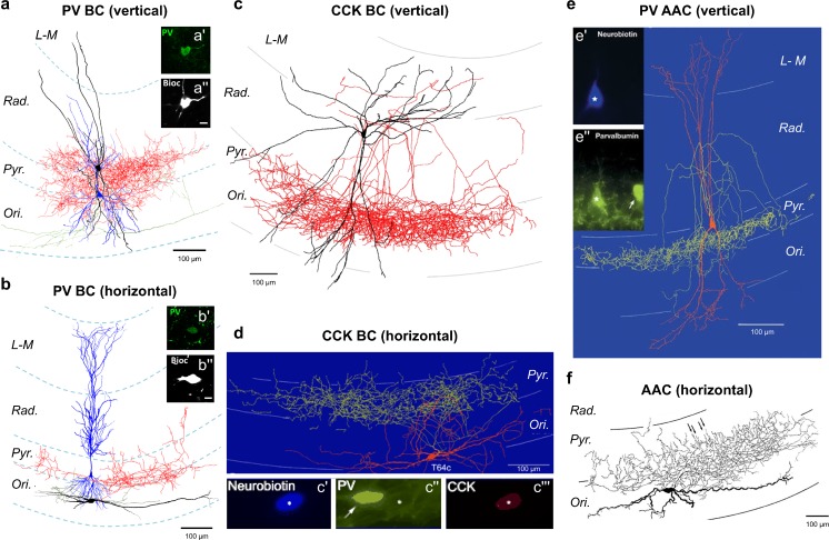
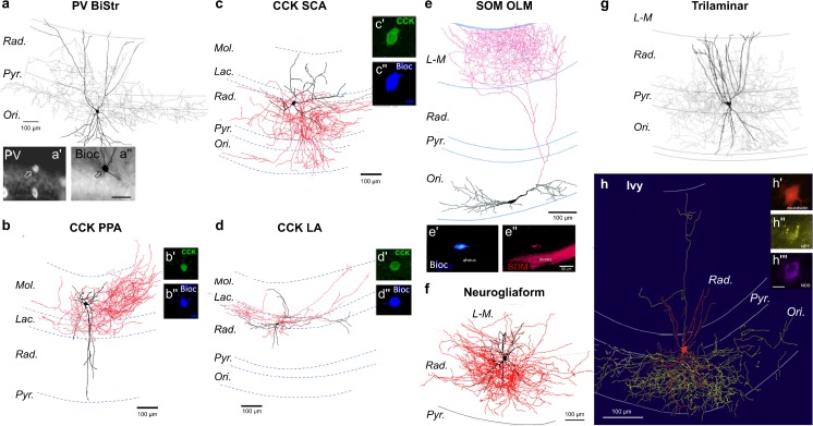
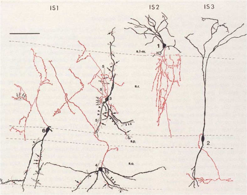
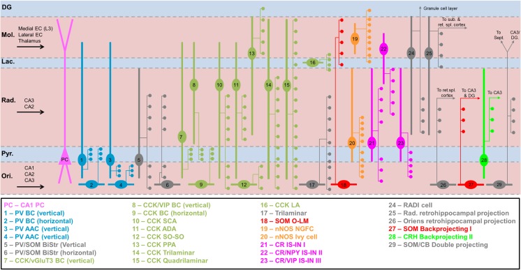
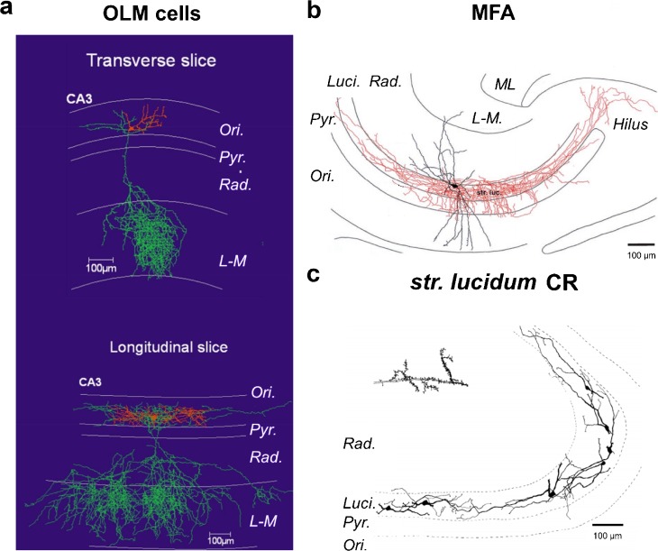
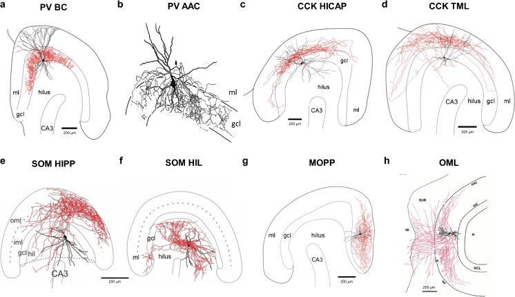
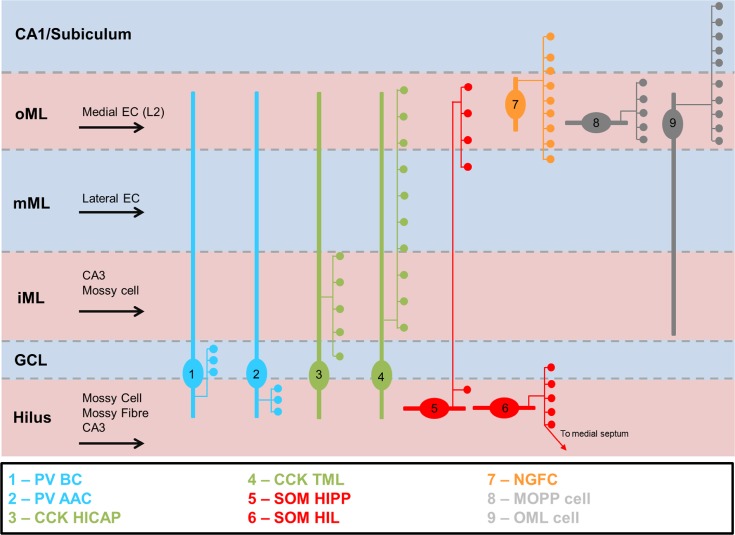
The mammalian forebrain is constructed from ensembles of neurons that form local microcircuits giving rise to the exquisite cognitive tasks the mammalian brain can perform. Hippocampal neuronal circuits comprise populations of relatively homogenous excitatory neurons, principal cells and exceedingly heterogeneous inhibitory neurons, the interneurons. Interneurons release GABA from their axon terminals and are capable of controlling excitability in every cellular compartment of principal cells and interneurons alike; thus, they provide a brake on excess activity, control the timing of neuronal discharge and provide modulation of synaptic transmission. The dendritic and axonal morphology of interneurons, as well as their afferent and efferent connections within hippocampal circuits, is central to their ability to differentially control excitability, in a cell-type- and compartment-specific manner. This review aims to provide an up-to-date compendium of described hippocampal interneuron subtypes, with respect to their morphology, connectivity, neurochemistry and physiology, a full understanding of which will in time help to explain the rich diversity of neuronal function.

Keywords: Connectivity; GABA; Hippocampus; Interneuron; Morphology.


Conflict of interest statement

The authors declare that they have no conflict of interest.

Figures

Fig. 1
Fig. 1
Perisomatic inhibitory INs of hippocampal subfield CA1. Example reconstructions of perisomatic inhibitory INs: PV BCs (a, b), CCK BCs (c, d) and axo-axonic cells (e, f) in the CA1 area, with either vertical (a, c, e) or horizontal (b, d, f) dendritic arbors. Soma and dendrites are shown as black (a, b, c, f) or red (d, e), while the axon is shown as red (ac), yellow (d, e), or gray (f). In a and b, CA1 PCs are shown for reference (blue). Immunohistochemical labeling for PV (a′, b′, c″, and e′) and CCK (c″′) are shown as insets. All reconstructions are shown with respect to CA1 layers: Ori, str. oriens; Pyr, str. pyramidale; Rad., str. radiatum; L-M, str. lacunosum-moleculare. Adapted with permission from: a, b—Booker et al. (2017); c—Vida et al. (1998); d—Klausberger et al. (2005); e—Klausberger et al. (2003); f—Ganter et al. (2004)
Fig. 2
Fig. 2
DI INs of hippocampal subfield CA1. Examples of CA1 DI INs. Soma and dendrites are shown as black (ag) or red (h), while the axon is shown as gray (a, g), red (bf), or yellow (h). Immunohistochemical labeling for PV (a′), CCK (b′, c′, d′), SOM (e″) and NPY (h″) and nNOS (h″′) is shown as insets. All reconstructions are shown with respect to CA1 layers: Ori, str. oriens; Pyr, str. pyramidale; Rad., str. radiatum; L-M, str. acunosum-moleculare. Adapted with permission from: a, g—Pawelzik et al. (2002); bd—Booker et al. (2016); e—Martina et al. (2000); f—Vida et al. (1998); h—Fuentealba et al. (2008)
Fig. 3
Fig. 3
IN-specific INs of hippocampal subfield CA1: distribution of CR immunoreactive IS-INs with respect to somatodendritic axis (black) and axonal arborisation (red). Reproduced with permission from Freund and Buzsáki (1996)
Fig. 4
Fig. 4
Summary of all described IN subtypes in hippocampal subfield CA. Schematic overview of known morphological and neurochemical IN subtypes in CA1. Somatodendritic domains (thick lines), axonal locations (thin lines) and major terminal fields (circles) are shown with respect to regional layers (thin dashed lines). Afferent inputs are indicated with black arrows. Layers: Ori, str. oriens; Pyr, str. pyramidale; Rad., str. radiatum; Lac., str. lacunosum; Mol., str. moleculare; DG, dentate gyrus
Fig. 5
Fig. 5
Divergent IN subtypes in hippocampal subfield CA3: reconstructions of CA3 INs showing divergence from CA1. Soma and dendrites are shown as red (a) or black (b, c), with the axon shown in green (a) or red (b). CR indicates calretinin immunoreactivity. Reconstructions are shown with respect to CA3 laminae: Ori., str. oriens; Pyr., str. pyramidale; Luci., str. lucidum; Rad., str. radiatum; L-M, str. lacunosum-moleculare; ML, molecular layer. Adapted with permission from: a—Tort et al. (2007); b—Vida and Frotscher (2000); c—Gulyás et al. (1992)
Fig. 6
Fig. 6
IN subtypes of the dentate gyrus: examples of morphological subtypes of identified IN in the DG of mice and rats. All panels except for b show soma and dendrites in black and axon in red. b Dendrites as thick black lines and axon as thin lines. Adapted from: a, c, d, g—Hosp et al. (2014); b—Soriano and Frotscher (1989); e, f—Yuan et al. (2017); h—Ceranik et al. (1997)
Fig. 7
Fig. 7
Summary of identified IN subtypes in the DG. The known morphological and neurochemical subtypes of IN in the DG are depicted, with typical localization of their somata, laminar distribution of their dendrites (thick lines) and axon distribution (thin lines) with major terminal fields (circles) with respect to DG layers (dashed lines): GCL, granule cell layer; iML, inner molecular layer; mML, medial molecular layer; oML, outer molecular layer; hippocampal fissure (thick dashed line)

References

    1. Acsády L, Arabadzisz D, Freund TF. Correlated morphological and neurochemical features identify different subsets of vasoactive intestinal polypeptide-immunoreactive interneurons in rat hippocampus. Neuroscience. 1996;73:299–315. - PubMed
    1. Acsády L, Görcs T, Freund T. Different populations of vasoactive intestinal polypeptide-immunoreactive interneurons are specialized to control pyramidal cells or interneurons in the hippocampus. Neuroscience. 1996;73:317–334. - PubMed
    1. Ali AB. Presynaptic inhibition of GABAA receptor-mediated unitary IPSPs by cannabinoid receptors at synapses between CCK-positive interneurons in rat hippocampus. J Neurophysiol. 2007;98:861–869. - PubMed
    1. Ali AB, Thomson AM. Facilitating pyramid to horizontal oriens-alveus interneurone inputs: dual intracellular recordings in slices of rat hippocampus. J Physiol. 1998;507:185–199. - PMC - PubMed
    1. Ali A, Todorova M. Asynchronous release of GABA via tonic cannabinoid receptor activation at identified interneuron synapses in rat CA1. Eur J Neurosci. 2010;31:1196–1207. - PubMed

LinkOut - more resources