. 2018 Nov;57(11):1640-1650.
doi: 10.1002/mc.22886. Epub 2018 Aug 26.

The Rb tumor suppressor regulates epithelial cell migration and polarity

Affiliations

The Rb tumor suppressor regulates epithelial cell migration and polarity

Tiziana Parisi et al. Mol Carcinog. 2018 Nov.

Abstract

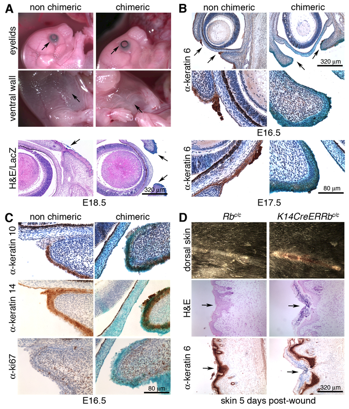
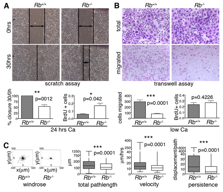
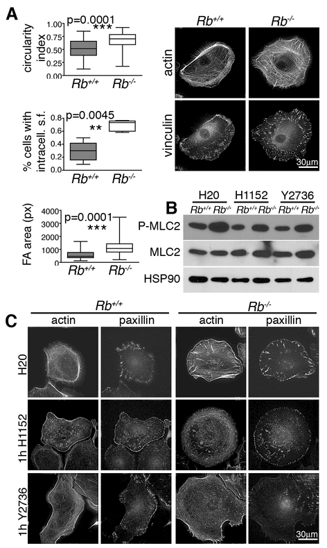
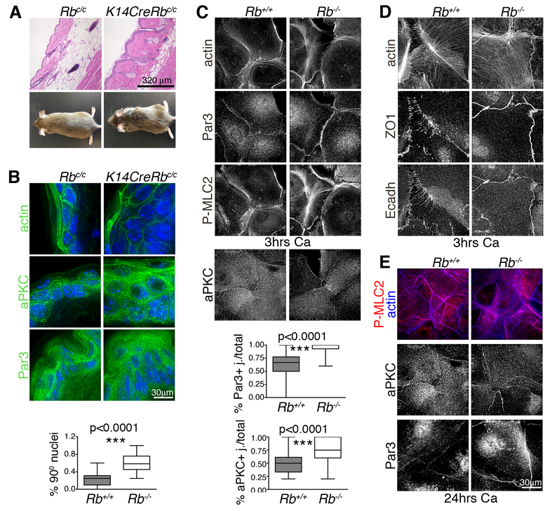
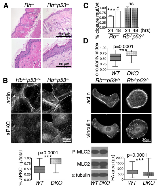
Altered cell polarity and migration are hallmarks of cancer and metastases. Here we show that inactivation of the retinoblastoma gene (Rb) tumor suppressor causes defects in tissue closure that reflect the inability of Rb null epithelial cells to efficiently migrate and polarize. These defects occur independently of pRB's anti-proliferative role and instead correlate with upregulation of RhoA signaling and mislocalization of apical-basal polarity proteins. Notably, concomitant inactivation of tp53 specifically overrides the motility defect, and not the aberrant polarity, thereby uncovering previously unappreciated mechanisms by which Rb and tp53 mutations cooperate to promote cancer development and metastases.

Keywords: PAR complex; RhoA Rock signaling; aPKC; eyes open phenotype; planar cell polarity.


Figures

Figure 1.
Figure 1.. Rb inactivation causes general tissue fusion defects.
(A) Representative images of E18.5 Rb null chimeric embryos displaying EOP and aberrant body wall fusion (upper panel). H&E counterstaining of whole mount LacZ stained sections showed co-occurrence of the EOP defect and Rb-/- (blue) cells at the eyelid end (lower panel). (B) Representative images of IHC of E16.5 (upper panel) and E17.5 (lower panel) embryos with α-k6 showed failure to form the tip in Rb-/- eyelids. (C) IHC for k10, k14 and Ki67 at E16.5 showed that the differentiated and basal layers are equally represented in Rb-/- chimeric mutant and non-chimeric eyelid epidermis, and that proliferation is negligible in both. (D) Representative images five days after wounding, showed a delay in wound closure in Rb-deficient skin, relative to the wildtype control (upper panel), despite displaying efficient k6 expression (lower panel).
Figure 2.
Figure 2.. Rb null keratinocytes have motility defects.
(A) Epithelial cell layers were generated by culturing wildtype and Rb-/- keratinocytes in high calcium for 24 hours. The Rb-/- keratinocytes had an impaired ability to repair scratches, as indicated by representative images and quantification (left graph; n=6 lines), and displayed ectopic proliferation, as assessed by BrdU incorporation (right graph; 3 lines/genotype, ~2000 cells. (B) Single Rb-/- keratinocytes (cultured in low calcium) showed impaired migration 12 hrs post-plating in Boyden chambers relative to wildtype controls, as judged by crystal violet staining of cells with representative images above and quantification in left graph (n=6 lines/genotype, plated in triplicate), without proliferation changes, which were assessed by BrdU incorporation (right graph; n=3 lines/genotype, ~500 cells). (C) Time-lapse microscopy was conducted for 15 hrs to track the path of individual wildtype and Rb-/- keratinocytes (n=3 lines and >350 cells per genotype). From left to right: windrose plots and graphs show that Rb-/- cells had significant reduced total pathlength, velocity, and persistence compared to their wildtype counterparts. Statistical significance was determined by Student’s t-test.
Figure 3.
Figure 3.. Rb loss causes changes in cell architecture that correlate with hyperactive RhoA.
(A) Compared to wildtype controls, single Rb-/- keratinocytes had a higher circularity index (n=3 lines/genotype, ~300 cells) indicating reduced polarization, and displayed more prominent non-peripheral stress fibers (phalloidin staining for F-actin; n=5 lines/genotype, >90 cells) and FAs that were larger and more localized within the cell body (α-vinculin IF; n= 3 lines/genotype, >150 cells). Significance was determined by Student’s t-test. (B) Wildtype and Rb-/- keratinocytes grown as single cells in low calcium with or without 1hr treatment with two different Rock inhibitors, H1152 and Y2673. Western blotting showed that Rb-deficiency caused increased levels of P-MLC2, which was reversed by the Rock inhibitors indicating that this is dependent on hyperactive Rho/Rock. (C) 1 hr treatment with H1152 and Y2673 (n= 3 lines/genotype, 80 cells) resulted in loss of the stress fibers (phalloidin staining for actin), including the aberrant fibers across the body of Rb-/- cells, and altered FAs (α-paxillin IF) by both reducing their size and eliminating the intracellular localization characteristic of Rb-/- cells.
Figure 4.
Figure 4.. Rb inactivation in the epidermis causes polarity defects.
Skin from mice with constitutive Rb mutation in the basal layer (n=4) showed: (A) disorganized skin architecture, misoriented hair and enlarged sebaceous glands (H&E sections) and sparse hair and rough coats (mouse pictures); and (B) aberrant tissue morphology (phalloidin staining of actin cytoskeleton) and nuclei orientation (DAPI), with Par3 and aPKC less restricted at the cell membrane and mislocalized within the epidermal layers (IF images). (C, D) Wildtype and Rb-/- keratinocytes were cultured in high calcium for 3 hours to trigger cell junction formation. (C) The Rb-/- keratinocytes (n>6 lines/genotype, ~ 500 cells) showed stress fibers (phalloidin staining) crossing the cells’ basal surface and aberrant phospho-myosin (P-MLC2) and actin rings at the cell-cell junctions. Additionally Par3 and aPKC were upregulated at the cell membrane (n>3 lines/genotype. The graphs show the number of Par3 and aPKC positive junctions calculated over the total number of touching cell sides (n=294 and 210 respectively). (D) Adherens junctions (E-cadherin staining) and tight junctions (ZO1 staining) were localized in continuous lines in Rb-/- cells versus punctae in wildtype controls. (E) Wildtype and Rb-/- keratinocytes were maintained in high calcium for 24 hours. The acto-myosin cytoskeletal defects, and Par3 and aPKC upregulation, persisted in Rb-/- keratinocytes at this time. Statistical significance was determined by Student’s t-test.
Figure 5.
Figure 5.. tp53 inactivation in Rb deficient epithelial cells rescues the migration but not the polarity defect.
(A) Representative H&E sections show that Rb and Rb;p53 deficient adult skins were thicker, had misoriented hairs and aberrant sebaceous glands (n=11/genotype). (B) Rb;p53 DKO keratinocytes, induced with high calcium for 3 hours, showed aPKC upregulation (n=222 cells) in a similar manner to Rb-/- (see Fig. 4C). (C) Rb;p53 DKO or Rb-/- cells were cultured in high calcium for 24 hours alongside their paired wildtype controls, to allow epithelial layer formation, and then subjected to scratch assays (n≥3 lines/genotype). The graphs (comparing healing to wildtype controls at 24 and 48 hours) showed that the Rb;p53 DKO cells closed the wound in a comparable manner to wildtypes, in contrast to the impaired healing of Rb-/- cells. (D) Wildtype and Rb;p53 DKO cells were cultured as single cells in low calcium. The graph, showing measurement of the circularity index establishes that the Rb;p53 DKO cells maintain the front back polarity defect (n=3 lines/genotype, 67 cells) present in the single Rb-/- keratinocytes (see Fig. 3A). In contrast, IF staining showed that the Rb;p53 DKO cells had smaller FAs (vinculin staining; n=3 lines/genotype, 174 cells) than their wildtype controls, which correlated with wildtype levels of P-MLC2 and total MLC2. Statistical significance was determined by Student’s t-test. ***p≤0.0001. *p≤0.01.

References

    1. Guilluy C, Garcia-Mata R, Burridge K. Rho protein crosstalk: another social network? Trends Cell Biol 2011;21(12):718–726. - PMC - PubMed
    1. Ridley AJ. Rho family proteins: coordinating cell responses. Trends Cell Biol 2001;11(12):471–477. - PubMed
    1. Etienne-Manneville S, Hall A. Cell polarity: Par6, aPKC and cytoskeletal crosstalk. Curr Opin Cell Biol 2003;15(1):67–72. - PubMed
    1. Goldstein B, Macara IG. The PAR proteins: fundamental players in animal cell polarization. Dev Cell 2007;13(5):609–622. - PMC - PubMed
    1. Vega FM, Ridley AJ. Rho GTPases in cancer cell biology. FEBS Lett 2008;582(14):2093–2101. - PubMed

Publication types