. 2018 Oct 15;24(20):5085-5097.
doi: 10.1158/1078-0432.CCR-18-0873. Epub 2018 Jul 3.

Mast Cell Activation and KSHV Infection in Kaposi Sarcoma

Affiliations

Mast Cell Activation and KSHV Infection in Kaposi Sarcoma

Leona W Ayers et al. Clin Cancer Res. .

Abstract

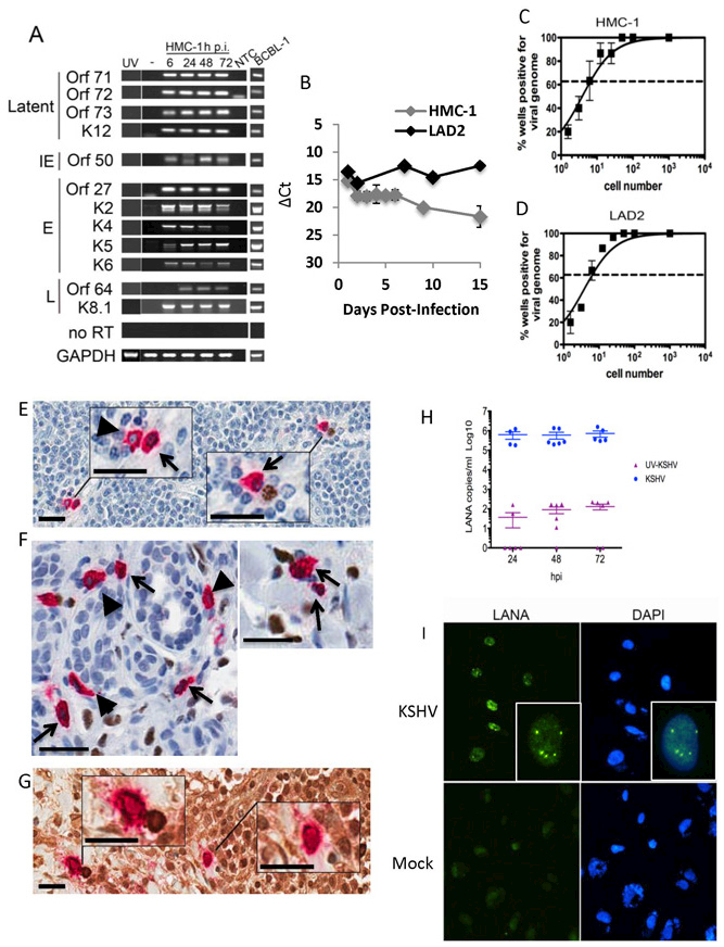
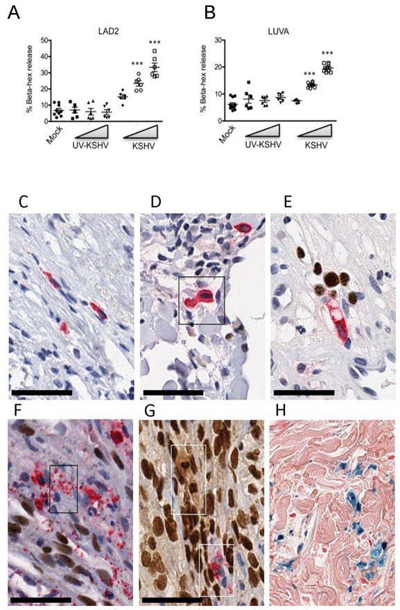
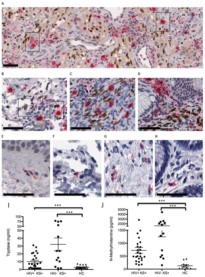
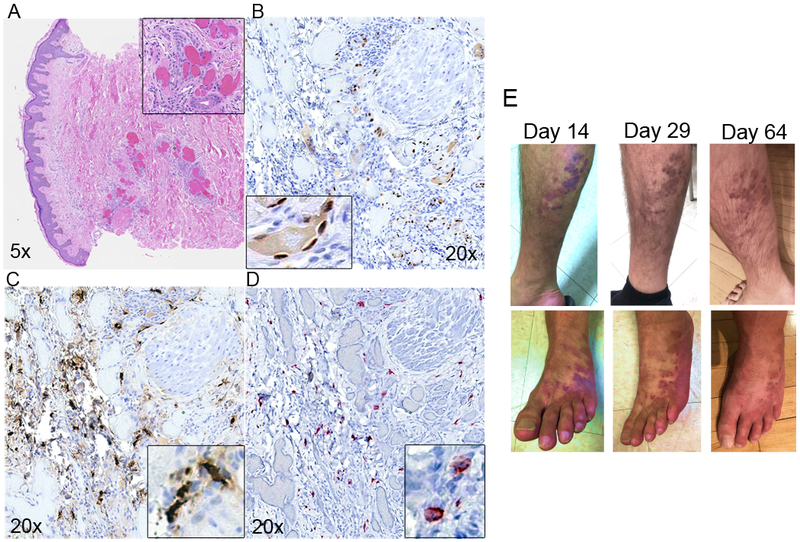
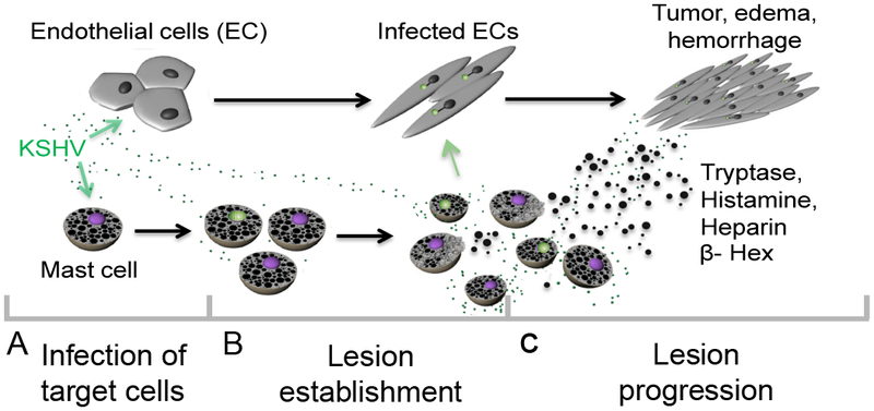
Purpose: Kaposi sarcoma (KS) is a vascular tumor initiated by infection of endothelial cells (ECs) with KS-associated herpesvirus (KSHV). KS is dependent on sustained proinflammatory signals provided by intralesional leukocytes and continued infection of new ECs. However, the sources of these cytokines and infectious virus within lesions are not fully understood. Here, mast cells (MCs) are identified as proinflammatory cells within KS lesions that are permissive for, and activated by, infection with KSHV.Experimental Design: Three validated MC lines were used to assess permissivity of MCs to infection with KSHV and to evaluate MCs activation following infection. Biopsies from 31 AIDS-KS cases and 11 AIDS controls were evaluated by IHC for the presence of MCs in KS lesions and assessment of MC activation state and infection with KSHV. Plasma samples from 26 AIDS-KS, 13 classic KS, and 13 healthy adults were evaluated for levels of MC granule contents tryptase and histamine.Results: In culture, MCs supported latent and lytic KSHV infection, and infection-induced MC degranulation. Within KS lesions, MCs were closely associated with spindle cells. Furthermore, MC activation was extensive within patients with KS, reflected by elevated circulating levels of tryptase and a histamine metabolite. One patient with clinical signs of extensive MC activation was treated with antagonists of MC proinflammatory mediators, which resulted in a rapid and durable regression of AIDS-KS lesions.Conclusions: Using complimentary in vitro and in vivo studies we identify MCs as a potential long-lived reservoir for KSHV and a source of proinflammatory mediators within the KS lesional microenvironment. In addition, we identify MC antagonists as a promising novel therapeutic approach for KS. Clin Cancer Res; 24(20); 5085-97. ©2018 AACR.


Conflict of interest statement

The authors declare they have no conflicts of interest.

Figures

Figure 1.
Figure 1.. Mast cells support KSHV infection in vitro and in patient lesions.
(A-D) HMC-1 and LAD2 cells were infected with KSHV and cultured for indicated times. (A) Mast cells express abundant KSHV genes. GAPDH was used as a loading control. BCBL-1 cells induced with valproic acid were used as a positive control for viral lytic gene expression. NTC- no template control. Data are representative of two separate experiments. (B) QRT-PCR gene expression indicated MCs express LANA in growing cultures over the entire culture period. Data are expressed as CT values normalized to GAPDH. Data are representative of three independent experiments performed in triplicate. (C-D) Limiting dilution qPCR analysis of infected (C) HMC-1 and (D) LAD2 demonstrated approximately 1 in 7 MCs were KSHV genome+ at 24 hpi.. Data are expressed as mean ±SEM, n=3. (E-G) HIV/KS tissue double stained for KSHV LANA (nucleus-brown) and MC-specific tryptase (cytoplasm-red) demonstrate: (E) LN shows (left upper inset) two tryptase+ MCs, one LANA (blue nucleus, arrow head) and the other LANA+ (brown nucleus, black arrow), (right lower inset) tryptase+ LANA+ MC (brown nucleus, black arrow) paired with a LANA+ plasma cell (brown nucleus); (F) dermal lesion shows tryptase+ LANA+ MCs (black arrows) and tryptase+ LANA MCs (arrow heads); (G) HIV-KS LN double stained for MC-specific tryptase (cytoplasm-red) and KSHV lytic antigen K8.1a (cytoplasm-brown) shows (left upper inset) a tryptase+ K8.1a+ MC paired with a KSHV K8.1a+ stained plasma cell cytoplasm and (right lower inset) a solo tryptase+ K8.1+ MC cytoplasm (brown). Scale 25 μm (inset scale, 10 μm). (H-I) HMC-1 cells were infected as described in the methods. (H) KSHV-infected MCs produce encapsulated, DNAse resistant, virus during infection. Data are from two independent experiments with three experimental replicates and two technical replicates each and expressed as mean ± SEM. Mock infected cultures gave no CTs. (I) Mast cell-derived-KSHV establishes latency in primary human endothelial cells. Cell-free supernatants were isolated from MCs uninfected or infected with KSHV at 24h p.i. and used to treat primary human ECs. 48 h post-treatment, primary ECs were fixed and stained as indicated in the Methods; LANA positive nuclei-localized staining demonstrated establishment of latency. Dapi was used to visualize nuclei. Magnification x630. Data are representative of 3 independent experiments.
Figure 2.
Figure 2.. KSHV virus activates mast cells in vitro and in vivo. (A-B)
Live KSHV virus induces a dose-dependent increase in β-hexosaminidase release from LAD2 (A) and LUVA (B) cells within 1 hr (n=3, mean ±SEM). Difference in percent β-hexosaminidase release was analyzed by using 1-way ANOVA, followed by Bonnferroni multiple comparison test. **p<0.01, ***p<0.001. (C-H) Activated, degranulating and degranulated MCs are associated with KSHV LANA+ KS cell nuclei and EC nuclei in vivo; HIV+ KSHV LANA+ KS tissues double stained for MC-specific tryptase (cytoplasm-red) and KSHV LANA (nucleus-brown) demonstrate: (C) A resting MC from HIV+ control tissue is shown for size comparison; (D) HIV+ early KS skin patch lesion demonstrates an enlarged, activated MC showing abundant tryptase filled cytoplasm and enlarged nucleus compared to (C); (E) MC directional tryptase degranulation. An activated, elongated and vacuolated MC produces a narrow stream of discrete tryptase+ granules (red) from the MC tip directed toward the enlarged intravascular KSHV LANA infected EC nuclei (brown); (F) MC mass tryptase granule degranulation (box) within a tissue bundle of proliferating KSHV infected spindle cells. Discrete tryptase+ granules flood the infected spindle cell bundle. (G) Partial and complete MC degranulation between compact KS spindle cells in developing KS nodular lesion. Note the MC ‘ghost’ lobulated LANA+ nucleus and foamy cytoplasm (upper box) and a partially degranulated MC (lower box); (H) Prussian blue hemosiderin granules are a persistent feature of MC degranulation in KS lesions. Scale bar 50 μm.
Figure 3.
Figure 3.. MCs associate with KSHV LANA+ KS spindle cells and are activated with degranulation of tryptase+ discrete granules within developing KS lesions.
(A-D) HIV+ patients with Kaposi’s sarcoma herpesvirus (KSHV) lesions double stained for MC-specific tryptase (cytoplasm-red) and KSHV LANA (nucleus-brown). (A) Skin- KS lesions with abundant LANA+ECs and activated tryptase+ MCs. A subset of activated MCs are LANA+ (right inset box). (B) Lung-LANA+ spindle cell nuclei and associated tryptase+ MCs. (C) lymph node- LANA+ spindle cell proliferation and associated tryptase+ activated, enlarged, MCs, and (D) gut with LANA+ spindle cell nuclei and abundant activated MCs with tryptase degranulation. (E-H) Control HIV+ KS negative tissues show small dendritic shaped MCs with tryptase+ cytoplasm. Note the broad distribution of MCs and their obscure appearance in control HIV+ tissues. Scale bar, 50μm. (I-J) Elevated mast cell tryptase (I) and N-methlyhistamine (J) in plasma samples from 26 AIDS-associated HIV+ KS patients, 13 classic HIV KS patients and 13 healthy comparators (HC). Data are expressed as mean ± SEM. Differences in tryptase and N-methylhistamine levels between KS patients and healthy comparators were assessed by 1-way ANOVA followed by Mann-Whitney two-tailed nonparametric test, ***p<0.001.
Figure 4.
Figure 4.. Regression of cutaneous KS lesions in a HIV+KS+ patient treated with MC-specific anti-inflammatory therapy.
Serial sections of lesion biopsy were stained (A-D); A) H&E, (B) IHC LANA staining indicating presence of KSHV, (C) MC specific tryptase shows extensive infiltration and activation, (D) MCs in lesions are CD117/c-Kit positive. (E) Visible lesion regression on right leg (top row) and left foot (bottom row) at Day 14, 29 and 64 post-initiation of anti-MC treatment.
Figure 5.
Figure 5.. Model for the role of mast cells in KSHV-induced Kaposi’s sarcoma.
MCs support both latent and lytic KSHV infection in vivo; in vitro our data suggest completion of the cycle with release infectious virus able to establish latency in primary infected ECs. By contrast, primary KSHV infection of EC results in the establishment of viral latency soon after infection and is characterized by very limited viral gene expression and no viral progeny production. Infection induces major EC actin cytoskeleton changes resulting in spindle formation. Spindle cells proliferate in response to local inflammatory mediators and during proliferation some ECs lose the viral episome and eventually die. KS lesion maintenance and expansion must involve both a source of infectious virus capable of “re-seeding” the lesion, and a potent source of required paracrine inflammatory effectors. KSHV virus induces significant MC activation, both in culture and in vivo, with release of pro-tumourigenic, pro-inflammatory and pro-angiogenic granule contents, including the highly abundant tryptase, histamine and heparin, and the lysosomal enzyme β-hexosaminidase, into the cellular environment promoting oncogenesis via proliferation and survival of the latently infected ECs that compose the bulk of the tumor. Furthermore, concomitant release of heparin upon release of tryptase likely induces the edema and hemorrhage that are prominent characteristics of KS lesions.

References

    1. Ganem D KSHV infection and the pathogenesis of Kaposi’s sarcoma. Annu Rev Pathol 2006;1:273–96 doi 10.1146/annurev.pathol.1.110304.100133. - DOI - PubMed
    1. Grundhoff A, Ganem D. Inefficient establishment of KSHV latency suggests an additional role for continued lytic replication in Kaposi sarcoma pathogenesis. J Clin Invest 2004;113(1):124–36 doi 10.1172/JCI17803. - DOI - PMC - PubMed
    1. Hagiwara K, Khaskhely NM, Uezato H, Nonaka S. Mast cell “densities” in vascular proliferations: a preliminary study of pyogenic granuloma, portwine stain, cavernous hemangioma, cherry angioma, Kaposi’s sarcoma, and malignant hemangioendothelioma. Journal of Dermatology 1999;26(9):577–86. - PubMed
    1. Maaninka K, Lappalainen J, Kovanen PT. Human mast cells arise from a common circulating progenitor. Journal of Allergy and Clinical Immunology 2013;132(2):463–9 e3 doi S0091–6749(13)00320–5 [pii] 10.1016/j.jaci.2013.02.011. - DOI - PubMed
    1. Galli SJ, Nakae S, Tsai M. Mast cells in the development of adaptive immune responses. Nat Immunol 2005;6(2):135–42 doi ni1158 [pii] 10.1038/ni1158. - DOI - PubMed

Publication types

MeSH terms