Review
. 2018 Sep 1;365(18):fny197.
doi: 10.1093/femsle/fny197.

Bacterial secretion chaperones: the mycobacterial type VII case

Affiliations
Review

Bacterial secretion chaperones: the mycobacterial type VII case

Trang H Phan et al. FEMS Microbiol Lett. .

Abstract

Chaperones are central players in maintaining the proteostasis in all living cells. Besides highly conserved generic chaperones that assist protein folding and assembly in the cytosol, additional more specific chaperones have evolved to ensure the successful trafficking of proteins with extra-cytoplasmic locations. Associated with the distinctive secretion systems present in bacteria, different dedicated chaperones have been described that not only keep secretory proteins in a translocation competent state, but often are also involved in substrate targeting to the specific translocation channel. Recently, a new class of such chaperones has been identified that are involved in the specific recognition of substrates transported via the type VII secretion pathway in mycobacteria. In this minireview, we provide an overview of the different bacterial chaperones with a focus on their roles in protein secretion and will discuss in detail the roles of mycobacterial type VII secretion chaperones in substrate recognition and targeting.


Figures

Figure 1.
Figure 1.
Crystal structures of representative T7S substrates showing conserved folds and the EspG binding site. Ribbon representations of EsxA/EsxB of M. tuberculosis (PDB 3FAV; Poulsen et al.2014), PE25-PPE41 in complex with their chaperone EspG5 of M. tuberculosis (PDB 4KXR; Korotkova et al.2014), and EspB from Mycobacterium smegmatis (4WJ1; Solomonson et al.2015). Notably, the C-terminal 11, 12, 8, and 20 residues of EsxA, EsxB, PE25, and PPE41, respectively, and the N-terminal 6 amino acids of PE25 are disordered in the structures. The structure of EspB lacks a C-terminal domain of 233 residues. Secretion signal motifs YxxxD/E (Daleke et al.2012) are shown in red. N, N-terminus; C, C-terminus.
Figure 2.
Figure 2.
Structures of (potential) T7S chaperones. Ribbon representations of the solved crystal structures of EspG5 of M. tuberculosis (selected from PDB 4KXR; Korotkova et al.2014), EspG3 of M. tuberculosis (4W4I; Ekiert and Cox 2014), and YbaB from H. influenzae (PDB 1J8B; Lim et al.2003), and predicted homologous models to YbaB of EspL (coverage of by 81% by residue 10-109; produced by SWISS-MODEL; Tian et al.2016), EspD (coverage of 38% by residue 65-134; produced by Phyre2) and EspH (coverage of 39% by residue 66-137; produced by Phyre2) of M. tuberculosis. Notably, although YbaB and EspL are able to form homodimers (Lim et al.; Tian et al.2016), this has not been observed for EspH (Phan, van Leeuwen et al., submitted). We therefore only show the monomeric structures for these proteins to emphasize the structural resemblance. N, N-terminus; C, C-terminus.
Figure 3.
Figure 3.
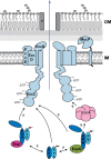
Working model for substrate recognition and targeting in type VII secretion. The three T7S substrate families, the Esp, Esx, and PE/PPE proteins, are generally exported as heterodimers by the T7S secretion machinery. While dimerization occurs via a conserved four-helix bundle fold, Esp, and PE/PPE proteins additionally contain highly variable C-terminal domains. Substrate recognition occurs via a C-terminal secretion motif on one of the dimer subunits (indicated by a red box). The cytosolic component EspG specifically recognizes a conserved hydrophobic helical tip in PPE proteins and possibly mediates targeting of PE/PPE substrates to the cognate membrane channel. Esp substrates also require binding of dedicated chaperones, possibly to a similar helical tip structure, to prevent their premature self-assembly. While the core membrane channel consists of the conserved membrane components EccB, EccC, EccD, and EccE (Houben et al.; Beckham et al.2017), the fifth conserved membrane component, the protease MycP, while essential for secretion, is not an integral part of this complex (van Winden et al. 2016). The three nucleotide binding domains of EccC are likely involved in energizing translocation of substrates through this channel. In this model, the T7S membrane complex mediates transport only across the inner membrane, while a so far unidentified separate channel mediates translocation across the outer membrane. The observed interaction between the secretion signal of an Esx pair with EccC in vitro (Champion et al.; Rosenberg et al.2015) is indicated by an arrow. Arrows with question marks indicate the potential interactions between the different substrate families, of the other substrates with EccC and of the postulated specific interaction of PE/PPE pairs with the cytosolic ATPase EccA. IM, inner membrane; OM, mycobacterial outer membrane.

References

    1. Abdallah AM, Verboom T, Hannes F et al. . A specific secretion system mediates PPE41 transport in pathogenic mycobacteria. Mol Microbiol 2006;62:667–79. - PubMed
    1. Abdallah AM, Verboom T, Weerdenburg EM et al. . PPE and PE_PGRS proteins of Mycobacterium marinum are transported via the type VII secretion system ESX-5. Mol Microbiol 2009;73:329–40. - PubMed
    1. Akopian D, Shen K, Zhang X et al. . Signal recognition particle: an essential protein-targeting machine. Annu Rev Biochem 2013;82:693–721. - PMC - PubMed
    1. Arbing MA, Kaufmann M, Phan T et al. . The crystal structure of the Mycobacterium tuberculosis Rv3019c-Rv3020c ESX complex reveals a domain-swapped heterotetramer. Protein Sci 2010;19:1692–703. - PMC - PubMed
    1. Ates LS, Dippenaar A, Ummels R et al. . Mutations in ppe38 block PE_PGRS secretion and increase virulence of Mycobacterium tuberculosis. Nat Microbiol 2018;3:181–8. - PubMed

Publication types

MeSH terms