. 2018 Sep 19;46(16):8471-8482.
doi: 10.1093/nar/gky701.

The human Obg protein GTPBP10 is involved in mitoribosomal biogenesis

Affiliations

The human Obg protein GTPBP10 is involved in mitoribosomal biogenesis

Elena Lavdovskaia et al. Nucleic Acids Res. .

Abstract

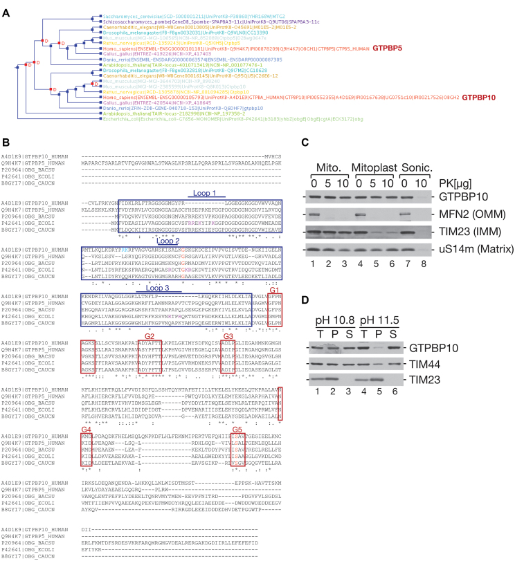
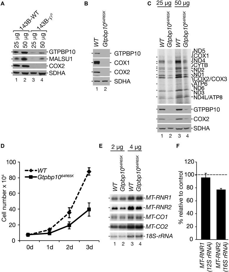
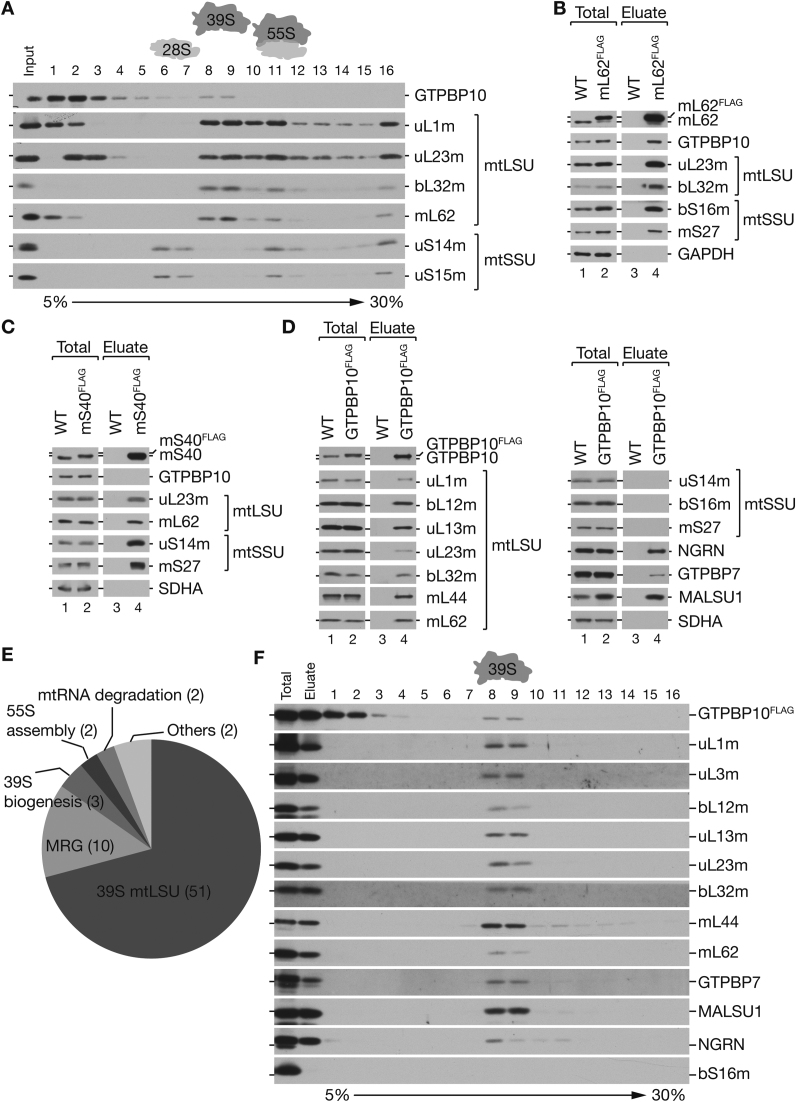
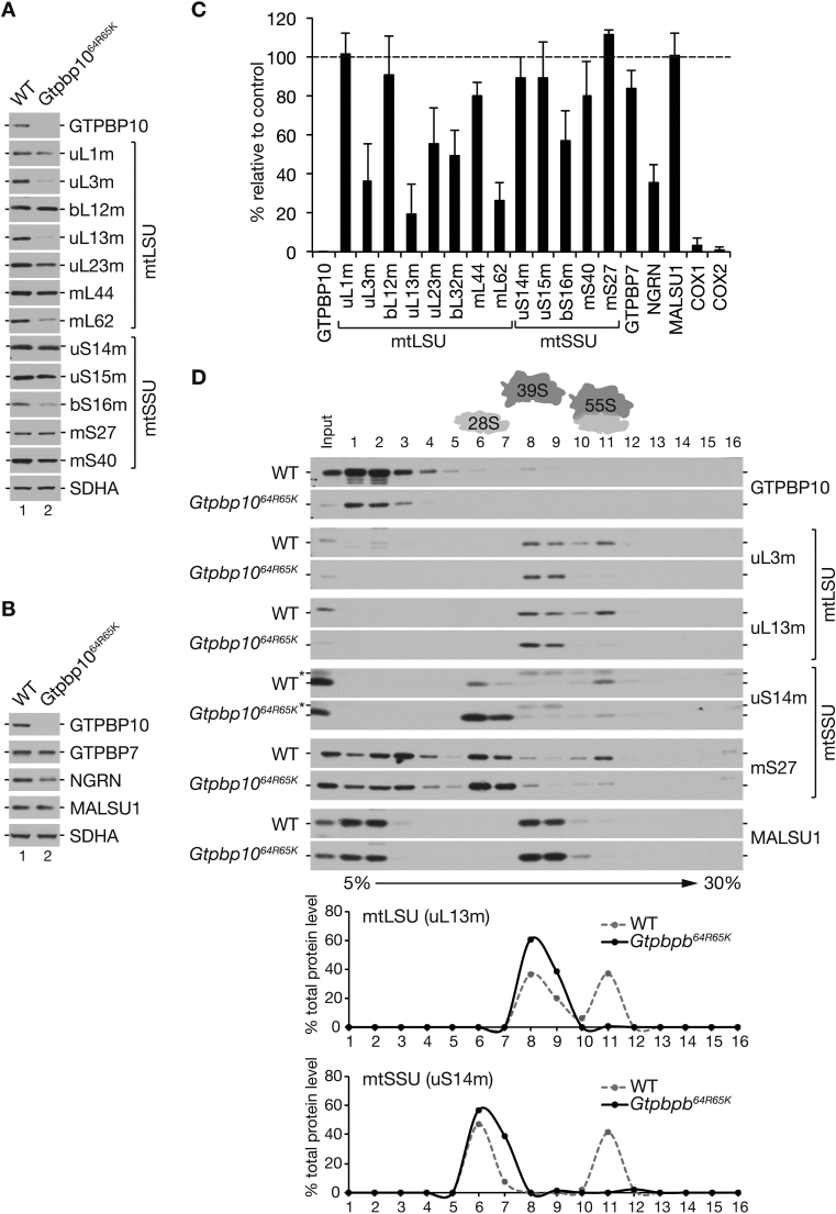
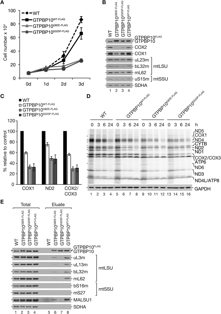
The human mitochondrial translation apparatus, which synthesizes the core subunits of the oxidative phosphorylation system, is of central interest as mutations in several genes encoding for mitoribosomal proteins or translation factors cause severe human diseases. Little is known, how this complex machinery assembles from nuclear-encoded protein components and mitochondrial-encoded RNAs, and which ancillary factors are required to form a functional mitoribosome. We have characterized the human Obg protein GTPBP10, which associates specifically with the mitoribosomal large subunit at a late maturation state. Defining its interactome, we have shown that GTPBP10 is in a complex with other mtLSU biogenesis factors including mitochondrial RNA granule components, the 16S rRNA module and late mtLSU assembly factors such as MALSU1, SMCR7L, MTERF4 and NSUN4. GTPBP10 deficiency leads to a drastic reduction in 55S monosome formation resulting in defective mtDNA-expression and in a decrease in cell growth. Our results suggest that GTPBP10 is a ribosome biogenesis factor of the mtLSU required for late stages of maturation.


Figures

Figure 1.
Figure 1.
GTPBP10 is a peripheral protein of the inner mitochondrial membrane belonging to the Obg-subfamily. (A) Cluster analysis of the Obg GTPase family based on P-POD – Princeton Protein Orthology Database (http://ppod.princeton.edu/). Human GTPBP5 and GTPBP10 are indicated in red. (B) Amino acid sequence alignment of GTPBP5 and GTPBP10 and their bacterial homologues. Blue and red boxes indicate Obg domain and the five motifs of the GTPase domain (G1–G5), respectively. Arginine 64 and lysine 65 (Gtpbp1064R65K), which were deleted in GTPBP10 using CRISPR/Cas9 (see below in the text), are labeled in blue. Amino acid substitutions for GTPBP10G82E-FLAG and GTPBP10S325P-FLAG are indicated in red. Purple shows amino acid substitutions in the E. coli ObgE protein, which abolish its function in bacterial ribosome assembly (9). (C) Localization of the GTPBP10. Isolated intact mitochondria (lanes 1-3), mitoplasts (lanes 4-6) and sonicated mitochondria (lanes 7,8) from HEK293T WT cells were treated with proteinase K as indicated. MFN2, TIM23 and uS14m were used as markers of the outer mitochondrial membrane (OMM), inner mitochondrial membrane (IMM) and matrix fraction, respectively. (D) GTPBP10 is a peripherally associated protein of the mitochondrial inner membrane. Carbonate extraction of mitochondrial membrane proteins at different pH from HEK293T WT cells. Fractions (T-total, P-pellet, S-supernatant) were analyzed by western blotting with specific antibodies as indicated.
Figure 2.
Figure 2.
GTPBP10 is involved in mitochondrial gene expression. (A) GTPBP10 is unstable in the absence of mtDNA. Protein steady state levels from 143B wild type (WT) or 143B-ρ0 cells were analyzed by western blotting. (B) Altered mitochondrial gene expression in Gtpbp1064R65K cells. Steady state levels of mtDNA-encoded proteins (COX1 and COX2) isolated from HEK293T WT or Gtpbp1064R65K cells. SDHA is used as a loading control. (C) Gtpbp1064R65K cells exhibit diminished mitochondrial translation. [35S]methionine de novo synthesized mtDNA-encoded proteins from HEK293T WT cells or Gtpbp1064R65K cells were visualized by autoradiography (upper panel) or with designated antibodies (lower panel). SDHA is used as a loading control (n = 3). (D) Ablation of GTPBP10 reduces growth rate. Equal numbers of HEK293T WT and Gtpbp1064R65K cells were seeded on day 0 (0d) and counted after 1 day (1d), 2 days (2d) and 3 days (3d) (mean ± SD, n = 3). (E and F) GTPBP10 is required for 16S rRNA stability. (E) Steady state levels of mtDNA-encoded RNAs extracted from HEK293T WT and Gtpbp1064R65K cells were analyzed by Northern blot with indicated probes. MT-RNR1: 12S rRNA; MT-RNR2: 16S rRNA; MT-CO1: mRNA encoding COX1; MT-CO2: mRNA encoding COX2. 18S-rRNA was used as a loading control. (F) MT-RNR1 and MT-RNR2 were quantified using ImageJ and normalized to 18S-rRNA (mean ± SEM; n = 3).
Figure 3.
Figure 3.
GTPBP10 interacts with the mtLSU and assembly factors. (A) GTPBP10 co-fractionates with mtLSU. Native protein complexes were isolated from HEK293T WT mitoplasts and separated by 5–30% sucrose gradient centrifugation. Fractions (1-16) were visualized by western blot with antibodies against mtLSU (uL1m, uL23m, bL32m, mL62) and mtSSU (uS14m, uS15m). (n = 4). (B-C) GTPBP10 associates specifically with mtLSU. Co-immunoprecipitation of FLAG-tagged mL62 (B) and mS40 (C). GAPDH and SDHA were used as negative controls for unspecific binding. Total, 3%; Eluate, 100% (n = 3). (D) Mitoribosomal proteins and biogenesis factors of the mtLSU co-purify with GTPBP10FLAG. Mitochondrial protein complexes containing GTPBP10 were co-purified via FLAG-tagged GTPBP10. Total, 3%; Eluate, 100% (n = 3). (E) The interactome of GTPBP10FLAG. Equal amounts of differentially labeled mitochondria from HEK293T WT and GTPBP10FLAG cells were mixed and applied to FLAG-immunoprecipitation. Native eluted complexes were analyzed by quantitative mass spectrometry. Diagram represents the results from four experiments (including label switch). P < 0.05; mean ratio ≥2 (n = 4). (F) GTPBP10 associates with mtLSU at a late assembly stage. FLAG-immunoprecipitation via GTPBP10FLAG was performed as in (D). Native eluate was subjected to 5–30% Sucrose gradients. Fractions were analyzed by western blot with specific antibodies against mitoribosomal proteins of the mtLSU and mtSSU, and assembly factors. Total (FLAG-immunoprecipitation input), 1%; Eluate (FLAG-immunoprecipitation eluate = gradient input), 10% (n = 3).
Figure 4.
Figure 4.
GTPBP10 is required for 55S monosome formation. (A-C) Gtpbp1064R65K leads to reduced protein levels of selected MRPs (A) and ribosome biogenesis factors (B). (C) Quantification of steady state analysis of MRPs and biogenesis factors in Gtpbp1064R65K cells relative to HEK293T WT control. SDHA was used as a loading control. (n = 3, mean ± SEM). (D) Ablation of GTPBP10 reduces monosome formation. Protein complexes from HEK293T WT and Gtpbp1064R65K mitoplasts were separated on 5–30% sucrose gradients and fractions (1-16) were analyzed by western blot with specific antibodies against mtSSU and mtLSU components and MALSU1. Protein distributions for uL13m (mtLSU) and uS14m (mtSSU) are presented as percentage of the total protein abundance. (*) indicates residual signals of bL32m.
Figure 5.
Figure 5.
Expression of Obg and GTPase domain mutants negatively affect the function of GTPBP10. (A) GTPBP10G80E and GTPBP10S325P affect cell growth. Equal numbers of HEK293T WT, GTPBP10G82E-FLAG, GTPBP10S325P-FLAG and GTPBP10WT-FLAG cells were seeded on day 0 (0d) and counted after 1 day (1d), 2 days (2d) or 3 days (3d) (n = 3, mean ± SD). (B) GTPBP10G80E and GTPBP10S325P affect the steady state level of mtDNA-encoded proteins. Proteins were extracted from indicated cell lines and analyzed by western blot. SDHA was used as a loading control. (C and D) Synthesis and stability of mtDNA-encoded proteins in GTPBP10 mutants. Cells were pulse labeled for 1h in the presence of [35S]Methionine (lanes 1, 5, 9 and 13) and chased for the indicated time points. Mitochondrial translation products from HEK293T WT cells or GTPBP10G82E-FLAG, GTPBP10S325P-FLAG and GTPBP10WT-FLAG cells were visualized by autoradiography (D, upper panel). GAPDH was used as a loading control. Newly synthesized COX1, ND2 and COX2/COX3 were quantified after 1h pulse labeling using ImageQuant TL (C) (mean ± SEM, n = 4). (E) GTPBP10 mutants show reduced mtLSU binding capacity. Lysed mitochondria from cell lines expressing FLAG-tagged GTPBP10G82E, GTPBP10S325P or wild type GTPBP10 were subjected to FLAG-immunoprecipitation. Samples were analyzed by western blot using indicated antibodies. SDHA was used as a negative control for unspecific binding. Total, 3%; Eluate, 10% (n = 2).

Similar articles

Cited by

References

    1. Boczonadi V., Horvath R.. Mitochondria: impaired mitochondrial translation in human disease. Int. J. Biochem. Cell Biol. 2014; 48:77–84. - PMC - PubMed
    1. Greber B.J., Ban N.. Structure and function of the mitochondrial ribosome. Annu. Rev. Biochem. 2016; 85:103–132. - PubMed
    1. Shajani Z., Sykes M.T., Williamson J.R.. Assembly of bacterial ribosomes. Annu. Rev. Biochem. 2011; 80:501–526. - PubMed
    1. Kint C., Verstraeten N., Hofkens J., Fauvart M., Michiels J.. Bacterial Obg proteins: GTPases at the nexus of protein and DNA synthesis. Crit. Rev. Microbiol. 2014; 40:207–224. - PubMed
    1. Jiang M., Sullivan S.M., Wout P.K., Maddock J.R.. G-protein control of the ribosome-associated stress response protein SpoT. J. Bacteriol. 2007; 189:6140–6147. - PMC - PubMed

Publication types

MeSH terms

LinkOut - more resources