

Liquid biopsy and PCR-free ultrasensitive detection systems in oncology (Review)
- PMID: 30085333
- PMCID: PMC6086621
- DOI: 10.3892/ijo.2018.4516
Liquid biopsy and PCR-free ultrasensitive detection systems in oncology (Review)
Abstract
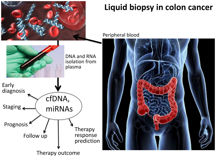
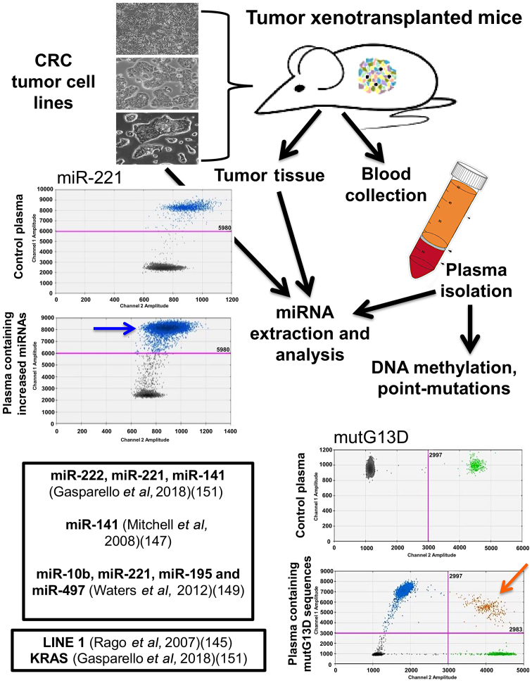
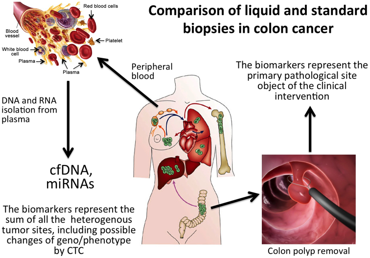
In oncology, liquid biopsy is used in the detection of next-generation analytes, such as tumor cells, cell-free nucleic acids and exosomes in peripheral blood and other body fluids from cancer patients. It is considered one of the most advanced non-invasive diagnostic systems to enable clinically relevant actions and implement precision medicine. Medical actions include, but are not limited to, early diagnosis, staging, prognosis, anticipation (lead time) and the prediction of therapy responses, as well as follow-up. Historically, the applications of liquid biopsy in cancer have focused on circulating tumor cells (CTCs). More recently, this analysis has been extended to circulating free DNA (cfDNA) and microRNAs (miRNAs or miRs) associated with cancer, with potential applications for development into multi-marker diagnostic, prognostic and therapeutic signatures. Liquid biopsies avoid some key limitations of conventional tumor tissue biopsies, including invasive tumor sampling, under-representation of tumor heterogeneity and poor description of clonal evolution during metastatic dissemination, strongly reducing the need for multiple sampling. On the other hand, this approach suffers from important drawbacks, i.e., the fragmentation of cfDNA, the instability of RNA, the low concentrations of certain analytes in body fluids and the confounding presence of normal, as well as aberrant DNAs and RNAs. For these reasons, the analysis of cfDNA has been mostly focused on mutations arising in, and pathognomonicity of, tumor DNA, while the analysis of cfRNA has been mostly focused on miRNA patterns strongly associated with neoplastic transformation/progression. This review lists some major applicative areas, briefly addresses how technology is bypassing liquid biopsy limitations, and places a particular emphasis on novel, PCR-free platforms. The ongoing collaborative efforts of major international consortia are reviewed. In addition to basic and applied research, we will consider technological transfer, including patents, patent applications and available information on clinical trials aimed at verifying the potential of liquid biopsy in cancer.
Figures
References
Publication types
MeSH terms
Substances
LinkOut - more resources
Full Text Sources
Other Literature Sources
