

Preoperative CT Findings and Interobserver Reliability of Fournier Gangrene
- PMID: 30085837
- PMCID: PMC6451931
- DOI: 10.2214/AJR.18.19683
Preoperative CT Findings and Interobserver Reliability of Fournier Gangrene
Abstract
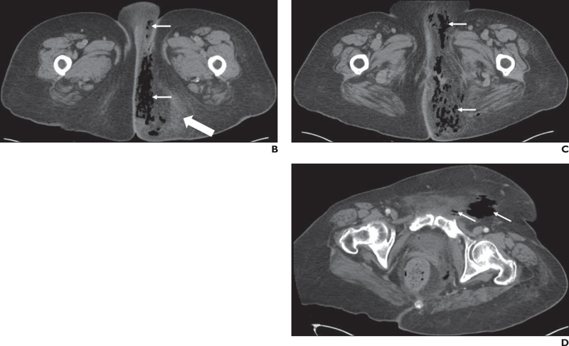
Objective: The objective of our study was to delineate CT findings and anatomic areas of involvement of surgically proven Fournier gangrene (FG) and determine interobserver reliability.
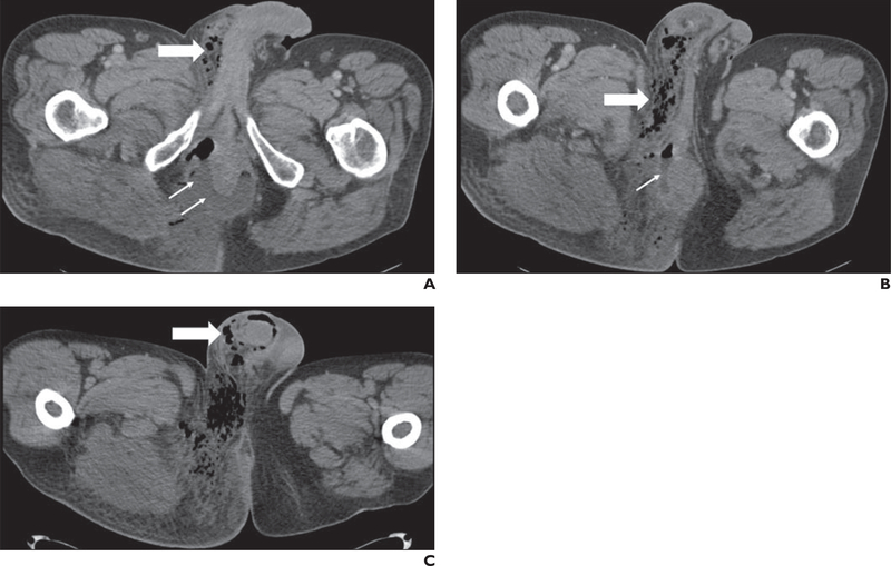
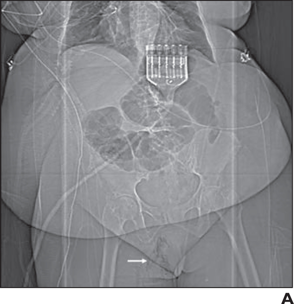
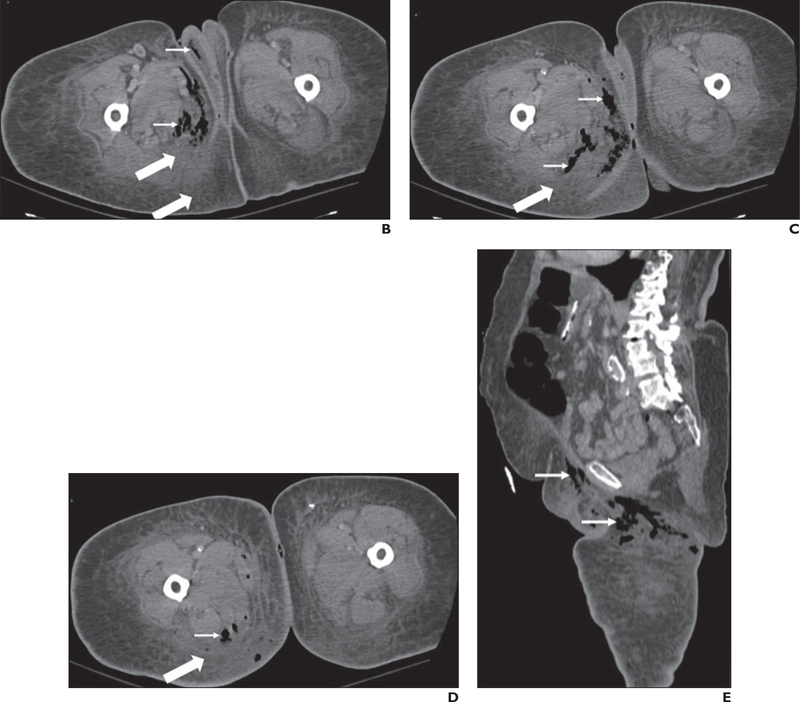
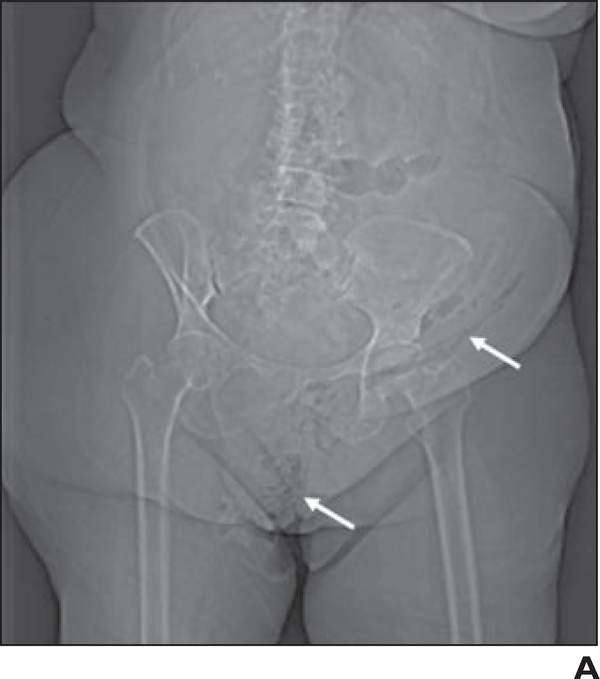
Materials and methods: This study was a single-center retrospective study of patients with FG who underwent CT before surgical débridement of FG during a 9-year period. Thirty-eight patients with FG, 17 male and 21 female patients, underwent preoperative CT. Two radiologists reviewed the CT studies and recorded findings and anatomic areas of involvement. CT findings were categorized according to a previously described CT scoring system for necrotizing fasciitis and included the presence or absence of fascial air, muscle or fascial edema, fluid tracking, lymphadenopathy, and subcutaneous edema. Cohen kappa was calculated for interobserver reliability.
Results: Mean body mass index (BMI [weight in kilograms divided by height in meters squared]) was 42, and 22 of 38 (58%) patients had diabetes. Mean BMI and proportion of patients with diabetes were significantly higher in female patients (mean BMI = 46; 16/21 with diabetes) than male patients (mean BMI = 36; 6/17 with diabetes). CT studies of most patients showed fascial air (36/38 [95%], both readers 1 and 2). Interobserver reliability was substantial to almost perfect for all CT findings except lymphadenopathy, for which it was fair (κ = 0.37). Genital, perineal, and ischiorectal involvement were seen in 87% (33/38), 87% (33/38), and 32% (12/38) of patients for reader 1 and 84% (32/38), 84% (32/38), and 26% (10/38) of patients for reader 2 (κ = 0.29, penis; κ = 0.65, scrotum; κ = 0.91, vulva and labia; κ = 0.68, perineal involvement; κ = 0.80, ischiorectal involvement).
Conclusion: Most CT findings of FG and anatomic areas of involvement showed good interobserver reliability. A high proportion of female patients with FG were observed in this study compared with prior series.
Keywords: CT; Fournier gangrene; acute care surgery; necrotizing fasciitis; radiology.
Figures
Similar articles
-
Fournier Gangrene in Men and Women: Appearance on CT, Ultrasound, and MRI and What the Surgeon Wants to Know.Can Assoc Radiol J. 2020 Feb;71(1):30-39. doi: 10.1177/0846537119888396. Epub 2020 Jan 28. Can Assoc Radiol J. 2020. PMID: 32063012 Free PMC article. Review.
-
Preoperative computed tomography in Fournier's gangrene does not delay time to surgery.Emerg Radiol. 2023 Dec;30(6):711-717. doi: 10.1007/s10140-023-02177-y. Epub 2023 Oct 19. Emerg Radiol. 2023. PMID: 37857761
-
Volumetric computed tomography measurements as predictors for outcomes in a cohort of Fournier's gangrene patients.Emerg Radiol. 2024 Aug;31(4):481-489. doi: 10.1007/s10140-024-02251-z. Epub 2024 Jun 6. Emerg Radiol. 2024. PMID: 38842620
-
Fournier gangrene: role of imaging.Radiographics. 2008 Mar-Apr;28(2):519-28. doi: 10.1148/rg.282075048. Radiographics. 2008. PMID: 18349455 Review.
-
CT of Gastric Volvulus: Interobserver Reliability, Radiologists' Accuracy, and Imaging Findings.AJR Am J Roentgenol. 2019 Jan;212(1):103-108. doi: 10.2214/AJR.18.20033. Epub 2018 Nov 7. AJR Am J Roentgenol. 2019. PMID: 30403524 Free PMC article.
Cited by
-
Fournier Gangrene in Men and Women: Appearance on CT, Ultrasound, and MRI and What the Surgeon Wants to Know.Can Assoc Radiol J. 2020 Feb;71(1):30-39. doi: 10.1177/0846537119888396. Epub 2020 Jan 28. Can Assoc Radiol J. 2020. PMID: 32063012 Free PMC article. Review.
-
Multimodality Imaging Review of Anorectal and Perirectal Diseases with Clinical, Histologic, Endoscopic, and Operative Correlation, Part II: Infectious, Inflammatory, Congenital, and Vascular Conditions.Curr Probl Diagn Radiol. 2019 Nov-Dec;48(6):563-575. doi: 10.1067/j.cpradiol.2018.07.013. Epub 2018 Jul 29. Curr Probl Diagn Radiol. 2019. PMID: 30154030 Free PMC article. Review.
-
Preoperative computed tomography in Fournier's gangrene does not delay time to surgery.Emerg Radiol. 2023 Dec;30(6):711-717. doi: 10.1007/s10140-023-02177-y. Epub 2023 Oct 19. Emerg Radiol. 2023. PMID: 37857761
-
Presence of Deep Fascial Fluid in Imaging Necrotizing Fasciitis: Importance of This Feature in Diagnosing Necrotizing Fasciitis without Imaging Manifestations of Soft Tissue Gas.Ultrasound Med Biol. 2019 Nov;45(11):3081. doi: 10.1016/j.ultrasmedbio.2019.08.004. Epub 2019 Aug 30. Ultrasound Med Biol. 2019. PMID: 31477369 Free PMC article. No abstract available.
-
Volumetric computed tomography measurements as predictors for outcomes in a cohort of Fournier's gangrene patients.Emerg Radiol. 2024 Aug;31(4):481-489. doi: 10.1007/s10140-024-02251-z. Epub 2024 Jun 6. Emerg Radiol. 2024. PMID: 38842620
References
-
- Eke N Fournier’s gangrene: a review of 1726 cases. Br J Surg 2000; 87:718–728 - PubMed
-
- Gürdal M, Yücebas E, Tekin A, Beysel M, Aslan R, Sengör F. Predisposing factors and treatment outcome in Fournier’s gangrene: analysis of 28 cases. Urol Int 2003; 70:286–290 - PubMed
-
- McGillicuddy EA, Lischuk AW, Schuster KM, et al. Development of a computed tomography: based scoring system for necrotizing soft-tissue infections. J Trauma 2011; 70:894–899 - PubMed
-
- Wong CH, Khin LW, Heng KS, Tan KC, Low CO. The LRINEC (Laboratory Risk Indicator for Necrotizing Fasciitis) score: a tool for distinguishing necrotizing fasciitis from other soft tissue infections. Crit Care Med 2004; 32:1535–1541 - PubMed
MeSH terms
Substances
Grants and funding
LinkOut - more resources
Full Text Sources
Other Literature Sources
Medical
