. 2018 Dec;194(3):283-294.
doi: 10.1111/cei.13196. Epub 2018 Sep 23.

Facilitated expansion of Th17 cells in lupus nephritis patients

Affiliations

Facilitated expansion of Th17 cells in lupus nephritis patients

B Jakiela et al. Clin Exp Immunol. 2018 Dec.

Abstract

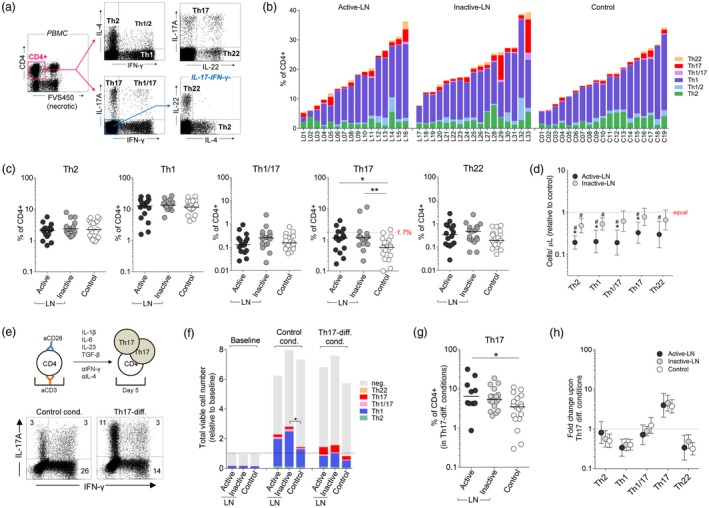
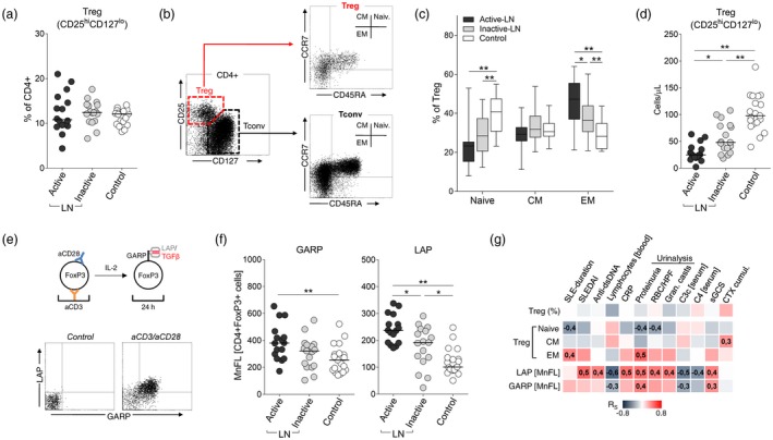
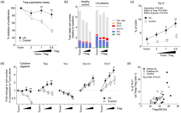
The objective of this study was to investigate the mechanisms of T helper type 17 (Th17) expansion in lupus nephritis (LN) patients, and to determine whether or not it is associated with impaired function of regulatory T cells (Treg ). Major effector subsets of peripheral blood CD4+ T cells were assessed by flow cytometry in 33 LN patients with different activity of the disease and 19 healthy controls. The percentage of circulating Th17 cells was increased in LN (median = 1·2% of CD4+ compared to 0·6% in the control group, P < 0·01), while Treg cells remained unchanged (12·3 versus 12·1% in controls), resulting in a significantly lower Treg /Th17 ratio. Th17 expansion in the patient group was not related to LN activity, renal histology or blood and urine inflammatory biomarkers, but has been associated with a higher cumulative dose of cyclophosphamide. Treg cells in LN displayed mainly effector memory phenotype and expressed higher levels of transforming growth factor (TGF)-β; however, their suppressant activity in lymphocyte proliferation assay was diminished compared to controls (~fourfold, P < 0·05). Co-culture of Treg and conventional CD4+ T cells resulted in marked suppression of the Th1 subset in both of the groups studied, but also in a potent expansion of Th17 cells, which in LN was twofold higher, as in controls (P < 0·05). In conclusion, our results demonstrate that Th17 expansion in LN is not increased during disease exacerbation, but is related to chronic immunosuppressive therapy. This immune signature is probably linked to the abnormal function of Treg cells, which were less suppressive in LN patients and even facilitated differentiation of Th17 cells.

Keywords: Th17 cells; lupus nephritis; regulatory T cells; systemic lupus erythematosus.


Figures

Figure 1
Figure 1
Cytokine production by in‐vitro activated CD4+ T cells. (a) Peripheral blood mononuclear cells (PBMC) were stimulated with phorbol myristate acetate (PMA)/ionomycin and intracellulary stained for T effector signature cytokines. Representative flow cytometry dot‐plots and gating strategy used to identify cytokine producing CD4+ T cells. (b) Graph showing cumulative percentages of major functional subsets of CD4+ T cells in individual lupus nephritis (LN) patients and controls. (c) The percentage of CD4+ T cells with certain functional phenotypes: T helper type 2 (Th2), Th1, Th1/17, Th17 (dashed line  =  90th percentile of the control group) and Th22 (Th1/2 cells not shown). Horizontal bars represent medians. *P < 0·05, **P < 0·01. (d) Absolute numbers of CD4+ T cell subtypes (peripheral blood). Data presented as fold difference in comparison to median value in the healthy controls group. #P < 0·05 in comparison to the control group; *P < 0·05 in comparison to inactive LN. (e) Th17‐expansion assay. Magnetically separated CD4+CD25 T cells (i.e. conventional CD4+ T cells) were stimulated in vitro with aCD3/aCD28 and cultured for 5 days in medium alone or in the presence of cytokines inducing Th17‐differentiation and blocking immunoglobulin (Ig)G (as indicated). Representative flow cytometry dot plots showing distribution of Th17 [interferon (IFN)‐γ IL‐17A+], Th1 (IFN‐γ+ IL‐17A‒) and Th1/17 (both positive) cells upon culture in Th17‐prone conditions [both graphs show 50 000 events (viable CD4+ gate)]. (f) The cumulative numbers (mean values) of cytokine positive CD4+ T cells (neg.  =  cytokine‐negative) in different culture conditions (data presented as relative to baseline, i.e. value 2 means twofold increase in the cell count after 5 days of culture). *P < 0·05 significant difference in the number of cytokine‐positive cells (all combined). (g) Percentage of Th17 cells after in‐vitro expansion in cytokine milieu [as in (e)]. *P < 0·05. (h) Comparable increase in the number of Th17 cells during in‐vitro cell culture of CD4+ Tconv cells from LN patients and controls. Data presented as mean change (95% confidence interval) in comparison to control conditions. There was a significant (P < 0·01) increase in the number of Th17 cells and a decrease of Th1, Th2 (except active LN) and Th22 cells in cytokine conditions (statistics symbols not shown for clarity).
Figure 2
Figure 2
Regulatory T cell (Treg) phenotype and function. (a) No difference in the percentage of peripheral blood CD25hiCD127lo CD4+ cells (Treg) between lupus nephritis (LN) patients and control subjects. (b) Flow cytometry (FC) gating strategy to identify memory (EM  =  effector memory, CM  =  central memory) subsets of Treg and conventional T cells (Tconv). (c) Decreased fraction of naive and increase in EM Treg cells (gated on CD4+CD25hiCD127lo) in LN patients. *P < 0·05, **P < 0·01. (d) Treg cell counts (peripheral blood) in LN patients and control subjects. (e) Transforming growth factor (TGF)‐β expression by in‐vitro activated Treg cells. Peripheral blood mononuclear cells (PBMC) were activated (aCD3/aCD28) for 24 h in the presence of interleukin (IL)‐2, stained for surface latency‐associated peptide (LAP) and glycoprotein A repetitions predominant (GARP) and intracellular forkhead box protein 3 (FoxP3), and analysed by flow cytometry (FC). Dot‐plots show expression of LAP and GARP in CD4+FoxP3+ cells in different culture conditions. (f) Higher surface expression of GARP and LAP by in‐vitro stimulated lymphocytes from active LN patients. Data presented as mean fluorescence intensity (MFI) in the FoxP3+CD4+ gate. *P < 0·05, **P < 0·01. (g) Matrix of correlation coefficients (R S – Spearman) in cross‐comparison of Treg phenotype [naive, CM and effector memory (EM) (% of Treg)], and expression of LAP/GARP, with major clinical and laboratory measures of LN. Data from active and inactive LN were combined. Numerical values indicate significant (P < 0·05) correlations.
Figure 3
Figure 3
Regulatory T cell (Treg) suppression in lupus nephritis (LN). (a) Suppression assay. Activated (aCD3/aCD28) CD4+CD25 (Tconv) and CD4+CD25+ (Treg) cells were cultured for 5 days at different ratios (triangle). Results are presented as % inhibition of proliferation (compared to Tconv alone). LN data were combined, as there was no difference between active and inactive patients. **P < 0·01 compared to control subjects. (b) Influence of Treg on CD4+ T cell proliferation and cytokine production. Stacked graph summarizes change in numbers of cytokine‐producing Tconv cells (neg.  =  cytokine negative) at day 5 compared to baseline (day 0). Experimental setup as in (a). (c) Percentage of T helper type 17 (Th17) cells in CD4+ T cell cultures (day 5) in LN patients (data combined) and control. Two‐way analysis of variance (anova) statistics and post‐hoc tests: *P < 0·05, **P < 0·01 in comparison to the control group. (d) Fold change in the number of cytokine producing CD4+ T cell subsets at different Tconv : Treg ratios [as in (a)]. Data were standardized to experimental control (Tconv alone). *P < 0·05, **P < 0·01 compared to control group (anova and post‐hoc test). (e) Correlation between the percentage of effector memory (EM) Treg cells and the fraction of Th17 cells in the cell culture at a 1 : 1 Tconv:Treg ratio.

References

    1. Kaul A, Gordon C, Crow MK et al. Systemic lupus erythematosus. Nat Rev Dis Primers 2016;2:16039. - PubMed
    1. Borchers AT, Leibushor N, Naguwa SM, Cheema GS, Shoenfeld Y, Gershwin ME. Lupus nephritis: a critical review. Autoimmun Rev 2012;12:174–94. - PubMed
    1. Krebs CF, Schmidt T, Riedel JH, Panzer U. T helper type 17 cells in immune‐mediated glomerular disease. Nat Rev Nephrol 2017;13:647–59. - PubMed
    1. Hemdan NY, Birkenmeier G, Wichmann G, Abu El‐Saad AM, Krieger T, Conrad K, Sack U. Interleukin‐17‐producing T helper cells in autoimmunity. Autoimmun Rev 2010;9:785–92. - PubMed
    1. Burkett PR, Meyer zu Horste G, Kuchroo VK. Pouring fuel on the fire: Th17 cells, the environment, and autoimmunity. J Clin Invest 2015;125:2211–9. - PMC - PubMed

Publication types

Substances