Review
. 2019 Feb;145(Pt B):230-246.
doi: 10.1016/j.neuropharm.2018.08.004. Epub 2018 Aug 4.

Pathophysiology and treatment of cerebral edema in traumatic brain injury

Affiliations
Review

Pathophysiology and treatment of cerebral edema in traumatic brain injury

Ruchira M Jha et al. Neuropharmacology. 2019 Feb.

Abstract

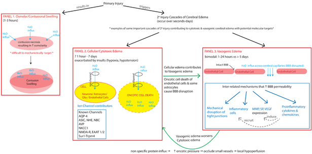
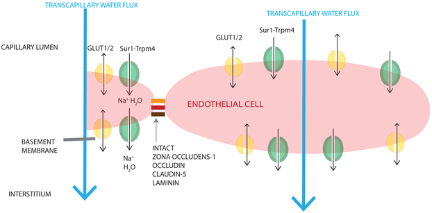
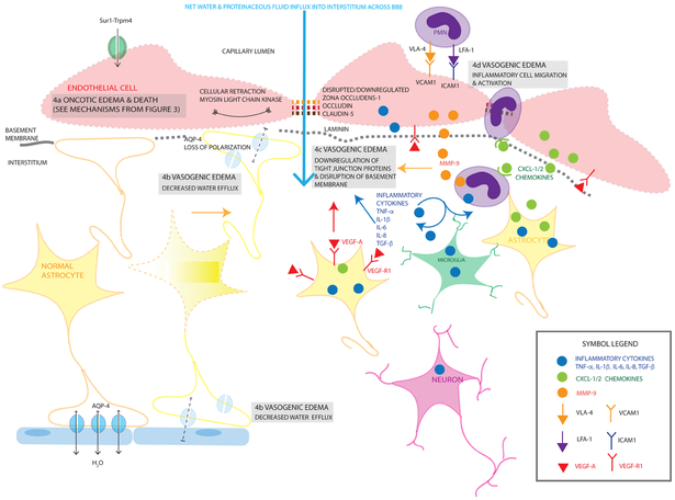
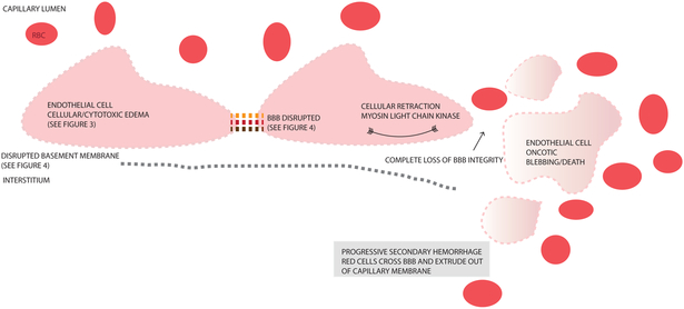
Cerebral edema (CE) and resultant intracranial hypertension are associated with unfavorable prognosis in traumatic brain injury (TBI). CE is a leading cause of in-hospital mortality, occurring in >60% of patients with mass lesions, and ∼15% of those with normal initial computed tomography scans. After treatment of mass lesions in severe TBI, an important focus of acute neurocritical care is evaluating and managing the secondary injury process of CE and resultant intracranial hypertension. This review focuses on a contemporary understanding of various pathophysiologic pathways contributing to CE, with a subsequent description of potential targeted therapies. There is a discussion of identified cellular/cytotoxic contributors to CE, as well as mechanisms that influence blood-brain-barrier (BBB) disruption/vasogenic edema, with the caveat that this distinction may be somewhat artificial since molecular processes contributing to these pathways are interrelated. While an exhaustive discussion of all pathways with putative contributions to CE is beyond the scope of this review, the roles of some key contributors are highlighted, and references are provided for further details. Potential future molecular targets for treating CE are presented based on pathophysiologic mechanisms. We thus aim to provide a translational synopsis of present and future strategies targeting CE after TBI in the context of a paradigm shift towards precision medicine. This article is part of the Special Issue entitled "Novel Treatments for Traumatic Brain Injury".

Keywords: Cerebral edema; Cytotoxic edema; Ionic edema; Traumatic brain injury; Vasogenic edema.


Figures

Figure 1.
Figure 1.. Schema of some key contributors to intracranial swelling after TBI
This diagram highlights different pathways that contribute to intracranial swelling and cerebral edema (CE). Panel 1: Immediately after primary injury, there may be osmolar/contusional swelling. As described by Katayama et al (1992), the necrotic core of the contusion has high osmolarity that drives water movement along this gradient (direction of water movement shown by light blue arrows). This results in acute swelling of the contusion. Primary injury simultaneously triggers many cascades of secondary injury including cellular/cytotoxic edema (CytE, Panel 2) and vasogenic edema (VasE, Panel 3). Some important examples of CytE (Panel 2) include the activation/upregulation of various ion channels (some known channels include AQP4, ASIC, NHE, NBC, AVP, NKCC1, NMDA-R, Sur1-Trpm4). These channels allow water influx into different cell types depending on the cell’s expression of the respective channel. Excessive ion-channel related water influx (light blue arrow) into a cell (yellow oval) may result in oncotic cell death. This occurs in neurons/astrocytes/glia and can occur within 1 hour −7 days. When this process occurs in cells contributing to blood-brain-barrier (BBB) integrity like endothelial cells/some astrocytes, it contributes to VasE (straight green arrow showing relationship between CytE in Panel 2 and VasE in Panel 3). Secondary injury cascades also include additional processes that disrupt the BBB, result in water movement across the disrupted BBB capillaries (light blue arrows) and cause VasE (Panel 3). These cascades include mechanical disruption, release of proinflammatory cytokines and chemokines that recruit migration/activation of inflammatory cells, increased expression and release of factors (such as MMP, SP, VEGF) that disrupt tight junctions and basement membrane proteins. VasE, with its proteinaceous fluid influx, may further raise oncotic pressure in the interstitium, occluding small vessels causing local hypoperfusion, which in turn may further exacerbate CytE development and upregulation of ion channels like Sur1-Trpm4. This relationship is shown by the curved green arrow between Panels 2 & 3). AQP-4 = aquaporin 4, ASIC =Acid sensing ion channel, NHE = Na+/H+ exchanger, NBC = Na+/HCO3 transporter family channel, AVP = arginine vasopressin, NKCC1 = Na+-K+-2Cl cotransporter, Sur1 = sulfonylurea receptor 1, Trpm4 = transient receptor potential cation channel subfamily M member 4, MMP = matrix metalloproteinase, SP = substance P, VEGF = vascular endothelial growth factor
Figure 2.
Figure 2.. Mechanisms contributing to ionic edema after TBI
The diagram is oriented such that the capillary lumen is superior to the endothelial cell, and the interstitium and neuronal/glial tissue is inferior. Ionic edema involves the transcapillary net water flux (large light blue arrow) from the capillary lumen to the brain interstitium without violation of the BBB or basement membrane. Like cellular/cytotoxic edema (CytE), water is transported through luminal ion channels (e.g. GLUT1/2 (shown in yellow), Sur1-Trpm4 (shown in green), NKCC1, NHE1/2, ASIC, NBC, EAAT1/2, SGLT1) into the endothelial cell; water is subsequently transported out of the endothelial cells by channels expressed abluminally (GLUT1/2 (yellow), Sur1-Trpm4 (green)) primarily due to an osmotic gradient created by CytE. Single arrows (black) are used for water co-transport, double headed arrows are used for passive water movement through a channel pore.
Figure 3.
Figure 3.. Mechanisms contributing to cellular/cytotoxic edema after TBI
3a Endothelial (light pink cells) cellular swelling results from net water influx (large light blue arrow) into an endothelial cell driven by ionic contributors including GLUT1/2 (yellow channel), Sur1-Trpm4 (green channel), NKCC1 (light green channel), NHE1/2 (pink channel), ASIC (pink channel), NBC (purple channel), EAAT1/2 (red channel), SGLT1 (orange channel)). Sur1-Trpm4 cotransports Na+ with water into the cell, NKCC1 transports 1 Na+, 1 K+, 2 Cl with 1 molecule of water into the cell, NHE 1/2 transports Na+ into the cell (with 1 molecule of water) and H+ out of the cell. NBC transports Na++ and HCO3 into the cell. EAAT1/2 transport 1 Na+ and 1 glutamate into the cell with 1 molecule of water. GLUT1/2 and SGLT1 transport glucose and Na+ into the cell respectively with water passively following. At this stage, the tight junctions are intact (solid orange/red/brown links between endothelial cells) as is the basement membrane (solid gray line under the endothelial layer). Cellular swelling/CytE in neurons (dark pink cells, 3b) and astrocytes (yellow cells, 3c) occurs by the same mechanisms with the same ion pumps and channels resulting in Na+ and water entering the cells causing cellular swelling (large light blue arrows). Eventually, this cellular swelling becomes cytotoxic and causes cell blebbing and oncotic death illustrated using lightened colors and dashed membrane outlines. Astrocytic endfeet also express AQP-4 at the blood-brain and blood-CSF junctions that allow passive water transport driven by an osmotic or pressure gradient. During cellular swelling, this gradient may direct water inwards. However, during vasogenic edema, these channels may be important in water egress from the cells as shown in Figure 4. Single arrows (black) are used for water co-transport, double headed arrows are used for passive water movement through a channel pore.
Figure 4.
Figure 4.. Mechanisms contributing to vasogenic edema after TBI
Vasogenic edema (VasE) results from BBB compromise resulting in net water and proteinaceous fluid influx into the interstitium (large light blue arrow). There are multiple contributors to this process including cellular retraction (via actin and myosin light chain kinase contraction of the cytoskeleton), cytotoxic edema in endothelial cells resulting in membrane disruption and eventual oncotic death (4a), decreased water efflux (4b), degradation of tight junction proteins (4c) and activation of inflammatory cells (4d). Mechanistic details of oncotic edema in endothelial cells (4a) are depicted earlier in Figure 3. Decreased water efflux (4b) also contributes to VasE; one potential mechanism illustrated here is by a loss of polarization of AQP-4 channels in the perivascular and peri-ependymal endfeet (depicted with a faded blue AQP4 channel). Additionally, as astrocytes undergo cell blebbing/oncotic cell death, their endfeet are no longer able to contribute towards sealing the BBB, furthering VasE formation. Another contributor to VasE is due to downregulation of tight junction proteins and disruption of the basement membrane (4c). Tight junction degradation involves proteins such as zona occludens, occludin, laminin shown by the interrupted links (orange/red/brown) between endothelial cells. Disrupted laminin/basement membrane is shown by the dashed-gray line under the endothelial cells. This occurs via multiple proposed mechanisms including MMP-9 (orange filled circles) produced primarily by neutrophils (purple cells) and endothelial cells, inflammatory cytokines (blue filled circles, TNF, IL-1β, IL-6, IL-8, TGF-β) produced by multiple cell types activated by injury including neurons, microglia, astrocytes, endothelial cells and migrating leukocytes, and VEGF-A (red filled triangle) and VEGF-R1 (red-Y shaped receptor) upregulation by multiple cell types including endothelial cells and astrocytes. Activation of and migration of inflammatory cells further exacerbates VasE (4d). PMNs adhere to the luminal endothelial cell surface via upregulated receptors/ligands such as VCAM-1 (orange Y-shaped receptor) and VLA-4 (orange filled arrowhead), ICAM-1 (purple Y-shaped receptor) and LFA-1 (purple filled arrowhead). Their migration into the interstitium is facilitated by chemokines (CXCL-1/2 for PMNs, green filled circles) secreted by astrocytes and other activated cells, and upregulated chemokine receptors. Each channel/cytokine/cell-type/chemokine/receptor is labelled once, and the same color and shape scheme is maintained for each item throughout the diagram with a legend provided in the inset panel. Cell types are labelled and color-coded in the figure: endothelial cells (light red), astrocytes (yellow), neurons (blue), polymorphonuclear leukocytes (PMN, purple), microglia (green). Single arrows (black) are used for water co-transport, double headed arrows are used for passive water movement through a channel pore.
Figure 5.
Figure 5.. Progressive Secondary Hemorrhage
This figure depicts consequences of blood brain barrier (BBB) breakdown with loss of tight junctions (interrupted orange/red/brown links between endothelial cells), disrupted basement membrane, retraction of endothelial cells, and oncotic cell blebbing/death of endothelial cells. This ultimately destroys all BBB integrity, allowing extravasation of blood components across the capillary membrane eventually resulting in progressive secondary hemorrhage (PSH). Details of the precursor CytE and VasE mechanisms that lead up to these processes and PSH are outlined in Figures 3 and 4.

References

    1. Allen CJ, Subhawong TK, Hanna MM, Chelala L, Bullock MR, Schulman CI, Proctor KG, 2018. Does Vasopressin Exacerbate Cerebral Edema in Patients with Severe Traumatic Brain Injury? Am. Surg 84, 43–50. - PubMed
    1. Alluri H, Wilson RL, Anasooya Shaji C, Wiggins-Dohlvik K, Patel S, Liu Y, Peng X, Beeram MR, Davis ML, Huang JH, Tharakan B, 2016. Melatonin Preserves Blood-Brain Barrier Integrity and Permeability via Matrix Metalloproteinase-9 Inhibition. PLoS One 11, e0154427. doi:10.1371/journal.pone.0154427 - DOI - PMC - PubMed
    1. Alperin NJ, Lee SH, Loth F, Raksin PB, Lichtor T, 2000. MR-Intracranial pressure (ICP): a method to measure intracranial elastance and pressure noninvasively by means of MR imaging: baboon and human study. Radiology 217, 877–885. doi:10.1148/radiology.217.3.r00dc42877 - DOI - PubMed
    1. Ameliorate JL, Ghabriel MN, Vink R, 2017. Magnesium enhances the beneficial effects of NK1 antagonist administration on blood-brain barrier permeability and motor outcome after traumatic brain injury. Magnes. Res 30, 88–97. doi:10.1684/mrh.2017.0427 - DOI - PubMed
    1. Amtul Z, Yang J, Nikolova S, Lee T-Y, Bartha R, Cechetto DF, 2018. The Dynamics of Impaired Blood-Brain Barrier Restoration in a Rat Model of Co-morbid Injury. Mol. Neurobiol doi:10.1007/s12035-018-0904-4 - DOI - PubMed

Publication types

MeSH terms

Substances