. 2018 Dec 15;24(24):6308-6318.
doi: 10.1158/1078-0432.CCR-17-3535. Epub 2018 Aug 7.

Loss of Function of Canonical Notch Signaling Drives Head and Neck Carcinogenesis

Affiliations

Loss of Function of Canonical Notch Signaling Drives Head and Neck Carcinogenesis

Patrick E Nyman et al. Clin Cancer Res. .

Abstract

Purpose: Head and neck squamous cell carcinoma (HNSCC), a common cancer worldwide, is etiologically associated with tobacco use, high alcohol consumption, and high-risk human papillomaviruses (HPV). The Notch signaling pathway, which is involved in cell differentiation decisions with differential downstream targets and effects depending on tissue type and developmental stage, has been implicated in human HNSCC. NOTCH1 is among the most frequently mutated genes in both HPV-positive and HPV-negative HNSCC. These mutations are predicted to inactivate the function of Notch. Other studies have argued the opposite-Notch signaling is increased in HNSCC.

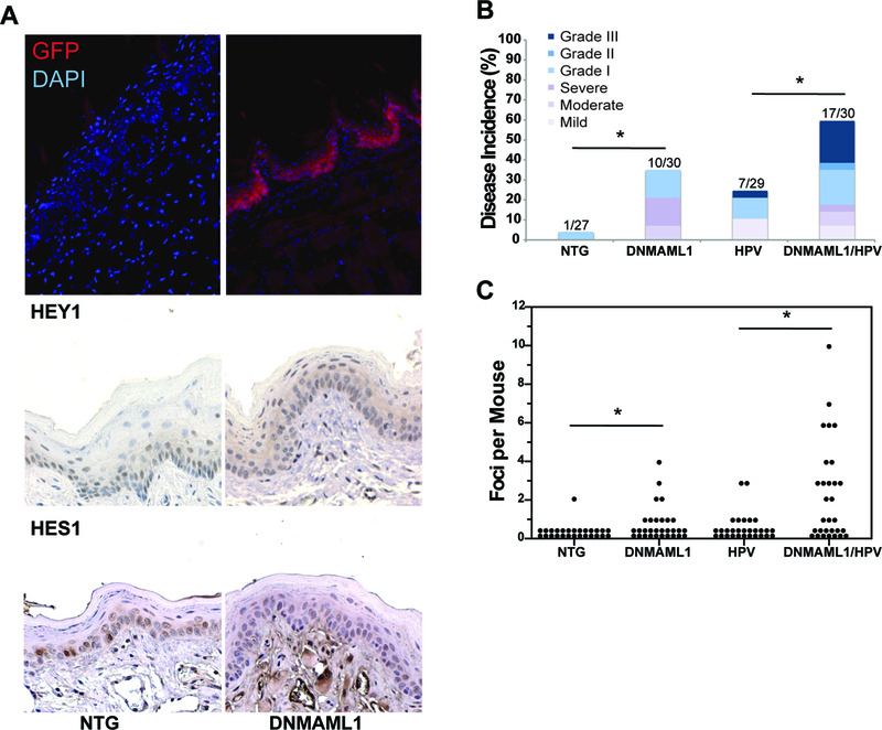
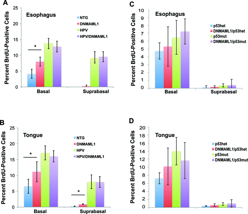
Experimental design: To assess the role of Notch signaling in HPV-positive and HPV-negative HNSCC, we utilized genetically engineered mouse (GEM) models for conventional keratinizing HNSCC, in which either HPV16 E6 and E7 oncoproteins or a gain-of-function mutant p53 are expressed, and in which we inactivated canonical Notch signaling via expression of a dominant negative form of MAML1 (DNMAML1), a required transcriptional coactivator of Notch signaling.

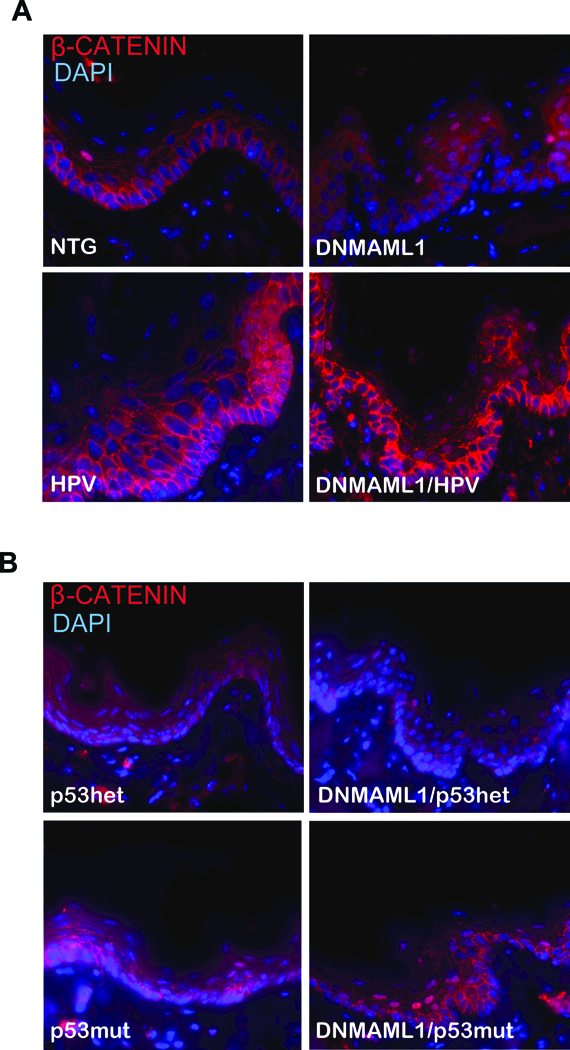
Results: Loss of canonical Notch signaling increased tumorigenesis in both contexts and also caused an increase in nuclear β-catenin, a marker for increased tumorigenic potential. When combined with loss of canonical Notch signaling, HPV oncogenes led to the highest frequency of cancers overall and the largest number of poorly differentiated (high-grade) cancers.

Conclusions: These findings inform on the contribution of loss of canonical Notch signaling in head and neck carcinogenesis.


Conflict of interest statement

The authors declare no potential conflicts of interest

Figures

Figure 1:
Figure 1:
A) Analysis of DNMAML1 gene expression upon K14CreER™ activation. To confirm DNMAML1 expression, immunofluorescence was performed against GFP (red), which is fused to the DNMAML1 gene, with a DAPI counterstain (blue). Expression of HEY1 and HES1, two Notch-dependent proteins, are shown via immunohistochemistry as evidence of lost Notch signaling upon DNMAML1 expression. Representative images of the tongue are shown (200x). B) Gross Tumor Incidence in the tongue and esophagus separated by histological grade of disease (mild-severe dysplasia and grade I-III carcinoma) for all mice in the HPV-positive cohort. Following 4NQO treatment, mice were sacrificed, and the tongue and esophagus were scored for grossly visible lesions. Incidence p-value via Fisher’s Exact Test: (NTG versus DNMAML1) = 0.04, (NTG versus HPV) = 0.052, (DNMAML1 versus DNMAML1/HPV) = 0.119, (HPV versus DNMAML1/HPV) = 0.017. C) Tumor Multiplicity in the tongue and esophagus for mice in the HPV-positive cohort. Tumor multiplicity was assessed by pathologist blinded to treatment groups as the number of discrete tumor foci separated by normal tissue. p-value via Wilcoxon Rank Sum Test: (NTG versus DNMAML1) = 0.006, (NTG versus HPV) = 0.039, (DNMAML1 versus DNMAML1/HPV) = 0.012, (HPV versus DNMAML1/HPV) = 0.001.
Figure 2:
Figure 2:
A) Gross Tumor Incidence in the tongue and esophagus separated by histological grade of disease for mice in the HPV-negative cohort (mild-severe dysplasia and grade I-sarcomatoid carcinoma). Incidence p-value via Fisher’s Exact Test: (p53mut versus DNMAML1/p53mut) = 0.010, (DNMAML1/p53het versus DNMAML1/p53mut) = 0.076. B) Tumor Multiplicity in the tongue and esophagus for all mice in the HPV-negative cohort. p-value via Wilcoxon Rank Sum Test: (p53mut versus DNMAML1/p53mut) = 0.002, (DNMAML1/p53het versus DNMAML1/p53mut) = 0.047. (C) Cancer Multiplicity for mice that developed cancer in the tongue and esophagus for HPV, DNMAML1/HPV, p53mut and DNMAML1/p53mut mice. Among the mice that developed cancer, DNMAML1/HPV mice had significantly more cancers than DNMAML1/p53mut mice (p value via Wilcoxon Rank Sum Test = 0.020), whereas there was no difference between HPV and p53mut mice.
Figure 3:
Figure 3:
Analysis of cellular proliferation with loss of Notch signaling on an HPV-positive or HPV-negative background. To determine basal and suprabasal cellular proliferation in the tongue and esophagus, mice were injected intraperitoneally with BrdU prior to sacrifice. The tissue was then stained via immunohistochemistry against anti-BrdU. A minimum of 3 mice from each genotype (NTG, DNMAML1/K14CreER, E6/E7, DNMAML1/K14CreER/E6/E7) were stained, and 8 to 10 sections per mouse were captured at 200 times magnification. Each section was scored for percentage of basal and suprabasal proliferation using ImageJ. A) Average Proliferation for the esophagus for the HPV-positive cohort. p-value via Student’s T-Test for basal NTG versus DNMAML1 = 0.009. B) Average proliferation for the tongue for the HPV-positive cohort. p-value via Student’s T-Test for basal NTG versus DNMAML1 = 0.042, for suprabasal NTG versus DNMAML1 < 0.001. C) Average proliferation for the esophagus for the HPV-negative cohort. D) Average proliferation for the tongue for the HPV-negative cohort. p53mut mice exhibited slightly higher proliferation in the tongue and esophagus with or without DNMAML1, but this difference was not significant. DNMAML1 did not increase cellular proliferation in the tongue or esophagus.
Figure 4:
Figure 4:
Analysis of MCM7 and HES1 expression in normal and cancerous epithelia. A) Normal or Cancerous tongue sections stained with anti-HES1 (brown) with a hematoxylin counterstain (200x). B) Normal or Cancerous tongue sections stained with anti-MCM7 (brown) with a hematoxylin counterstain (200x).
Figure 5:
Figure 5:
Analysis of β-CATENIN in the tongue epithelium. Immunofluorescence was performed against β-CATENIN (red) with a DAPI (blue) counterstain. Representative images of the tongue are shown for each genotype of A) the HPV-positive cohort and B) the HPV-negative cohort at a high magnification (400x).

References

    1. Cancer Facts and Figures. In: American Cancer Society Edited by Society AC. Atlanta, GA; 2011.
    1. Gillison ML, Koch WM, Capone RB, Spafford M, Westra WH, Wu L et al.: Evidence for a causal association between human papillomavirus and a subset of head and neck cancers. Journal of the National Cancer Institute 2000, 92(9):709–720. - PubMed
    1. Stein AP, Saha S, Yu M, Kimple RJ, Lambert PF: Prevalence of human papillomavirus in oropharyngeal squamous cell carcinoma in the United States across time. Chemical research in toxicology 2014, 27(4):462–469. - PMC - PubMed
    1. Franceschi S, Talamini R, Barra S, Baron AE, Negri E, Bidoli E et al.: Smoking and drinking in relation to cancers of the oral cavity, pharynx, larynx, and esophagus in northern Italy. Cancer research 1990, 50(20):6502–6507. - PubMed
    1. Agrawal N, Frederick MJ, Pickering CR, Bettegowda C, Chang K, Li RJ et al.: Exome sequencing of head and neck squamous cell carcinoma reveals inactivating mutations in NOTCH1. Science 2011, 333(6046):1154–1157. - PMC - PubMed

Publication types

MeSH terms