. 2018 Aug 7;19(8):2312.
doi: 10.3390/ijms19082312.

CTLA-4 Mediates Inhibitory Function of Mesenchymal Stem/Stromal Cells

Affiliations

CTLA-4 Mediates Inhibitory Function of Mesenchymal Stem/Stromal Cells

Timo Gaber et al. Int J Mol Sci. .

Abstract

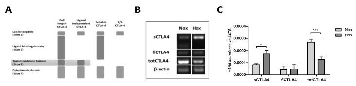
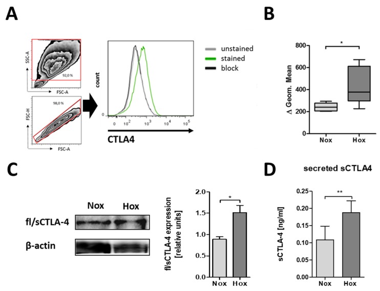
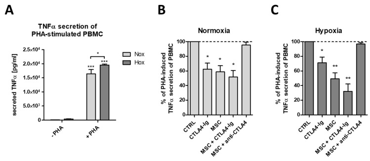
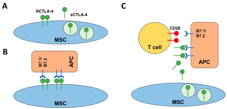
Mesenchymal stem/stromal cells (MSCs) are stem cells of the connective tissue, possess a plastic phenotype, and are able to differentiate into various tissues. Besides their role in tissue regeneration, MSCs perform additional functions as a modulator or inhibitor of immune responses. Due to their pleiotropic function, MSCs have also gained therapeutic importance for the treatment of autoimmune diseases and for improving fracture healing and cartilage regeneration. However, the therapeutic/immunomodulatory mode of action of MSCs is largely unknown. Here, we describe that MSCs express the inhibitory receptor CTLA-4 (cytotoxic T lymphocyte antigen 4). We show that depending on the environmental conditions, MSCs express different isoforms of CTLA-4 with the secreted isoform (sCTLA-4) being the most abundant under hypoxic conditions. Furthermore, we demonstrate that the immunosuppressive function of MSCs is mediated mainly by the secretion of CTLA-4. These findings open new ways for treatment when tissue regeneration/fracture healing is difficult.

Keywords: CTLA-4; fracture healing; hypoxia; immune modulation; mesenchymal stem/stromal cells; regeneration.


Conflict of interest statement

The authors declare that they have no conflict of interest.

Figures

Figure 1
Figure 1
Mesenchymal stem/stromal cells (MSCs) differentially express distinct splice variants of cytotoxic T lymphocyte antigen 4 (CTLA-4) with regard to oxygen availability. (A) Scheme of the documented splice variants of total CTLA-4 (totCTLA-4)—full length CTLA-4 (flCTLA-4), ligand-independent CTLA-4, soluble CTLA-4 (sCTLA-4), and 1/4 CTLA-4—which were detectable in (B) conventional RT-PCR and (C) quantitative RT-PCR under normoxia (Nox; 18% O2) and hypoxia (Hox; 1% O2). Data in (C) are given as the mean ± SEM (statistical analysis: two-way ANOVA with Bonferroni’s post hoc test: * p < 0.05, *** p < 0.001).
Figure 2
Figure 2
MSCs differentially express CTLA-4 protein with regard to oxygen availability. (A) Representative histogram of CTLA-4 surface expression on human bone marrow-derived MSCs and surface CTLA-4 staining of human MSCs as analyzed by flow cytometry. (B) Fluorescence intensities are depicted as box plots of delta of geometric means normalized to an antibody block (n = 5; * p < 0.05; paired t-test). (C,D) Intracellular CTLA-4 expression and secretion by human MSCs was confirmed by immunoblotting after native PAGE and ELISA. (C) Intracellular fl/sCTLA-4 (n = 4; * p < 0.05; paired t-test) and (D) secreted sCTLA-4 was enhanced in MSCs cultured under hypoxic conditions (Hox; 1% O2) as compared to normoxic conditions (Nox; 18% O2) (n = 6; * p < 0.05; paired t-test).
Figure 3
Figure 3
Human MSCs reduce tumor necrosis factor alpha (TNFα) secretion of mitogen-stimulated peripheral blood leukocytes in a CTLA-4-dependent manner. (A) Phytohaemagglutinin (PHA)-induced TNFα secretion of peripheral blood mononuclear cells (PBMCs) (n = 4; * p < 0.05; *** p < 0.001; two-way ANOVA with Bonferroni’s post hoc test). (B,C) PHA-induced TNFα secretion by PBMCs was significantly reduced by CTLA4-Ig (Abatacept), by human bone marrow-derived MSCs, and their combination (n = 4; * p < 0.05; ** p < 0.01; one-sample t-test). MSC-mediated reduction of PHA-induced TNFα secretion was blocked by anti-CTLA-4 antibody under both (A) normoxic (18% O2) and (B) hypoxic (1% O2) conditions.
Figure 4
Figure 4
Schematic representation of the mode of action of CTLA-4 mediated inhibition of, e.g., T cell activation. (A) MSCs are able to express two different isoforms of CTLA-4. The flCTLA-4 is a membrane-bound dimer while sCTLA-4 is a secreted monomeric isoform of CTLA-4. (B) Under normoxic conditions, flCTLA-4 may mediate cell contact-dependent inhibition of T cell responses by direct interaction with B7.1/B7.2 on the surface of APCs. (C) Under hypoxic conditions, MSC may switch to a cell contact-independent mechanism where sCTLA-4 is able to bind B7.1/B7.2 on APCs, thereby blocking CD28 mediated co-stimulation of T cells.

References

    1. Alexander T., Arnold R., Hiepe F., Radbruch A. Resetting the immune system with immunoablation and autologous haematopoietic stem cell transplantation in autoimmune diseases. Clin. Exp. Rheumatol. 2016;34:53–57. - PubMed
    1. Freitag J., Berod L., Kamradt T., Sparwasser T. Immunometabolism and autoimmunity. Immunol. Cell Biol. 2016;94:925–934. doi: 10.1038/icb.2016.77. - DOI - PubMed
    1. Schett G. Autoimmunity as a trigger for structural bone damage in rheumatoid arthritis. Mod. Rheumatol. 2017;27:193–197. doi: 10.1080/14397595.2016.1265907. - DOI - PubMed
    1. Mauri C., Blair P.A. Regulatory B cells in autoimmunity: Developments and controversies. Nat. Rev. Rheumatol. 2010;6:636–643. doi: 10.1038/nrrheum.2010.140. - DOI - PubMed
    1. Sakaguchi S., Miyara M., Costantino C.M., Hafler D.A. FOXP3+ regulatory T cells in the human immune system. Nat. Rev. Immunol. 2010;10:490–500. doi: 10.1038/nri2785. - DOI - PubMed