. 2018 Aug 7;8(8):145.
doi: 10.3390/brainsci8080145.

Clinical and Functional Characterization of the Recurrent TUBA1A p.(Arg2His) Mutation

Affiliations

Clinical and Functional Characterization of the Recurrent TUBA1A p.(Arg2His) Mutation

Jennifer F Gardner et al. Brain Sci. .

Abstract

The TUBA1A gene encodes tubulin alpha-1A, a protein that is highly expressed in the fetal brain. Alpha- and beta-tubulin subunits form dimers, which then co-assemble into microtubule polymers: dynamic, scaffold-like structures that perform key functions during neurogenesis, neuronal migration, and cortical organisation. Mutations in TUBA1A have been reported to cause a range of brain malformations. We describe four unrelated patients with the same de novo missense mutation in TUBA1A, c.5G>A, p.(Arg2His), as found by next generation sequencing. Detailed comparison revealed similar brain phenotypes with mild variability. Shared features included developmental delay, microcephaly, hypoplasia of the cerebellar vermis, dysplasia or thinning of the corpus callosum, small pons, and dysmorphic basal ganglia. Two of the patients had bilateral perisylvian polymicrogyria. We examined the effects of the p.(Arg2His) mutation by computer-based protein structure modelling and heterologous expression in HEK-293 cells. The results suggest the mutation subtly impairs microtubule function, potentially by affecting inter-dimer interaction. Based on its sequence context, c.5G>A is likely to be a common recurrent mutation. We propose that the subtle functional effects of p.(Arg2His) may allow for other factors (such as genetic background or environmental conditions) to influence phenotypic outcome, thus explaining the mild variability in clinical manifestations.

Keywords: TUBA1A; cerebellar hypoplasia; p.(Arg2His), R2H; polymicrogyria; tubulin; tubulinopathy.


Conflict of interest statement

The authors declare no conflict of interest.

Figures
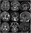
Figure 1
Figure 1
Magnetic resonance images from patients with the recurrent p.(Arg2His) TUBA1A mutation. T2-weighted axial and T1-weighted midline sagittal brain images for Patient 1 at age three years (AC), Patient 2 at age six months (DF), and Patient 3 at age 19 months (GI). The images demonstrate hypoplasia and dysplasia of the cerebellar vermis (yellow arrows), thinning or partial agenesis of the corpus callosum (red arrows), globular basal ganglia with incomplete formation of the anterior limb internal capsule (white arrows), and bilateral perisylvian polymicrogyria (blue arrows). The pons is similar in size to the midbrain which suggests the pons is relatively small (C,F,I).
Figure 2
Figure 2
Neuropathology from Patient 4. (A) The medial aspect of right cerebral hemisphere showing a thin corpus callosom with absent rostrum. (B) Midline sagittal section of brain stem and cerebellum showing mild hypoplasia of the cerebellar vermis. (C) Coronal section of the cerebral hemispheres. The corpus callosum is thinned and there is thickening of the cortex around the sylvian fissures. (D) Stained section of the right cerebral hemisphere revealing abnormal folding of the cortical ribbon around the sylvian fissure. (E) A magnified view of (D) demonstrating polymicrogyria around the sylvian fissure.
Figure 3
Figure 3
In silico modelling and in vitro functional analysis of the p.(Arg2His) mutation. (A) Ribbon models of alpha-tubulin (green) and beta-tubulin (blue) subunits aligned in a microtubule polymer. The position of Arg2 is shown (arrow) close to the inter-dimer interface (between alpha-tubulin and the beta-tubulin of an adjacent heterodimer). The mutation is on the opposite side of TUBA1A from the binding site of guanosine-5′-triphosphate (GTP, orange). (B) A close-up view of the Arg2 residue (arrow) with the mutant (purple ribbon, red side chain) and wild type (green ribbon and side chain) proteins superimposed. Only mild confirmation changes are predicted around the Arg2 residue. However, additional conformational changes are predicted between residues 38 and 51 (bracket). These may affect the interaction between heterodimers. (C) HEK-293 cells expressing FLAG-tagged TUBA1A-R2H. The cells are stained with DAPI (4′,6-diamidino-2-phenylindole, blue), anti-FLAG- (red), and anti-alpha-tubulin (green) antibodies. The microtubules appear yellow due to the colocalisation of endogenous (green) and FLAG-tagged transgenic (red) tubulin. The arrows indicate diffuse patches of transgenic mutant tubulin (red) in the cytoplasm between the microtubules. (D) Control cells expressing FLAG-tagged wild-type TUBA1A have less staining for the transgenic tubulin in the cytoplasm between the microtubules.

References

    1. Gloster A., Wu W., Speelman A., Weiss S., Causing C., Pozniak C., Reynolds B., Chang E., Toma J.G., Miller F.D. The T alpha 1 alpha-tubulin promoter specifies gene expression as a function of neuronal growth and regeneration in transgenic mice. J. Neurosci. 1994;14:7319–7330. doi: 10.1523/JNEUROSCI.14-12-07319.1994. - DOI - PMC - PubMed
    1. Bamji S.X., Miller F.D. Comparison of the expression of a T alpha 1:nlacZ transgene and T alpha 1 alpha-tubulin mRNA in the mature central nervous system. J. Comp. Neurol. 1996;374:52–69. doi: 10.1002/(SICI)1096-9861(19961007)374:1<52::AID-CNE4>3.0.CO;2-M. - DOI - PubMed
    1. Keays D.A., Tian G., Poirier K., Huang G.-J., Siebold C., Cleak J., Oliver P.L., Fray M., Harvey R.J., Molnár Z., et al. Mutations in alpha-tubulin cause abnormal neuronal migration in mice and lissencephaly in humans. Cell. 2007;128:45–57. doi: 10.1016/j.cell.2006.12.017. - DOI - PMC - PubMed
    1. Jansen A.C., Oostra A., Desprechins B., De Vlaeminck Y., Verhelst H., Régal L., Verloo P., Bockaert N., Keymolen K., Seneca S., et al. TUBA1A mutations: from isolated lissencephaly to familial polymicrogyria. Neurology. 2011;76:988–992. doi: 10.1212/WNL.0b013e31821043f5. - DOI - PubMed
    1. Cushion T.D., Dobyns W.B., Mullins J.G.L., Stoodley N., Chung S.-K., Fry A.E., Hehr U., Gunny R., Aylsworth A.S., Prabhakar P., et al. Overlapping cortical malformations and mutations in TUBB2B and TUBA1A. Brain. 2013;136:536–548. doi: 10.1093/brain/aws338. - DOI - PubMed

LinkOut - more resources