. 2019 Jan;38(2):194-208.
doi: 10.1038/s41388-018-0426-6. Epub 2018 Aug 7.

ATP in the tumour microenvironment drives expression of nfP2X7, a key mediator of cancer cell survival

Affiliations

ATP in the tumour microenvironment drives expression of nfP2X7, a key mediator of cancer cell survival

S M Gilbert et al. Oncogene. 2019 Jan.

Abstract

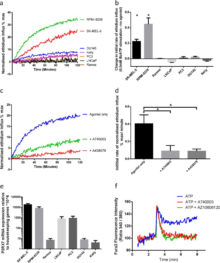
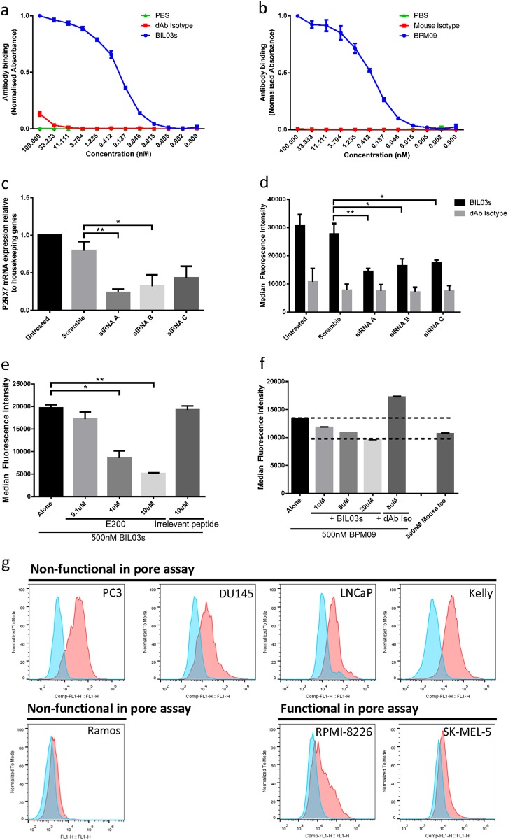
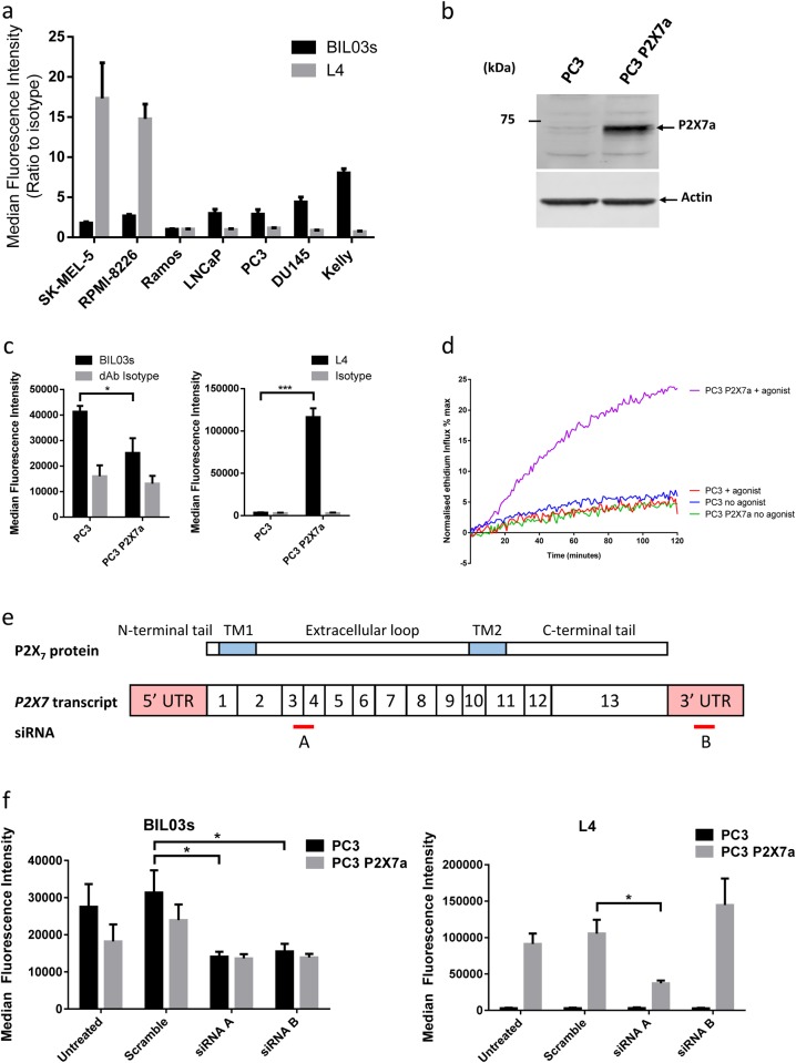
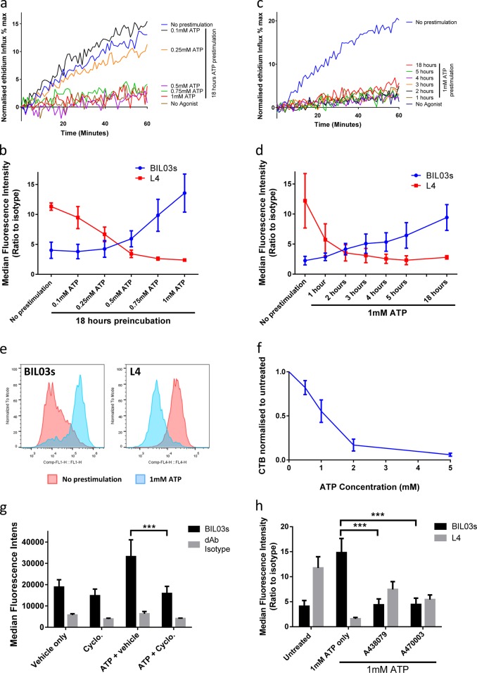
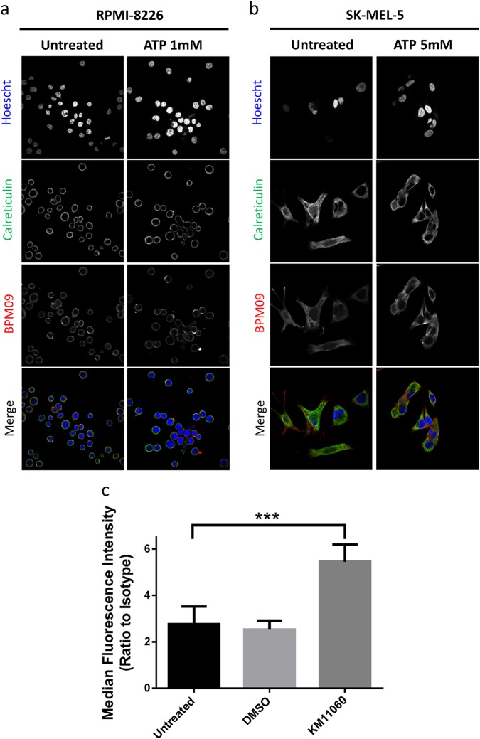
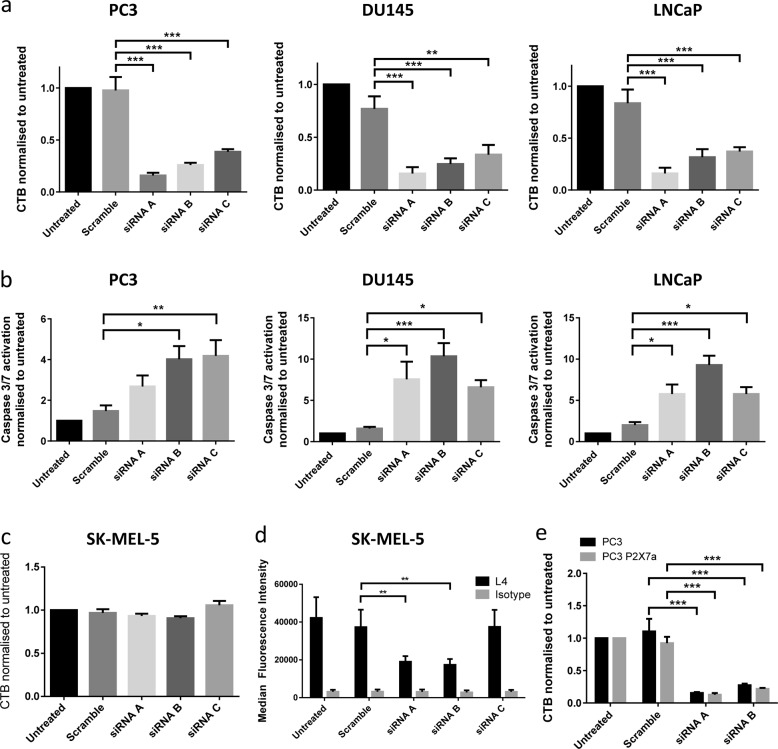
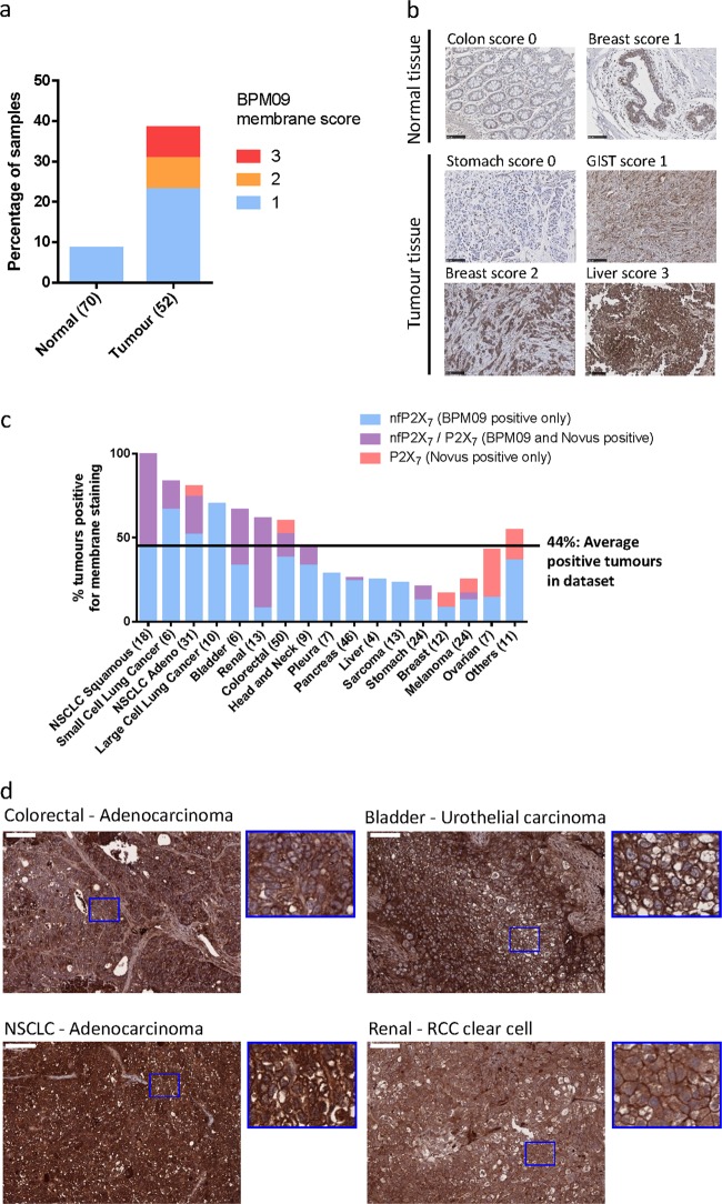
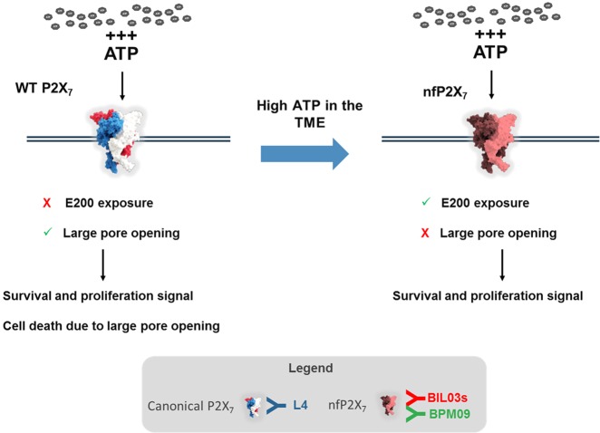
The ATP-gated receptor P2X7 is expressed in multiple malignant tumours including neuroblastoma, melanoma, prostate, lung and breast. P2X7 has a significant role in mediating diverse cell responses, which upon dysregulation are associated with tumour initiation and development. The rapid, ATP-mediated activation of P2X7 induces a fast-inward cation current in cells. However, prolonged ATP-mediated activation of P2X7 leads to formation of a pore that increases membrane permeability and eventually causes cell death. This presents a potential paradox, as the tumour microenvironment contains extracellular ATP at levels sufficient to activate the P2X7 pore and trigger cell death. However, P2X7 expression is associated with enhanced cancer cell survival, proliferation and metastatic potential. At least one distinct conformational form of P2X7, termed non-pore functional P2X7 (nfP2X7), has been described, which is not able to form a functional pore. We demonstrate for the first time in this study that exposure to a high ATP concentration, equivalent to those measured in the tumour microenvironment, drives nfP2X7 expression and also that nfP2X7 is essential for tumour cell survival. We show that monoclonal antibodies raised against a P2X7 amino acid sequence (200-216), whose conformation is distinct from that of wild-type (WT) P2X7, bind specifically to nfP2X7 expressed on the surface of tumour cells. We also show that nfP2X7 is broadly expressed in patient-derived tumour sections from a wide range of cancers. Therefore, antibodies raised against E200 provide tools that can differentiate between forms of the P2X7 receptor that have a key role in cancer.


Conflict of interest statement

SMG, CJO, SM and RL are employees of Biosceptre (UK) Limited, a company commercialising nfP2X7-targeted therapies. ALP and PB were contracted by Biosceptre (UK) Limited as part of Charles River Discovery Research Services Germany GmbH (formerly named Oncotest GmbH). SH is BBSRC CASE PhD student partly funded by Biosceptre (UK) Limited. FDV serves as a member of Biosceptre International Limited’s Scientific Advisory Board. SF has no conflict of interest.

Figures

Fig. 1
Fig. 1
P2X7 mRNA is expressed in multiple cancer cell lines which do not show pore function. a Normalised ethidium influx in response to 0.5 mM BzATP stimulation in a panel of cancer cell lines. Mean of three independent experiments is shown. b Quantification of initial rate (5–40 min) of ethidium influx in 0.5 mM BzATP-treated cells above rate of increase in untreated cells. Mean and SEM from three independent experiments are shown. Two-tailed Student’s T-test was used to test whether agonist stimulation caused a significant increase in ethidium influx relative to untreated in each cell line. c Normalised ethidium influx in RPMI-8226 cells treated with 0.5 mM BzATP alone or in combination with P2X7 inhibitors A740003 or A438079 are shown. d Quantification of initial rate (5–30 min) of ethidium influx. Mean and SEM from three independent experiments are shown. One-way ANOVA with Dunnett’s post test was used to test significance. e P2RX7 mRNA quantification by qPCR in a panel of cancer cell lines. Mean and SEM from three independent experiments are shown. f Fura-2-loaded PC3 cells were pre-incubated with A740003 or AZ10606120, incubated in a fluorimeter cuvette in standard saline solution and challenged with 3 mM ATP. *P < 0.05
Fig. 2
Fig. 2
E200-targeted antibodies specifically bind nfP2X7. a, b ELISA assay of BIL03s (a) and BPM09 (b) binding to E200 peptide compared with PBS and isotype controls. Results were normalised to maximum binding. Mean and SEM from three independent experiments are shown. c Quantification of P2RX7 transcript expression relative to a panel of housekeeping genes in PC3 cells 72 h after transfection with P2RX7-targeted siRNA. Mean and SEM from four independent experiments are shown. Each experiment was normalised to untreated control. One-way ANOVA with Dunnett’s post test was used to test significance. d BIL03s binding to live PC3 cells 72 h after transfection with P2RX7-targeted siRNA was tested by flow cytometry. Mean and SEM from four independent experiments are shown. Two-way ANOVA with Dunnett’s post test was used to test significance. e BIL03s binding to live PC3 cells, measured by flow cytometry, can be competed by E200 peptide. Mean and SEM from three independent experiments are shown. One-way ANOVA with Dunnett’s post test was used to test significance. f Flow cytometry analysis shows that BPM09 binding to live PC3 cells can be competed with increasing amounts of BIL03s. Representative plot from three experiments are shown. g Representative flow cytometry plots from three experiments showing BIL03s binding to live cells in a panel of cancer cell lines. *P < 0.05, **P < 0.01
Fig. 3
Fig. 3
nfP2X7 is molecularly distinct from functional P2X7. a BIL03s and L4 binding relative to isotype control was measured by flow cytometry in a panel of cancer cell lines. Mean and SEM from at least three independent experiments are shown. b Western blotting of lysate from PC3 cells or PC3 cells overexpressing P2X7a. Representative images from three independent experiments are shown. c PC3 cells overexpressing P2X7a show increased L4 binding relative to untransfected PC3 but no change in BIL03s binding. Mean and SEM from three independent experiments are shown. Two-way ANOVA with Sidak’s multiple comparison test was used. d Ethidium influx into PC3 cells or PC3 cells overexpressing P2X7a in response to 0.5 mM BzATP or no agonist stimulation. Mean of three independent experiments shown. e Representation of the P2RX7 transcript and the target sequence location for siRNA. a, b, f The effect of P2RX7-targeted siRNA A and B on BIL03s and L4 binding in PC3 cells and PC3 cells overexpressing P2X7a was measured by flow cytometry. Mean and SEM from three independent experiments are shown. Two-way ANOVA with Dunnett’s post test was used to test significance. *P < 0.05, ***P < 0.001
Fig. 4
Fig. 4
nfP2X7 expression can be induced by mimicking the high ATP concentration present in the tumour microenvironment. a, b RPMI-8226 cells were treated with ATP at the indicated concentration for 18 h. After treatment, a normalised ethidium influx in response to 0.5 mM BzATP stimulation was measured (mean of three independent experiments is shown) and b flow cytometry was used to quantify binding of BIL03s and L4 to live RPMI-8226 cells (mean and SEM from three independent experiments are shown). c, d RPMI-8226 cells were treated with 1 mM ATP for the indicated length of time. After treatment, c normalised ethidium influx in response to 0.5 mM BzATP stimulation was measured (mean from three independent experiments is shown) and d flow cytometry was used to quantify binding of BIL03s and L4 to live RPMI-8226 cells (mean and SEM from three independent experiments are shown). e Representative flow cytometry plots showing BIL03s and L4 binding to live RPMI-8226 cells before and after 1 mM ATP treatment for 18 h. f RPMI-8226 cells were treated with ATP at indicated concentration for 18 h before measurement of the number of live cells using CellTitre-Blue (CTB) assay. CTB fluorescence was normalised to untreated cells. Mean and SEM from three independent experiments are shown. g RPMI-8226 cells were treated with 250 μg/ml Cycloheximide, 1 mM ATP or both for 4 h. Flow cytometry analysis showed that increased BIL03s binding following 1 mM ATP treatment was abrogated in the presence of Cycloheximide. Mean and SEM from three independent experiments are shown. Two-way ANOVA with Dunnett’s post test was used to test significance. h RPMI-8226 cells were treated with 1 mM ATP alone or in the presence of P2X7 inhibitors A438079 or A740003 for 18 h. Flow cytometry analysis showed that the ATP-induced increase in BIL03s binding was significantly reduced by P2X7 inhibitors. Mean and SEM from three independent experiments are shown. Two-way ANOVA with Dunnett’s post test was used to test significance. ***P < 0.001
Fig. 5
Fig. 5
nfP2X7 is present intracellularly and addressed to the membrane upon ATP stimulation. a, b RPMI-8226 and SK-MEL-5 cells were treated with 1 mM ATP and 5 mM ATP, respectively, before performing immunofluorescence staining for BPM09 (7.6 μg/ml) and calreticulin (Abcam ab2907 2 μg/ml). Hoescht was used to stain the nucleus. Representative images from three independent experiments are shown. c RPMI-8226 cells were treated with 10 µM KM11060 or DMSO vehicle control for 16 h before analysing BIL03s and its isotype control binding by flow cytometry. Mean and SEM of three independent experiments are shown. One-way ANOVA with Dunnett’s post test was used to test significance. ***P < 0.001
Fig. 6
Fig. 6
nfP2X7 is necessary for cancer cell survival. a, b PC3, DU145 and LNCaP non-functional cell lines were transfected with P2RX7-targeted siRNA. After 72 h, a number of live cells were measured using CellTitre-Blue (CTB) assay. b Induction of apoptosis was measured using an Apo One caspase 3/7 activation assay. Mean and SEM from at least four independent experiments shown. One-way ANOVA with Dunnett’s post test was used to test significance. c, d SK-MEL-5 functional cells were transfected with P2RX7-targeted siRNA. After 72 h, c no effect on cell viability was measured by CTB assay. d Surface expression of P2X7 as measured by L4 flow cytometry binding was significantly reduced. Mean and SEM from at least four independent experiments shown. Two-way ANOVA with Dunnett’s post test was used to test significance. e PC3 cells or PC3 cells overexpressing P2X7a were transfected with P2RX7-targeted siRNA. After 72 h, number of live cells was quantified using a CTB assay. P2RX7-targeted siRNA caused a reduction in cell numbers even in the presence of overexpressed P2X7a. Mean and SEM from at least four independent experiments shown. Two-way ANOVA with Dunnett’s post test was used to test significance. *P < 0.05, **P < 0.01, ***P < 0.001
Fig. 7
Fig. 7
nfP2X7 is present in a broad range of tumour samples. a Tissue sections from 70 normal biopsies and 52 tumour biopsies were stained with BPM09 and scored for membrane staining. b Representative images for each score of normal and tumour tissue section stained with BPM09. Scale bar represents 100 µM. c Tissue sections from 290 Patient-derived xenograft (PDX) models were stained with BPM09 and a polyclonal antibody raised against part of the extracellular domain of P2X7a. Tissue sections were scored for membrane staining. For each tumour type the percentage of PDX with positive staining is shown with the number of PDX examined in brackets. d Representative images of colorectal (adenocarcinoma), bladder (urothelial carcinoma), non-small cell lung cancer (NSCLC—adenocarcinoma) and renal cancer (RCC clear cell) tissue sections with positive BPM09 staining together with higher magnification section showing BPM09 membrane staining. Scale bar represents 100 µm
Fig. 8
Fig. 8
Illustration of the WT P2X7 to nfP2X7 switch and its impact on cell fate. L4 antibody binds specifically to WT P2X7 while BIL03s and BPM09 bind specifically to nfP2X7

References

    1. Burnstock G. Purinergic signalling. Br J Pharmacol. 2006;147(Suppl 1):S172–181. - PMC - PubMed
    1. North RA. Molecular physiology of P2X receptors. Physiol Rev. 2002;82:1013–67. doi: 10.1152/physrev.00015.2002. - DOI - PubMed
    1. North RA, Surprenant A. Pharmacology of cloned P2X receptors. Annu Rev Pharmacol Toxicol. 2000;40:563–80. doi: 10.1146/annurev.pharmtox.40.1.563. - DOI - PubMed
    1. Surprenant A, Rassendren F, Kawashima E, North RA, Buell G. The cytolytic P2Z receptor for extracellular ATP identified as a P2X receptor (P2X7) Science. 1996;272:735–8. doi: 10.1126/science.272.5262.735. - DOI - PubMed
    1. Auger R, Motta I, Benihoud K, Ojcius DM, Kanellopoulos JM. A role for mitogen-activated protein kinase(Erk1/2) activation and non-selective pore formation in P2X7 receptor-mediated thymocyte death. J Biol Chem. 2005;280:28142–51. doi: 10.1074/jbc.M501290200. - DOI - PubMed

Publication types

MeSH terms