. 2018 Jul 10;2(8):949-964.
doi: 10.1210/js.2018-00131. eCollection 2018 Aug 1.

Long-Lasting Androgen-Induced Cardiometabolic Effects in Polycystic Ovary Syndrome

Affiliations

Long-Lasting Androgen-Induced Cardiometabolic Effects in Polycystic Ovary Syndrome

Edgar D Torres Fernandez et al. J Endocr Soc. .

Abstract

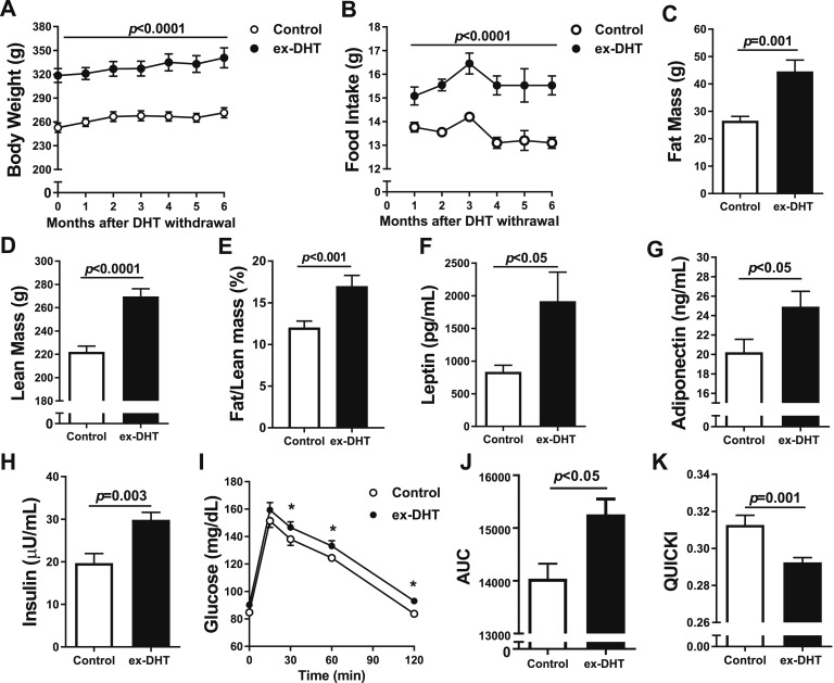
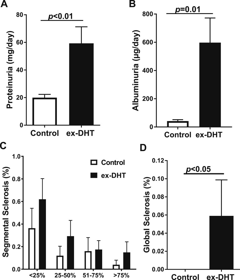
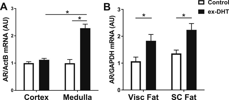
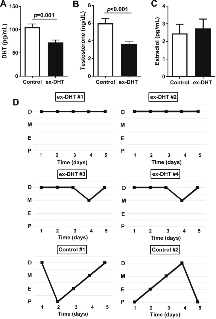
Polycystic ovary syndrome (PCOS), the most common endocrine disorder in women of reproductive age, is characterized by androgen excess and ovarian dysfunction and presents with increased cardiometabolic risk factors such as obesity, insulin resistance, and elevated blood pressure (BP). We previously reported that administration of dihydrotestosterone (DHT) to female rats elicits cardiometabolic derangements similar to those found in women with PCOS. In this study, we tested the hypothesis that the DHT-mediated cardiometabolic derangements observed in PCOS are long lasting despite DHT withdrawal. Four-week-old female Sprague Dawley rats were treated with DHT (7.5 mg/90 days) or placebo for 6 months. DHT was discontinued (ex-DHT), and rats were followed for 6 additional months. After 6 months of DHT withdrawal, food intake, body weight, fat and lean mass, fasting plasma insulin, leptin, and adiponectin were elevated in ex-DHT rats. BP remained significantly elevated, and enalapril, an angiotensin-converting enzyme (ACE) inhibitor, normalized BP in ex-DHT rats. Expression of components of the intrarenal renin-angiotensin system was increased in ex-DHT rats. The cardiometabolic features found in ex-DHT rats were associated with lower plasma androgen levels but increased expression of renal and adipose tissue androgen receptors. In summary, androgen-induced cardiometabolic effects persisted after DHT withdrawal in a PCOS experimental model. Activation of intrarenal renin-angiotensin system plays a major role in the androgen-mediated increase in BP in ex-DHT. Upregulation of the renal and adipose tissue androgen receptor may explain the long-lasting effects of androgens. In clinical scenarios characterized by hyperandrogenemia in women, prompt normalization of androgen levels may be necessary to prevent their long-lasting cardiometabolic effects.

Keywords: androgen receptor; androgens; blood pressure; cardiometabolic risk factors; polycystic ovary syndrome; renin angiotensin system.


Figures

Figure 1.
Figure 1.
Body composition and metabolic parameters in ex-DHT and control rats. (A) Body weight and (B) food intake were elevated in ex-DHT rats and sustained throughout the 6 months after DHT withdrawal. (C) Fat mass, (D) lean mass, and (E) fat/lean mass ratio were increased in ex-DHT rats. (F) Leptin, (G) adiponectin, and (H) fasting insulin levels were higher in ex-DHT rats. (I–K) Insulin resistance measured by AUC (I and J) during oral glucose tolerance test was increased, whereas results obtained with the quantitative insulin sensitivity check index (QUICKI) (K) were decreased in ex-DHT rats. Determinations were performed at 13 months of age (6 months after DHT withdrawal). AUC, area under the curve. *P < 0.05.
Figure 2.
Figure 2.
Blood pressure, heart rate, and effect of enalapril in ex-DHT and control rats. (A and B) ex-DHT rats had elevated MAP during light and dark cycles compared with controls. (C) ex-DHT rats had decreased heart rate compared with controls under both baseline and enalapril treatment. *P < 0.05, ex-DHT vs control. &P < 0.05 vs baseline ex-DHT rats, same treatment. #P < 0.05 vs baseline control rats, same treatment.
Figure 3.
Figure 3.
Renal function and histology in ex-DHT and control rats. (A) ex-DHT rats had increased urinary protein excretion compared with control rats. (B) ex-DHT rats had elevated urinary albumin excretion compared with control rats. (C and D) Histological analysis of glomerular injury showed that segmental sclerosis at all grades and global sclerosis was increased in ex-DHT rats.
Figure 4.
Figure 4.
Expression of intrarenal renin-angiotensin system components in ex-DHT and control rats. (A) Renal cortical and medullary renin mRNA expression had a tendency to be lower in ex-DHT rats but did not reach statistical significance. (B) Renal cortical expression of ACE was significantly decreased in ex-DHT rats; however, no differences were observed in the renal medulla. (C and D) Medullary mRNA expression of angiotensinogen (AGTN) and angiotensin II type 1 receptor (AT1R) was significantly increased in ex-DHT rats. (E) No differences were observed cortically nor medullary in mineralocorticoid receptor (MR) mRNA expression among the groups. *P < 0.05. ActB, β-actin; AU, arbitrary units.
Figure 5.
Figure 5.
Expression of androgen receptor in ex-DHT and control rats. (A) Renal medullary androgen receptor (AR) mRNA expression was increased in ex-DHT rats; however, no differences were observed in the renal cortex. (B) Androgen receptor mRNA expression was upregulated in both visceral (Visc fat) and subcutaneous (SC fat) adipose tissue in ex-DHT rats. ActB, β-actin; AU, arbitrary units; GAPDH, glyceraldehyde 3-phosphate dehydrogenase. *P < 0.05.
Figure 6.
Figure 6.
(A and B) Sex hormones and (C) plasma DHT and testosterone levels were decreased in ex-DHT rats. Plasma estradiol levels were similar among groups. (D) Vaginal cytology was performed daily for 5 consecutive days at the end of the study. The figure shows the pattern of four representative ex-DHT rats (#1 to #4) and two controls (#5 and #6). D, diestrus; E, estrus; M, metestrus; P, proestrus.

References

    1. Bozdag G, Mumusoglu S, Zengin D, Karabulut E, Yildiz BO. The prevalence and phenotypic features of polycystic ovary syndrome: a systematic review and meta-analysis. Hum Reprod. 2016;31(12):2841–2855. - PubMed
    1. Knochenhauer ES, Key TJ, Kahsar-Miller M, Waggoner W, Boots LR, Azziz R. Prevalence of the polycystic ovary syndrome in unselected black and white women of the southeastern United States: a prospective study. J Clin Endocrinol Metab. 1998;83(9):3078–3082. - PubMed
    1. Azziz R, Carmina E, Chen Z, Dunaif A, Laven JS, Legro RS, Lizneva D, Natterson-Horowtiz B, Teede HJ, Yildiz BO. Polycystic ovary syndrome. Nat Rev Dis Primers. 2016;2:16057. - PubMed
    1. McCartney CR, Marshall JC. Polycystic ovary syndrome. N Engl J Med. 2016;375(1):54–64. - PMC - PubMed
    1. Escobar-Morreale HF. Polycystic ovary syndrome: definition, aetiology, diagnosis and treatment. Nat Rev Endocrinol. 2018;14(5):270–284. - PubMed

LinkOut - more resources