

Deep dense multi-path neural network for prostate segmentation in magnetic resonance imaging
- PMID: 30088208
- PMCID: PMC6177294
- DOI: 10.1007/s11548-018-1841-4
Deep dense multi-path neural network for prostate segmentation in magnetic resonance imaging
Abstract
Purpose: We propose an approach of 3D convolutional neural network to segment the prostate in MR images.
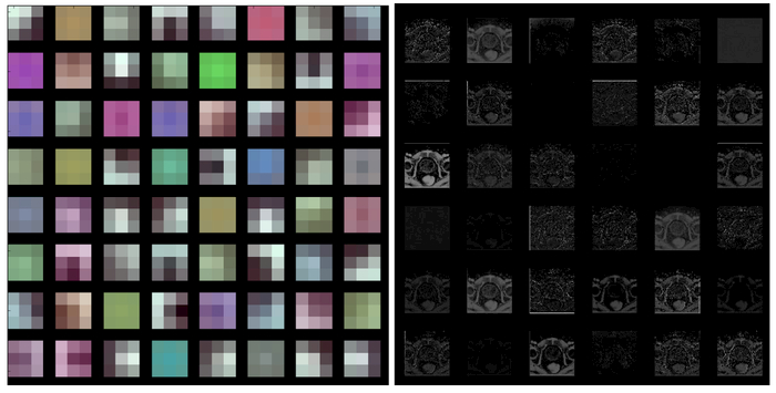
Methods: A 3D deep dense multi-path convolutional neural network that follows the framework of the encoder-decoder design is proposed. The encoder is built based upon densely connected layers that learn the high-level feature representation of the prostate. The decoder interprets the features and predicts the whole prostate volume by utilizing a residual layout and grouped convolution. A set of sub-volumes of MR images, centered at the prostate, is generated and fed into the proposed network for training purpose. The performance of the proposed network is compared to previously reported approaches.
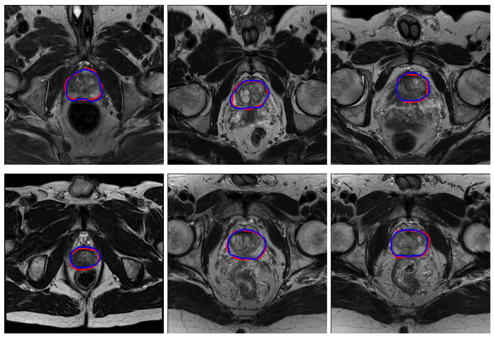
Results: Two independent datasets were employed to assess the proposed network. In quantitative evaluations, the proposed network achieved 95.11 and 89.01 Dice coefficients for the two datasets. The segmentation results were robust to variations in MR images. In comparison experiments, the segmentation performance of the proposed network was comparable to the previously reported approaches. In qualitative evaluations, the segmentation results by the proposed network were well matched to the ground truth provided by human experts.
Conclusions: The proposed network is capable of segmenting the prostate in an accurate and robust manner. This approach can be applied to other types of medical images.
Keywords: Deep learning; Dense connections; Grouped convolution; Magnetic resonance imaging; Prostate segmentation.
Figures


References
-
- Siegel RL, Miller KD, and Jemal A, “Cancer statistics,”. CA: a cancer journal for clinicians, 66(1): p. 7–30, (2016). - PubMed
-
- Fei B, Kemper C, and Wilson DL, “A comparative study of warping and rigid body registration for the prostate and pelvic MR volumes.” Comput Med Imaging Graph, 27(4): p. 267–81, (2003). - PubMed
-
- Fei B, et al., “Slice-to-volume registration and its potential application to interventional MRI-guided radio-frequency thermal ablation of prostate cancer.” IEEE Trans Med Imaging, 22(4): p. 515–525, (2003). - PubMed
-
- Wu Qiu JY, Ukwatta Eranga, Sun Yue, Rajchl Martin, and Fenster Aaron, “Prostate Segmentation: An Efficient Convex Optimization Approach With Axial Symmetry Using 3-D TRUS and MR Images.” IEEE Transactions on Medical Imaging, 33(4): p. 947–960, (2014). - PubMed
-
- Litjens G, et al., “Computer-aided detection of prostate cancer in MRI.” IEEE transactions on Medical Imaging, 33(5): p. 1083–1092, (2014). - PubMed
MeSH terms
Grants and funding
LinkOut - more resources
Full Text Sources
Other Literature Sources
Medical
