. 2018 Aug 8;14(8):e1007572.
doi: 10.1371/journal.pgen.1007572. eCollection 2018 Aug.

Heterochromatin and RNAi regulate centromeres by protecting CENP-A from ubiquitin-mediated degradation

Affiliations

Heterochromatin and RNAi regulate centromeres by protecting CENP-A from ubiquitin-mediated degradation

Jinpu Yang et al. PLoS Genet. .

Abstract

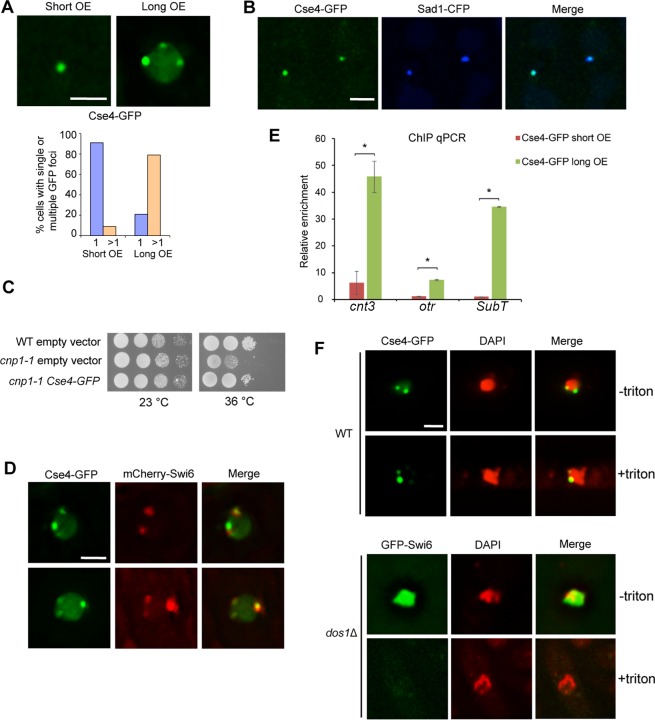
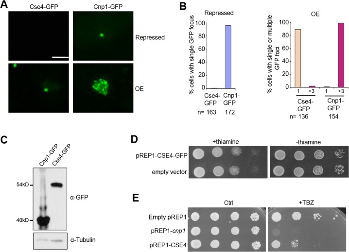
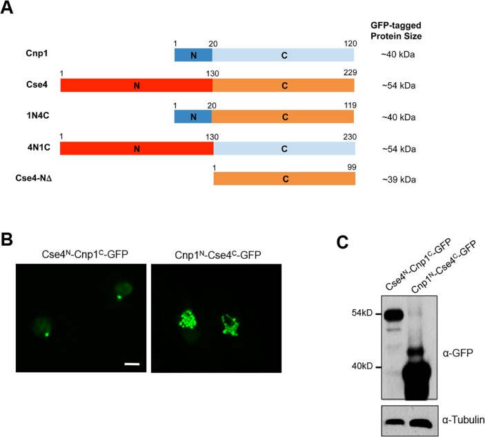
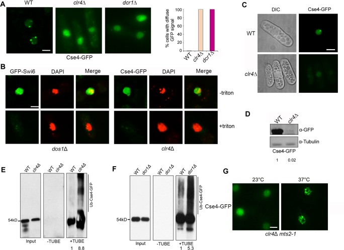
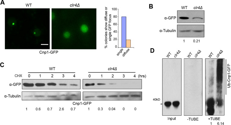
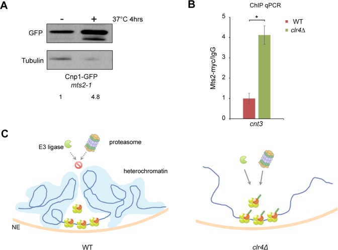
Centromere is a specialized chromatin domain that plays a vital role in chromosome segregation. In most eukaryotes, centromere is surrounded by the epigenetically distinct heterochromatin domain. Heterochromatin has been shown to contribute to centromere function, but the precise role of heterochromatin in centromere specification remains elusive. Centromeres in most eukaryotes, including fission yeast (Schizosaccharomyces pombe), are defined epigenetically by the histone H3 (H3) variant CENP-A. In contrast, the budding yeast Saccharomyces cerevisiae has genetically-defined point centromeres. The transition between regional centromeres and point centromeres is considered as one of the most dramatic evolutionary events in centromere evolution. Here we demonstrated that Cse4, the budding yeast CENP-A homolog, can localize to centromeres in fission yeast and partially substitute fission yeast CENP-ACnp1. But overexpression of Cse4 results in its localization to heterochromatic regions. Cse4 is subject to efficient ubiquitin-dependent degradation in S. pombe, and its N-terminal domain dictates its centromere distribution via ubiquitination. Notably, without heterochromatin and RNA interference (RNAi), Cse4 fails to associate with centromeres. We showed that RNAi-dependent heterochromatin mediates centromeric localization of Cse4 by protecting Cse4 from ubiquitin-dependent degradation. Heterochromatin also contributes to the association of native CENP-ACnp1 with centromeres via the same mechanism. These findings suggest that protection of CENP-A from degradation by heterochromatin is a general mechanism used for centromere assembly, and also provide novel insights into centromere evolution.


Conflict of interest statement

The authors have declared that no competing interests exist.

Figures

Fig 1
Fig 1. Cse4 preferentially targets to centromeres and functionally substitutes Cnp1 in fission yeast.
A, Cells carrying pREP1-CSE4-GFP that were induced for 24 hours show a single GFP focus (short). Multiple foci (commonly 3–6) were detected for 28-hour induction (long). (Bottom) Percentage of cells containing single or multiple focus. OE, overexpression. B, The single Cse4-GFP focus colocalizes with Sad1-CFP. C, Cse4-GFP partially rescues cnp1-1 growth defects at 36°C. Serial dilutions of indicated strains were plated in minimal medium without thiamine. Dilution = 10. D, Multiple GFP foci in cells overexpressing Cse4-GFP colocalize with mCherry-Swi6. E. ChIP qPCR showing relative enrichment of Cse4-GFP to centromeric region (cnt3), peri-centromeric region (otr) and sub-telomeric region (subT). ChIP was repeated in triplicate. Error bar indicates SEM. *, p<0.01. F, in situ chromatin-binding assay for wild type cells expressing Cse4-GFP. The heterochromatin mutant dos1Δ carrying GFP-Swi6 was used as a control (Bottom). GFP-Swi6 is dissociated from heterochromatin in dos1Δ, and was readily washed away by Triton X-100 as expected. Scale bars: 2 μm.
Fig 2
Fig 2. Cse4 is expressed at a significantly low level in the fission yeast.
A, Distribution patterns of Cse4-GFP and Cnp1-GFP at repressed (Top) and overexpressed (Bottom) conditions (24-hour induction). B, Quantification of the percentage of cells showing a single focus when repressed (Left), and a single or multiple GFP foci (>3) in overexpressed condition (Right). C, Western blot analysis of cells expressing indicated proteins using an anti-GFP antibody. Tubulin was used as a loading control. D, Cells harboring pREP1-CSE4-GFP were grown in minimal medium with thiamine to suppress its expression (Left) or lacking thiamine to induce overexpression (Right). Wild type cells carrying an empty vector were used as a control. E, Overexpression of Cse4 results in minor chromosome segregation defects. Cells carrying indicated plasmids were plated in minimal medium that contains 15μg/ml TBZ but no thiamine. Dilution = 10. Ctrl, a control plate without TBZ. Scale bar: 2 μm.
Fig 3
Fig 3. Cse4 is subject to efficient ubiquitin-dependent degradation in the fission yeast.
A, RT-PCR analysis of cells expressing Cnp1-GFP or Cse4-GFP. Total RNA extracted from cells overexpressing Cnp1-GFP or Cse4-GFP was used. Cnp1-GFP or Cse4-GFP transcripts were analyzed with primers specific for GFP. Actin was used as an internal control. B, Lysates from cells collected at indicated time points (hrs) following cycloheximide treatment were analyzed by western blotting with an anti-GFP antibody. C, Cse4 level is enhanced after proteasome inactivation in fission yeast. Cells overexpressing Cse4-GFP in wild type or mts2-1 background were incubated at 37°C for 4 hours, and were subject to western blot analysis using an anti-GFP antibody. Tubulin was used as a loading control. D, Extracts from cells expressing indicated proteins were split, and subject to TUBE pull-down and reverse pull-down assays, respectively. For TUBE pull-down assays, extracts were immunoprecipitated with tandem ubiquitin-binding entities (+TUBE), or control Argarose beads (-TUBE), followed by western blot analysis using an anti-GFP antibody. For reverse pull-down assays (right panel), extracts were immunoprecipitated with an anti-GFP antibody, then analyzed by western blotting using a pan ubiquitin antibody. Induction time: 20 hours for Cnp1-GFP; 24 hours for Cse4-GFP.
Fig 4
Fig 4. N-terminus of Cse4 is important for its distribution and stability.
A, Schematic diagram of the domain organizations of Cnp1N-Cse4C (1N4C) and Cse4N-Cnp1C (4N1C) proteins. Full-length Cnp1, Cse4 and N-terminal deleted Cse4 (Cse4-NΔ) are also shown. B, Distribution patterns of overexpressed Cse4N-Cnp1C-GFP and Cnp1N-Cse4C-GFP. Induction times: 24 hours. Scale bar: 2 μm. C, Western blot analysis of cells expressing indicated proteins using an anti-GFP antibody. Tubulin was used as a loading control.
Fig 5
Fig 5. Heterochromatin protects Cse4 at centromeres from ubiquitin-mediated degradation.
A, Cse4-GFP in clr4Δ and dcr1Δ cells at 28-hour induction show total diffuse pattern. (Right) Percentage of cells exhibiting a diffuse GFP signal. B, in situ chromatin-binding assay for clr4Δ cells overexpressing Cse4-GFP. (Left) dos1Δ mutant carrying GFP-Swi6 was used as a control. C, Distribution pattern of Cse4-GFP in WT and clr4Δ at 24-hour induction. D, Western blot analysis of Cse4-GFP in WT and clr4Δ. Induction time: 28 hours. E, F, Extracts from indicated cells were analyzed by TUBE assays. Induction time: 24 hours for Cse4-GFP and 28 hours for Cse4-GFP clr4Δ. G, Distribution pattern of overexpressed Cse4-GFP in the clr4Δ mts2-1 double mutant incubated at 37°C for 4 hours. Cells were induced for 40 hours at 23°C, then switched to permissive or restrictive temperature for additional 4 hours. Scale bars: 2 μm.
Fig 6
Fig 6. Heterochromatin promotes centromeric targeting of Cnp1 by preventing ubiquitin-mediated degradation.
A, clr4Δ colonies expressing Cnp1-GFP under its native promoter obtained by crossing exhibited a diffuse GFP signal or single focus. (Right) Percentage of colonies displaying diffuse GFP or a single focus, and quantifications were based on random colony analysis. Scale bar: 2 μm. B, Western blot analysis of clr4Δ cells expressing Cnp1-GFP. WT was used as control. C, Stability assays for WT and clr4Δ cells expressing Cnp1-GFP. D, Extracts from indicated cells were analyzed by TUBE assays. C,D, Induction time:20 hours for WT and 22 hours for clr4Δ.
Fig 7
Fig 7. Heterochromatin restricts access of proteasome to centromeric regions.
A, Cnp1-GFP level is increased in mts2-1 cells after incubation at 37°C for 4 hours. Western blotting was performed using an anti-GFP antibody. B, The association of proteasome with centromeres is increased in clr4Δ. ChIP assays were conducted with indicated cells expressing Mts2-myc using an anti-myc antibody. Data from ChIP with the myc antibody were normalized against those from IgG mock ChIP. cnt3, centromeric region. n = 8, error bar represents SEM. *, p<0.01. C, Model: heterochromatin mediates centromere specificity by blocking the access of ubiquitin-proteosome machinery to centromeres to prevent the degradation of CENP-A.

Similar articles

Cited by

References

    1. Allshire RC, Karpen GH. Epigenetic regulation of centromeric chromatin: old dogs, new tricks? Nat Rev Genet. 2008;9(12):923–37. Epub 2008/11/13. 10.1038/nrg2466 [pii]. ; PubMed Central PMCID: PMC2586333. - DOI - PMC - PubMed
    1. Black BE, Jansen LE, Foltz DR, Cleveland DW. Centromere identity, function, and epigenetic propagation across cell divisions. Cold Spring Harb Symp Quant Biol. 2010;75:403–18. 10.1101/sqb.2010.75.038 ; PubMed Central PMCID: PMC3140419. - DOI - PMC - PubMed
    1. Kursel LE, Malik HS. Centromeres. Curr Biol. 2016;26(12):R487–90. 10.1016/j.cub.2016.05.031 . - DOI - PubMed
    1. Le HD, Donaldson KM, Cook KR, Karpen GH. A high proportion of genes involved in position effect variegation also affect chromosome inheritance. Chromosoma. 2004;112(6):269–76. 10.1007/s00412-003-0272-2 ; PubMed Central PMCID: PMC3116012. - DOI - PMC - PubMed
    1. Ishii K, Ogiyama Y, Chikashige Y, Soejima S, Masuda F, Kakuma T, et al. Heterochromatin integrity affects chromosome reorganization after centromere dysfunction. Science. 2008;321(5892):1088–91. Epub 2008/08/23. 321/5892/1088 [pii] 10.1126/science.1158699 . - DOI - PubMed

Publication types

MeSH terms