. 2018 Aug 8;13(8):e0201858.
doi: 10.1371/journal.pone.0201858. eCollection 2018.

Proteasome inhibition induces IKK-dependent interleukin-8 expression in triple negative breast cancer cells: Opportunity for combination therapy

Affiliations

Proteasome inhibition induces IKK-dependent interleukin-8 expression in triple negative breast cancer cells: Opportunity for combination therapy

Mohammad M Uddin et al. PLoS One. .

Abstract

Triple negative breast cancer (TNBC) cells express increased levels of the pro-inflammatory and pro-angiogenic chemokine interleukin-8 (IL-8, CXCL8), which promotes their proliferation and migration. Because TNBC patients are unresponsive to current targeted therapies, new therapeutic strategies are urgently needed. While proteasome inhibition by bortezomib (BZ) or carfilzomib (CZ) has been effective in treating hematological malignancies, it has been less effective in solid tumors, including TNBC, but the mechanisms are incompletely understood. Here we report that proteasome inhibition significantly increases expression of IL-8, and its receptors CXCR1 and CXCR2, in TNBC cells. Suppression or neutralization of the BZ-induced IL-8 potentiates the BZ cytotoxic and anti-proliferative effect in TNBC cells. The IL-8 expression induced by proteasome inhibition in TNBC cells is mediated by IκB kinase (IKK), increased nuclear accumulation of p65 NFκB, and by IKK-dependent p65 recruitment to IL-8 promoter. Importantly, inhibition of IKK activity significantly decreases proliferation, migration, and invasion of BZ-treated TNBC cells. These data provide the first evidence demonstrating that proteasome inhibition increases the IL-8 signaling in TNBC cells, and suggesting that IKK inhibitors may increase effectiveness of proteasome inhibitors in treating TNBC.


Conflict of interest statement

The authors have declared that no competing interests exist.

Figures
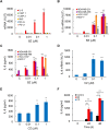
Fig 1
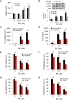
Fig 1. Proteasome inhibition upregulates IL-8 expression in TNBC cells.
(A) Real time RT-PCR of mRNA levels of different NFκB-dependent genes measured in MDA-MB-231 cells treated with increasing BZ concentrations for 24 hours. (B) RT-PCR of IL-8 mRNA in MDA-MB-231, MDA-MB-468, HCC-1937, and MCF-7 cells treated 24 h with increasing BZ. (C) IL-8 release measured by ELISA in cell culture supernatants of MDA-MB-231, MDA-MB-468, HCC-1937, and MCF-7 cells treated 24 h with increasing BZ. (D) RT-PCR of IL-8 mRNA in MDA-MB-231 cells treated 24 h with increasing CZ concentrations. (E) IL-8 release measured by ELISA in cell culture supernatants of MDA-MB-231 cells treated 24 h with increasing CZ. (F) IL-8 release in cell culture supernatants of MDA-MB-231 cells treated with control DMSO, 100 nM BZ, or 100 nM CZ for 0, 24, and 48 h. The values represent the mean +/− SE of four experiments; asterisks denote a statistically significant change compared to control untreated (UT) cells (* p<0.05; ** p<0.01; *** p<0.001).
Fig 2
Fig 2. Suppression of BZ-induced IL-8 augments BZ cytotoxic and anti-proliferative effect in TNBC cells.
(A) RT-PCR of CXCR1 and CXCR2 mRNA levels in MDA-MB-231 cells treated with increasing BZ for 24 h. (B) Western analysis of CXCR1, CXCR2, and control actin protein levels in whole cell extracts (WCE) of MDA-MB-231 cells treated with increasing BZ for 24 h. The bottom panel represents densitometric evaluation of CXCR1 and CXCR2 protein levels shown in the top panel. The CXCR1/2 densities were normalized to actin, and expressed relative to untreated cells. (C) RT-PCR of IL-8 mRNA in MDA-MB-231 cells transfected with control or IL-8 siRNA and incubated 24 h with increasing BZ. (D) IL-8 release measured by ELISA in supernatant of MDA-MB-231 cells transfected with control or IL-8 siRNA and incubated 24 h with increasing BZ. (E) Viability of MDA-MB-231 cells transfected with control or IL-8 siRNA, and incubated 24 h with increasing BZ, measured be Trypan Blue exclusion. (F) Proliferation of MDA-MB-231 cells transfected with control or IL-8 siRNA, and incubated 24 h with BZ, measured by CellTiter 96 One Solution Cell Proliferation Assay. (G) Viability of MDA-MB-231 cells incubated 24 h with increasing BZ in the presence of control pre-immune IgG or IL-8 neutralizing monoclonal antibody. (H) Proliferation of MDA-MB-231 cells incubated 24 h with increasing BZ in the presence of control pre-immune IgG or IL-8 neutralizing monoclonal antibody. The values represent the mean +/− SE of four experiments; asterisks denote a statistically significant change compared to control.
Fig 3
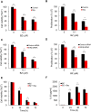
Fig 3. BZ-induced IL-8 expression in TNBC cells is mediated by IKK.
(A) RT-PCR of IL-8 mRNA in MDA-MB-231 cells pre-treated 12 h with control DMSO, Bay-117082 (10 or 40 μM), or SC-514 (10 or 40 μM), and incubated 24 h with 100 nM BZ. (B) Western analysis of IKKα, IKKβ, and IKKε in WCE of MDA-MB-231 cells transfected with control, IKKα, IKKβ, and IKKε siRNA, and treated with BZ (100 nM, 24 h). The bottom panel represents densitometric evaluation of IKKα, IKKβ, and IKKε protein levels shown in the top panel; the IKKs densities were normalized to actin, and expressed relative to cells transfected with control siRNA. (C) RT-PCR of IL-8 mRNA in MDA-MB-231 cells transfected with control, IKKα, IKKβ, or IKKε siRNA, and incubated 24 h with 100 nM BZ. (D) IL-8 release measured by ELISA in MDA-MB-231 cells transfected with control, IKKα, IKKβ, or IKKε siRNA and incubated 24 h with 100 nM BZ. The values represent the mean +/− SE of four experiments; asterisks denote a statistically significant change compared to control.
Fig 4
Fig 4. BZ increases nuclear p65 accumulation, and IKK-dependent p65 recruitment to IL-8 promoter in TNBC cells.
(A) Western blotting of cytoplasmic (CE) and nuclear extracts (NE) prepared from MDA-MB-231 cells treated 24 h with BZ, and analyzed by using p65 and IκBα antibodies. The presence of cytoplasmic proteins in nuclear fraction was evaluated by re-probing the membrane with lactate dehydrogenase (LDH) antibody. Nuclear contamination in the cytoplasmic fraction was assessed using histone H3 specific antibody. To confirm equal protein loading, the membranes were stripped and re-probed with actin antibody. Each lane corresponds to approximately 5×10⁴ cells. (B) Densitometric evaluation of p65, IκBα, and histone H3 levels in nuclear extracts of BZ-treated MDA-MB-231 cells, shown in panel A. The densities of nuclear p65, IκBα, and histone H3 were normalized to the densities of nuclear actin. The values for untreated cells were arbitrarily set to 1, and the other values are presented relative to these values. (C) Recruitment of p65 to endogenous IL-8 promoter analyzed by ChIP and quantified by real time PCR in MDA-MB-231 cells treated with 100 nM BZ for 0, 3, 6 and 24 h. (D) ChIP of p65 recruitment to IL-8 promoter in MDA-MB-231 cells pre-incubated 12 h with control DMSO, Bay-117082 (10 μM), or SC-514 (10 μM), and then incubated 24 h with 100 BZ. The data are presented as the change in occupancy over the human IGX1A (Qiagen) sequence control and represent the mean +/− SE of three experiments. Asterisks denote a statistically significant change compared to control.
Fig 5
Fig 5. Suppression of IKK activity enhances BZ cytotoxic and anti-proliferative effect in TNBC cells.
(A) Viability of MDA-MB-231 cells treated 24 h with BZ in the presence or absence of Bay-117082, analyzed by using Trypan Blue exclusion. (B) Proliferation of MDA-MB-231 cells treated 24 h with BZ in the presence or absence of Bay-117082, analyzed by CellTiter 96 One Solution Cell Proliferation Assay. (C) Viability of MDA-MB-231 cells transfected with control or IKKβ siRNA and incubated 24 h with BZ. (D) Proliferation of MDA-MB-231 cells transfected with control or IKKβ siRNA, and incubated 24 h with BZ. (E) Viability and (F) IL-8 release in MDA-MB-231 cells concomitantly incubated with 100 nM BZ with and without 10 μM Bay-117082 for up to 72 h. The data are expressed as the percentage compared to untreated cells. The values represent the mean +/−SE of four experiments; asterisks denote a statistically significant change compared to control.
Fig 6
Fig 6. IKK and proteasome inhibitors have synergistic effect in reducing migration of TNBC cells.
(A) MDA-MB-231 cells were incubated with 10 μM Bay-117082 and/or 10 nM BZ as indicated, and cell migration was evaluated by wound-healing assay as described in “Materials and methods”. (B) Images from panel A were quantified using ImageJ software, and the data are expressed as wound width at 24 h compared to the corresponding wound width at 0 h. (C) IL-8 release measured in cell supernatants in the wound-healing assay shown in panel A. The values represent the mean +/−SE of three experiments; asterisks denote a statistically significant change compared to control.
Fig 7
Fig 7. IKK inhibition decreases invasion of BZ-treated TNBC cells.
(A) MDA-MB-231 cells were incubated with 10 μM Bay-117082 and/or 10 nM BZ as indicated, and cell invasion was measured as described in “Materials and methods”. (B) Quantification of images from panel A using ImageJ software. (C) IL-8 release measured in cell supernatants in the invasion assay shown in panel A. The values represent the mean +/−SE of three experiments; asterisks denote a statistically significant change compared to control.

References

    1. Waugh DJ, Wilson C. The interleukin-8 pathway in cancer. Clin Cancer Res. 2008;14: 6735–6741. 10.1158/1078-0432.CCR-07-4843 - DOI - PubMed
    1. Lazennec G, Richmond A. Chemokines and chemokine receptors: new insights into cancer-related inflammation. Trends Mol Med. 2010;16: 133–144. 10.1016/j.molmed.2010.01.003 - DOI - PMC - PubMed
    1. Freund A, Chauveau C, Brouillet JP, Lucas A, Lacroix M, Licznar A, et al. IL-8 expression and its possible relationship with estrogen-receptor-negative status of breast cancer cells. Oncogene. 2003;22: 256–265. 10.1038/sj.onc.1206113 - DOI - PMC - PubMed
    1. Freund A, Jolivel V, Durand S, Kersual N, Chalbos D, Chavey C, et al. Mechanisms underlying differential expression of interleukin-8 in breast cancer cells. Oncogene. 2004;23: 6105–6114. 10.1038/sj.onc.1207815 - DOI - PMC - PubMed
    1. Rody A, Karn T, Liedtke C, Pusztai L, Ruckhaeberle E, Hanker L, et al. A clinically relevant gene signature in triple negative and basal-like breast cancer. Breast Cancer Res. 2011;13: R97 10.1186/bcr3035 - DOI - PMC - PubMed

Publication types

MeSH terms

LinkOut - more resources