. 2018 Aug 8;8(1):11875.
doi: 10.1038/s41598-018-30087-4.

Antenatal IL-1-dependent inflammation persists postnatally and causes retinal and sub-retinal vasculopathy in progeny

Affiliations

Antenatal IL-1-dependent inflammation persists postnatally and causes retinal and sub-retinal vasculopathy in progeny

Alexandra Beaudry-Richard et al. Sci Rep. .

Erratum in

Abstract

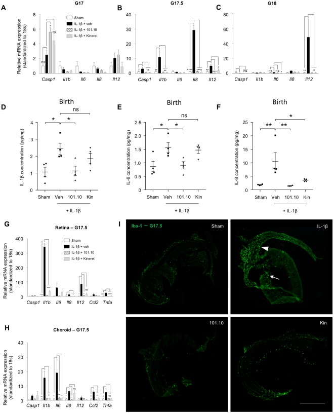
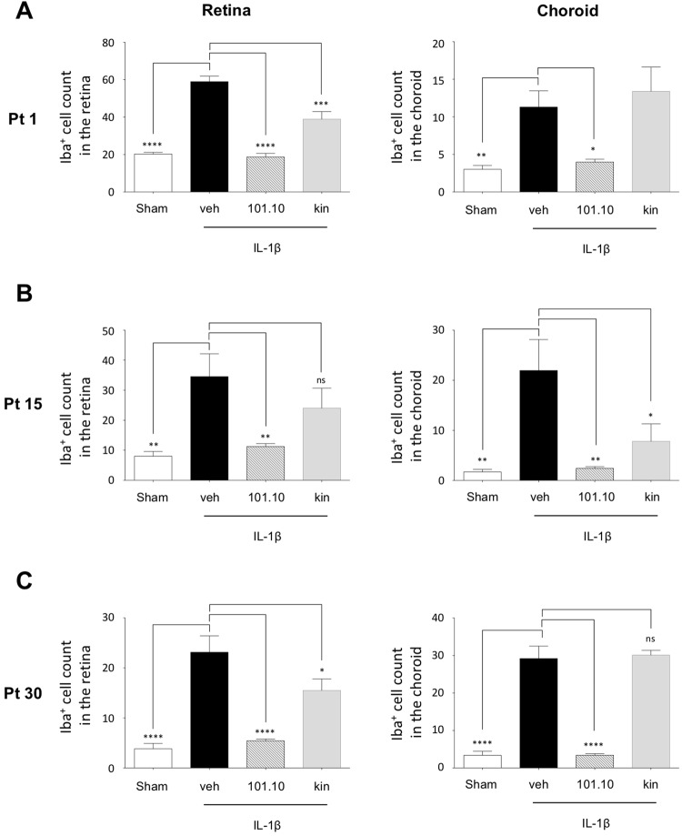
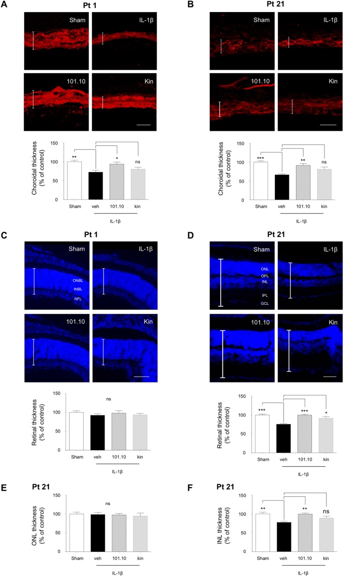
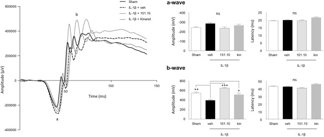
Antenatal inflammation as seen with chorioamnionitis is harmful to foetal/neonatal organ development including to eyes. Although the major pro-inflammatory cytokine IL-1β participates in retinopathy induced by hyperoxia (a predisposing factor to retinopathy of prematurity), the specific role of antenatal IL-1β associated with preterm birth (PTB) in retinal vasculopathy (independent of hyperoxia) is unknown. Using a murine model of PTB induced with IL-1β injection in utero, we studied consequent retinal and choroidal vascular development; in this process we evaluated the efficacy of IL-1R antagonists. Eyes of foetuses exposed only to IL-1β displayed high levels of pro-inflammatory genes, and a persistent postnatal infiltration of inflammatory cells. This prolonged inflammatory response was associated with: (1) a marked delay in retinal vessel growth; (2) long-lasting thinning of the choroid; and (3) long-term morphological and functional alterations of the retina. Antenatal administration of IL-1R antagonists - 101.10 (a modulator of IL-1R) more so than Kineret (competitive IL-1R antagonist) - prevented all deleterious effects of inflammation. This study unveils a key role for IL-1β, a major mediator of chorioamnionitis, in causing sustained ocular inflammation and perinatal vascular eye injury, and highlights the efficacy of antenatal 101.10 to suppress deleterious inflammation.


Conflict of interest statement

The authors declare no competing interests.

Figures

Figure 1
Figure 1
Murine model of inflammation-induced PTB employed for the study. (A) IL-1β (1 µg) was administered intrauterine at G16.5, whereas 101.10 (1 mg/Kg/12 h), Kineret (4 mg/Kg/12 h) or vehicle were administered subcutaneously for 2 consecutive days, with the first dose administered 30 min prior to IL-1β. (B) Intrauterine injection of IL-1β was made between two foetal membranes, without penetrating the amniotic cavity.
Figure 2
Figure 2
Inflammatory response in the retina and sub-retina of the foetus and newborn. (AC) Foetal eyes were collected at G17 (A), G17.5 (B) and G18 (C) after in utero exposure to IL-1β to perform quantitative PCR. Results are relative to 18 S and plotted as fold change vs. the control groups. n = 3–8 dams/group; 4 foetal eyes per sample. (DF) Cytokine levels in eyes of newborns exposed to the indicated treatments in utero (Fig. 1). n = 4 dams/group; 4 eyes per sample. (GH) Quantitative PCR of IL-1, IL-6 and IL-8 performed on isolated retina (G) and choroid (H) collected on foetuses at G17.5; n = 3 dams/group; 10 retinas or sub-retinas per sample. (I) Iba-1-stained cryosections of retina from foetuses at G17.5 exposed to the indicated treatments in utero; n = 3 foetuses/group. Kin refers to Kineret. Scale bar, 300 μm. Values are presented as mean ± S.E.M. *p < 0.05, **p < 0.01, ***p < 0.001 by one-way ANOVA with Dunnett’s post-analysis.
Figure 3
Figure 3
Infiltration of immune cells in eyes during development of the pups. (AC) Quantification of iba-1+ cells observed on retinal and choroidal flat mounts at Pt 1 (A), Pt 15 (B) and Pt 30 (C). n = 3–6 pup/group for each time point. Values are presented as mean ± S.E.M. *p < 0.05, **p < 0.01, ***p < 0.001, ****p < 0.0001 by one-way ANOVA with Dunnett’s post-analysis.
Figure 4
Figure 4
Delay of retinal vessel growth in progeny. (A–C) Lectin-stained flat-mounts of retinas from pups at Pt 1 (A), Pt 4 (B) and Pt 8 (C) previously exposed in utero to the indicated treatments (Fig. 1). Images are representative of 3 to 5 separate pups per treatment group. Dotted lines depict the vascular front. Scale bar for A, 1500 μm; scale bar for B, 2500 μm; scale bar for C, 3000 μm. Right panels show quantification of the vascular area, n = 3–5 dams/group. (D) Magnification of lectin-stained flat-mounts of retinas from pups at Pt15 showing vascular density. Images are representative of 5 to 8 separate pups per treatment group. Scale bar, 150 μm. Right panel shows quantification of the vascular area at Pt8 and Pt15, n = 5–8 dams/group. Values are presented as mean ± S.E.M. *p < 0.05, ***p < 0.001 by one-way ANOVA with Dunnett’s post-analysis.
Figure 5
Figure 5
Choroidal thinning precedes retinal degeneration and thinning of the inner nuclear layer. (A–B) Representative images (top panels) and quantification (bottom panels) of lectin-stained cross-sections of choroids from pups at Pt 1 (A) and Pt 21 (B) previously exposed to the indicated treatments in utero (Fig. 1). Vertical bars represent the average choroidal thickness. Scale bar for A, 30 μm; scale bar for B, 50 μm. n = 3–4 pups/group. (C,D) Representative images (top panels) and quantification (bottom panels) of DAPI-stained cross-sections of retinas from pups at Pt1 (C) and Pt21 (D) previously exposed to the indicated treatments in utero (Fig. 1); n = 3–4 pups/group. Vertical bars represent the average retinal thickness. Scale bar for C, 250 μm; scale bar for B, 500 μm. (E,F) Quantification of DAPI-stained cross-sections of the ONL (E) and INL (F) from the same retinas measured in D. Values are presented as mean ± S.E.M. *p < 0.05, **p < 0.01, ***p < 0.001 by one-way ANOVA with Dunnett’s post-analysis. ONBL, outer neuroblastic layer; INBL, inner neuroblastic layer; NFL, nerve fibre layer; ONL, outer nuclear layer; OPL, outer plexiform layer; INL, inner nuclear layer; IPL, inner plexiform layer; GCL, ganglion cell layer.
Figure 6
Figure 6
Electroretinogram readings after completion of ocular development. (A) Representative ERG response of pups at Pt 30 previously exposed to the indicated treatments in utero (Fig. 1). (B,C) a-wave amplitude (B) and latency (C) of scotopic ERGs of corresponding animals. (D,E) b-wave amplitude (D) and latency (E) of scotopic ERGs of corresponding animals. n = 8–22 pups/group. Values are presented as mean ± S.E.M. *p < 0.05, **p < 0.01, ***p < 0.001 by one-way ANOVA with Dunnett’s post-analysis.

References

    1. Institute of Medicine Committee on Understanding Premature, B. & Assuring Healthy, O. In Preterm Birth: Causes, Consequences, and Prevention (eds Behrman, R. E. & Butler, A. S.) (National Academies Press (US) National Academy of Sciences., 2007). - PubMed
    1. Liu L, et al. Global, regional, and national causes of child mortality in 2000–13, with projections to inform post-2015 priorities: an updated systematic analysis. Lancet (London, England) 2015;385:430–440. - PubMed
    1. Verstraeten BS, Mijovic-Kondejewski J, Takeda J, Tanaka S, Olson DM. Canada’s pregnancy-related mortality rates: doing well but room for improvement. Clinical and investigative medicine. Medecine clinique et experimentale. 2015;38:E15–22. - PubMed
    1. Blencowe H, et al. Born too soon: the global epidemiology of 15 million preterm births. Reproductive health. 2013;10(Suppl 1):S2. - PMC - PubMed
    1. ACOG committee opinion no. 561 Nonmedically indicated early-term deliveries. Obstetrics and gynecology. 2013;121:911–915. - PubMed

Publication types

Substances

Grants and funding