. 2015 Dec 2;5(1):116-125.
doi: 10.1039/c5tx00291e. eCollection 2016 Jan 1.

Sodium tanshinone IIA sulfonate suppresses pulmonary fibroblast proliferation and activation induced by silica: role of the Nrf2/Trx pathway

Affiliations

Sodium tanshinone IIA sulfonate suppresses pulmonary fibroblast proliferation and activation induced by silica: role of the Nrf2/Trx pathway

Zhonghui Zhu et al. Toxicol Res (Camb). .

Abstract

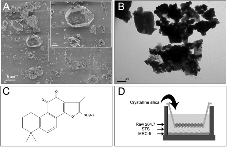
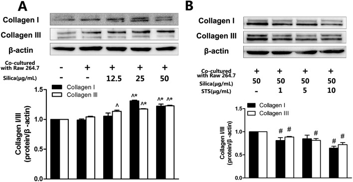
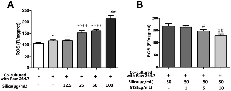
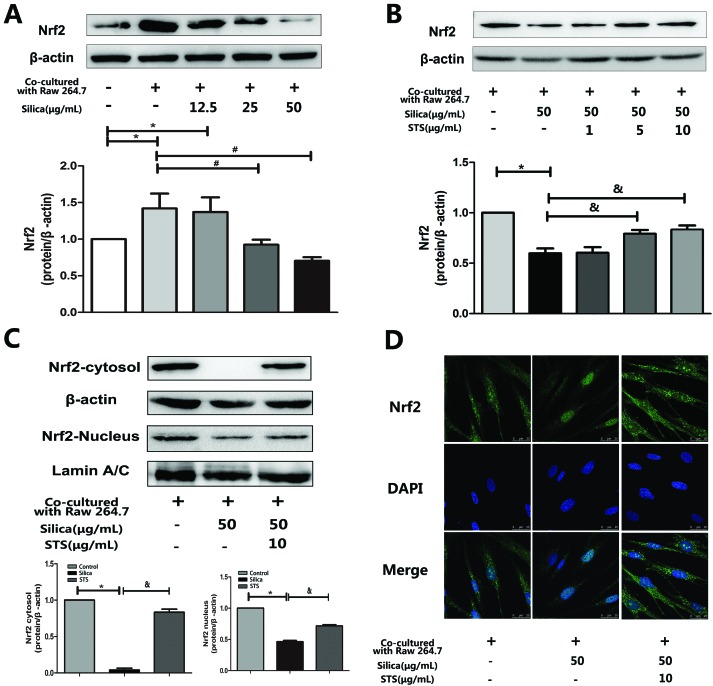
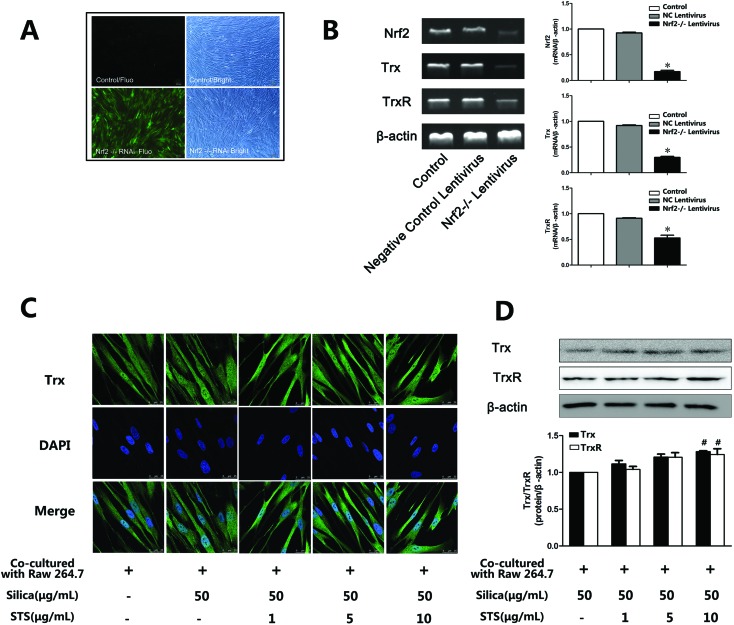
Alveolar macrophages are believed to induce oxidative stress via reactive oxygen species (ROS) when silica particles are inhaled. This process can contribute to the pathogenesis of silicosis, but the mechanism is unclear. A traditional Chinese herbal derivative, sodium tanshinone IIA sulfonate (STS), displays significant antioxidant effects. Here, we determine whether STS can attenuate the oxidative stress induced by silica. Traditionally, studies on the toxic effects of silica have focused on monocultures of macrophages or fibroblasts. A coculture model of macrophages (Raw 264.7) and pulmonary fibroblasts (MRC-5) was used in this study to mimic a more in vivo-like environment. We investigated the protective effects of STS on the abnormal proliferation of MRC-5 fibroblasts in an in vitro model. The results showed that fibroblast viability increased with the accumulation of intracellular ROS induced by cocultured Raw 264.7 cells after silica exposure. Treatment with STS markedly ameliorated the silica-induced cell proliferation and oxidative stress. Western blotting and immunofluorescence analysis of the Nrf2 and thioredoxin (Trx) system were conducted, and the results confirmed that treatment with STS enhanced nuclear Nrf2 accumulation and mediated antioxidant Trx system expression. These findings suggest that silica exposure might induce some level of oxidative stress in fibroblasts and that STS might augment antioxidant activities via up-regulation of the Nrf2 and Trx system pathways in MRC-5 cells in vitro.


Figures

Fig. 1
Fig. 1. SEM (A) and TEM (B) images of silica showing an overview of the scale structure and dispersion, with scale bars of 5 μm or 0.5 μm individually. (C) The molecular structure of STS. (D) Macrophage/fibroblast coculture system. Macrophages (Raw 264.7) were cultured on the apical side of polyester Transwell inserts (0.4 μm pore size), while lung fibroblast cells (MRC-5) were cultured in the basolateral chamber of the Transwell. Macrophages were exposed to silica particles at various doses without or with STS treatment, and the effects on the underlying fibroblasts were assayed.
Fig. 2
Fig. 2. Effects of STS on silica/H2O2 induced fibroblast proliferation. Raw 264.7 and MRC-5 cells were grown in coculture, followed by exposure of different concentrations of silica/H2O2, as well as STS (1, 5, 10, 25, or 50 μg mL–1) addition. After 24 h, cell proliferation was determined with the MTT assay. The experiments were repeated five times with reproducible results. *P < 0.05 vs. negative control group; #P < 0.05, ##P < 0.01 vs. the control group. &P < 0.05 vs. silica/H2O2 treatment group.
Fig. 3
Fig. 3. Effects of STS on silica-induced collagen type I/III expression. Collagen type I/III expression (A & B) was determined by Western blotting. The experiments were repeated three times with reproducible results. ^P < 0.05 vs. control; *P < 0.05 vs. negative control group. #P < 0.05, ##P < 0.01 vs. silica treatment group.
Fig. 4
Fig. 4. Effects of STS on the silica-induced production of ROS. Raw 264.7 and MRC-5 cells were grown in coculture, followed by exposure of Raw 264.7 cells to various doses of silica without or with STS (1, 5, 10, 25, or 50 μg mL–1). After 24 h, DCFH-DA fluorescence was measured by a fluorospectrophotometer (A&B). The experiments were repeated three times with reproducible results. ^P < 0.05, ^^P < 0.01 vs. control; *P < 0.05, **P < 0.01 vs. negative control group. #P < 0.05, ##P < 0.01 vs. silica treatment group.
Fig. 5
Fig. 5. Effects of silica and STS on the expression of antioxidative proteins. (A & B) Western blot results are shown: one for Nrf2 in silica-treated groups (left) and one for Nrf2 in STS-treated groups (right). Silica decreased the amount of Nrf2 in whole cells, and the high dose of STS up-regulated the expression of Nrf2 in whole cells. *P < 0.05 vs. control; #P < 0.05 vs. negative control group. &P < 0.05 vs. silica treatment group. (C) Total form Nrf2 in the nucleus and cytoplasm were prepared and determined using Western blot. (D) Immunofluorescence staining of Nrf2 plus or minus silica and/or STS. With exposure to 50 μg mL–1 silica particles for 24 h, Nrf2 immunofluorescence (green) is barely observed in the cytoplasm but is clearly present with added STS. The magnification (630×) in all panels is the same. Scale bar = 25 μm. Nuclei are stained blue with DAPI.
Fig. 6
Fig. 6. TrxR and Trx gene and protein expression in MRC-5 cells was inhibited by Nrf2 knockdown and increased by STS treatment. (A) Transfection efficiency detected by fluorescence microscopy. GFP-emitted green fluorescence (left) and same fields of vision under an optical microscope (right). More than 80% of cells were infected with lent-GFP at an MOI of 10 after 72 h of treatment. (B) Antioxidant mRNA levels by RT-PCR. The Lent-GFP-Nrf2-/-lentivirus significantly decreased the Nrf2 mRNA level and the Trx/TrxR mRNA level in MRC-5 cells. The effects of STS on Trx and TrxR protein expression, examined using immunofluorescence microscopy (C) and/or western blotting (D).

References

    1. Cox Jr. L. A. Risk Anal. 2011;31(10):1543–1560. - PubMed
    1. Gilberti R. M., Joshi G. N., Knecht D. A. Am. J. Respir. Cell Mol. Biol. 2008;39(5):619–627. - PMC - PubMed
    1. Shen H. M., Zhang Z., Zhang Q. F., Ong C. N. Am. J. Physiol.: Lung Cell. Mol. Physiol. 2001;280(1):L10–LL7. - PubMed
    1. Sundaresan M., Yu Z. X., Ferrans V. J., Sulciner D. J., Gutkind J. S., Irani K. Biochem. J. 1996;318(Pt 2):379–382. - PMC - PubMed
    1. Krieger-Brauer H. I., Kather H. Biochem. J. 1995;307(Pt 2):549–556. - PMC - PubMed

LinkOut - more resources