. 2016 Jun 6;5(5):1335-1345.
doi: 10.1039/c6tx00111d. eCollection 2016 Sep 1.

Salvianolic acid B protects against doxorubicin induced cardiac dysfunction via inhibition of ER stress mediated cardiomyocyte apoptosis

Affiliations

Salvianolic acid B protects against doxorubicin induced cardiac dysfunction via inhibition of ER stress mediated cardiomyocyte apoptosis

Rongchang Chen et al. Toxicol Res (Camb). .

Abstract

Salvia miltiorrhiza Bunge is a well-known medicinal plant in China. Salvianolic acid B (Sal B) is the most abundant bioactive compound extracted from the root of S. miltiorrhiza. The present study investigates the effect of Sal B on cardiac function and cardiomyocyte apoptosis in doxorubicin (DOX)-treated mice. After pretreatment with Sal B (2 mg kg-1 iv) for 7 d, male BALB/c mice were injected with a single dose of DOX (20 mg kg-1 ip). The cardioprotective effect of Sal B was observed on the 7th day after DOX treatment. DOX caused retarded body growth, apoptotic damage, and Bcl-2 expression disturbance. In contrast, Sal B pretreatment (2 mg kg-1 iv before DOX administration) attenuated the DOX induced apoptotic damage in heart tissues. Further study indicated that Sal B protected against DOX induced cardiotoxicity, at least, partially, by inhibiting endoplasmic reticulum stress, and by being involved in the PI3K/Akt pathway. These findings clarified the potential of Sal B as a promising reagent for treating DOX induced cardiotoxicity.


Figures

Fig. 1
Fig. 1. Effects of Sal B and DOX on body weight. (A) Molecular structure of Sal B. (B) Body weights and heart weights (C) of mice were measured 7 d after DOX injection. (D) Relative heart weight index (heart weight-to-body weight ratio) was determined. Data are presented as mean ± SE, *P < 0.05 versus Control (Cont) group; #P < 0.05 versus DOX group.
Fig. 2
Fig. 2. Effects of Sal B and DOX on cardiac function. (A) Representative M-mode ECG images are shown. (B) ECG values are expressed as mean ± SE. Data are presented as mean ± SE, *P < 0.05 versus Control group; #P < 0.05 versus DOX group.
Fig. 3
Fig. 3. Effects of Sal B on DOX induced myocardial injury. (A) Effects of Sal B and DOX on AST, LDH and CK activities. (B) Effects of Sal B and DOX on histological changes in mice hearts by HE staining (scale bar = 10 μm). (C) Effects of Sal B and DOX on ultrastructure changes in mice hearts observed under the electron microscope (scale bar = 200 pm). (D) Effects of Sal B and DOX on the mice's ECG pattern. (E) Effects of Sal B and DOX on the heart rate of mice. Data are presented as mean ± SE, *P < 0.05 versus Control group; #P < 0.05 versus DOX group.
Fig. 4
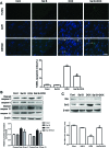
Fig. 4. Effects of Sal B and DOX on heart apoptosis and apoptosis related proteins. (A) Representative images of TUNEL and DAPI staining of myocardium tissue and quantification of TUNEL positive cells (scale bar = 10 μm). Arrowheads in the pictures indicate the nuclei of apoptotic cells and the blue color represents cell nuclei that were counterstained with DAPI. (B) Effects of Sal B and DOX on protein expression of cleaved cas-3, cas-3, cleaved cas-12 and cas-12. (C) Effects of Sal B and DOX on protein expression of Bcl-2 and Bax. Data are presented as mean ± SE, *P < 0.05 versus Control group; #P < 0.05 versus DOX group.
Fig. 5
Fig. 5. Effects of Sal B and DOX on ER stress sensors and ER stress related apoptotic protein expression in heart tissues. (A) Western blot analysis of GRP78 and CHOP. (B) Western blot analysis of p-IRE1, IRE-1, p-JNK, JNK, ATF-6, p-PERK and PERK. Data are presented as mean ± SE, *P < 0.05 versus Control group; #P < 0.05 versus DOX group.
Fig. 6
Fig. 6. Effects of Sal B and DOX on protein expression of the PI3K/Akt signaling pathway. (A) Protein levels of p-Akt, Akt, p-GSK3β and GSK3β in the myocardium examined by western blot analysis. (B) Effects of pharmacological inhibitor WM (a selective PI3K antagonist) on levels of p-Akt, p-GSK3β, GRP78, CHOP, in the myocardium of Sal B and DOX co-treated mice. Data are presented as mean ± SE, *P < 0.05 versus Control; #P < 0.05 versus DOX treated mice; $P < 0.05 versus Sal B and DOX co-treated mice.
Fig. 7
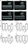
Fig. 7. Involvement of PI3K/Akt signaling in DOX induced cardiac dysfunction. (A) Cardiac function was examined by echocardiography 7 d after DOX administration. Representative M-mode echocardiography images are shown. (B) Echocardiography values are expressed as mean ± SE. Data are presented as mean ± SE, *P < 0.05 versus Control; #P < 0.05 versus DOX treated mice; $P < 0.05 versus Sal B and DOX co-treated mice.

References

    1. AlGhamdi S., Leoncikas V., Plant K. E., Plant N. J. Toxicol. Res. 2015;4:1479–1487. - PMC - PubMed
    1. Church R. J., McDuffie J. E., Sonee M., Otieno M., Ma J. Y., Liu X., Watkins P. B., Harrill A. H. Toxicol. Res. 2014;3:384–394.
    1. Yamanaka S., Tatsumi T., Shiraishi J., Mano A., Keira N., Matoba S., Asayama J., Fushiki S., Fliss H., Nakagawa M. J. Am. Coll. Cardiol. 2003;41:870–878. - PubMed
    1. Jirkovská-Vávrová A., Roh J., Lenčová-Popelová O., Jirkovský E., Hrušková K., Potůčková-Macková E., Jansová H., Hašková P., Martinková P., Eisner T., Kratochvíl M., Šůs J., Macháček M., Vostatková-Tichotová L., Geršl V., Kalinowski D. S., Muller M. T., Richardson D. R., Vávrová K., Štěrba M., Šimůnek T. Toxicol. Res. 2015;4:1098–1114.
    1. Arun R., Dhivya S., Abrahamb S. K., Premkumar K. Toxicol. Res. 2016;5:547–556. - PMC - PubMed