. 2016 Nov 2;6(1):89-97.
doi: 10.1039/c6tx00361c. eCollection 2017 Jan 1.

A DEHP plasticizer alters synaptic proteins via peroxidation

Affiliations

A DEHP plasticizer alters synaptic proteins via peroxidation

Shaohui Wang et al. Toxicol Res (Camb). .

Abstract

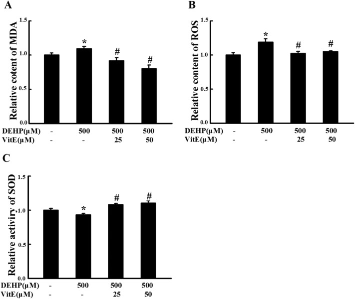
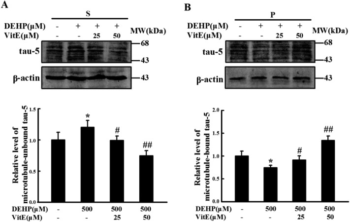
Di-(2-ethylhexyl) phthalate (DEHP) is a widely used commercial plasticizer. DEHP exposure has a negative impact on brain development and cognition, but the mechanisms responsible for DEHP-induced neurotoxicity are not well understood. Here we showed that DEHP exposure increased maleic dialdehyde and reactive oxygen species contents and decreased endogenous superoxide dismutase activity in a mouse neuroblastoma cell line (N2a cell line). DEHP exposure not only induced reduction of neurite outgrowth, but also led to microtubule-associated protein tau hyperphosphorylation and dissociation from microtubules. Furthermore, DEHP exposure decreased the levels of synapsin-1 and postsynaptic density protein 95 (PSD95), which play critical roles in synaptic function. Antioxidant vitamin E pretreatment prevented DEHP-induced abnormalities in the cells. These results indicate that DEHP exposure could induce abnormal action of proteins including tau, synapsin-1 and PSD95, which play critical roles in the synaptic structure and function, and that these alterations might be mediated by peroxidative damage.


Figures

Fig. 1
Fig. 1. MTT-assay of N2a cells treated with different doses of DEHP (125, 250, 500 and 1000 μM). Cell viability was determined by MTT assay. Results are represented as mean ± SD, n = 3 independent observations.
Fig. 2
Fig. 2. N2a cells were treated with DEHP (125, 250, 500 and 1000 μM) for 24 h. The relative content of MDA (A) and ROS (B), and the relative activity of SOD (C) were expressed as fold changes over DEHP-untreated cells and are represented as mean ± SD (n = 5). *p < 0.05, **p < 0.01 vs. control group.
Fig. 3
Fig. 3. Vitamin E pretreatment prevented DEHP-induced oxidative damage in N2a cells. 25 and 50 μM vitamin E pretreatment for 1 h protected N2a cells against DEHP-induced MDA (A) and ROS (B) deficits and SOD activity deficits (C). All data are expressed as fold changes over control cells and are represented as mean ± SD (n = 5). *p < 0.05 vs. control group; #p < 0.05, ##p < 0.01 vs. DEHP treatment group.
Fig. 4
Fig. 4. N2a cell neurite changes following DEHP, or vitamin E plus DEHP (VitE + DEHP) treatment. A, The morphology of N2a cells in the control group (a), DEHP exposure group (b), 25 μM vitamin E (c) and 50 μM vitamin E (d) pretreatment plus DEHP exposure group. B, The quantification of neurite length. Scale bar = 50 μm. n = 30 cells. *p < 0.05 vs. control group; #p < 0.05 vs. DEHP treatment group.
Fig. 5
Fig. 5. Vitamin E pretreatment mitigates DEHP-induced tau-microtubule binding function impairment in N2a cells. Immunostaining of unbound tau in (A) the supernatant fraction (S) and (B) microtubule-bound tau in the pellet fraction (P) were detected by western blot analysis. Values were normalized to those of the control group. The protein levels were expressed as fold changes over untreated cells and are represented as mean ± SD, *p < 0.05 vs. control group; #p < 0.05, ##p < 0.01 vs. DEHP treatment group.
Fig. 6
Fig. 6. Tau protein was hyperphosphorylated at pT231 and pS262 sites after DEHP treatment. (A) 500 μM DEHP exposure for 24 h hyperphosphorylated tau at pT231 and pS262 sites in N2a cells; 25 and 50 μM vitamin E (VitE) pretreatment prevented DEHP-induced tau hyperphosphorylation in N2a cells in a dose-dependent manner. The total level of tau had no observable change. (B) Quantification of the total tau and phosphorylated tau levels. The protein levels were expressed as fold changes over untreated cells and are represented as mean ± SD, *p < 0.05, **p < 0.01 vs. control cells; #p < 0.05, ##p < 0.01 vs. DEHP treatment cells.
Fig. 7
Fig. 7. Vitamin E pretreatment mitigated DEHP induced GSK-3β activation. (A) The level of phosphorylated GSK-3β at the Ser9 site and the total level of GSK-3β were detected by western blot analysis. (B) Quantification of the relative intensity of phosphorylated GSK-3β at the Ser9 site. The values were expressed as fold changes over control and are represented as mean ± SD. Significance is denoted as **p < 0.01 vs. control group, and #p < 0.05, ##p < 0.01 vs. DEHP treatment group.
Fig. 8
Fig. 8. DEHP treatment decreased the levels of synapsin-1 and PSD95. (A) The expressions of synapsin-1 and PSD95 were detected by western blot analysis. (B) Quantification of the relative intensity of synapsin-1 and PSD95; the values were expressed as fold changes over control and are represented as mean ± SD. Significance is denoted as **p < 0.01 vs. control group, and ##p < 0.01 vs. DEHP treatment group.

References

    1. Anderson D., Yu T. W., Hincal F. Teratog., Carcinog., Mutagen. 1999;19:275–280. - PubMed
    1. Rubin R. J., Schiffer C. A. Transfusion. 1976;16:330–335. - PubMed
    1. Hashizume K., Nanya J., Toda C., Yasui T., Nagano H., Kojima N. Biol. Pharm. Bull. 2002;25:209–214. - PubMed
    1. Rudel R. A., Camann D. E., Spengler J. D., Korn L. R., Brody J. G. Environ. Sci. Technol. 2003;37:4543–4553. - PubMed
    1. Chen T., Yang W., Li Y., Chen X., Xu S. Toxicol. Lett. 2011;201:34–41. - PubMed

LinkOut - more resources