. 2018 Jul 9:10:165-178.
doi: 10.1016/j.omtm.2018.07.002. eCollection 2018 Sep 21.

Cerebrospinal Fluid-Directed rAAV9-rsATP7A Plus Subcutaneous Copper Histidinate Advance Survival and Outcomes in a Menkes Disease Mouse Model

Affiliations

Cerebrospinal Fluid-Directed rAAV9-rsATP7A Plus Subcutaneous Copper Histidinate Advance Survival and Outcomes in a Menkes Disease Mouse Model

Marie Reine Haddad et al. Mol Ther Methods Clin Dev. .

Abstract

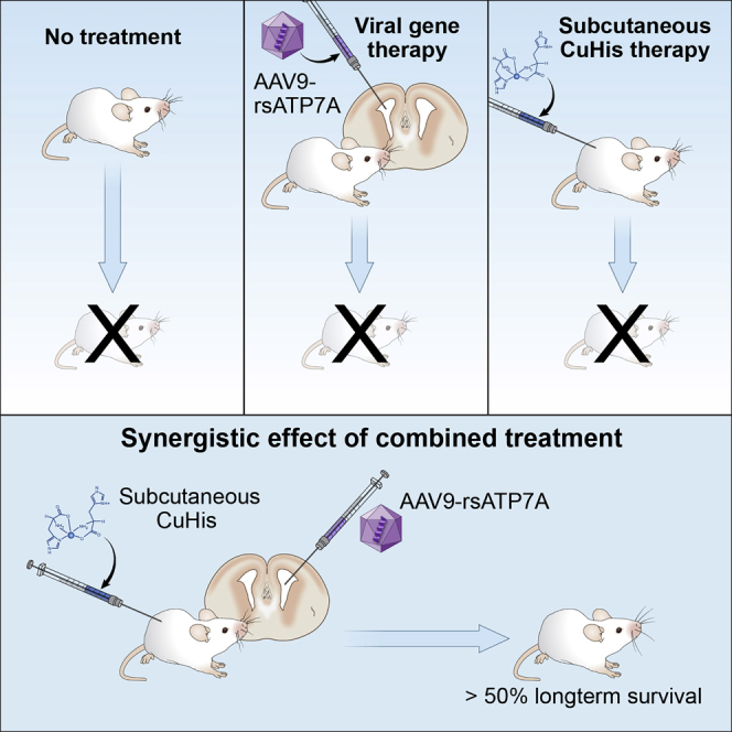
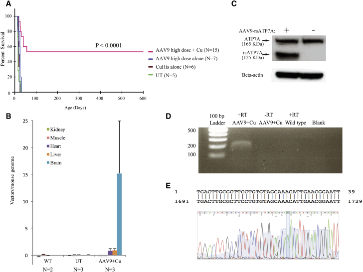
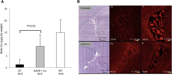
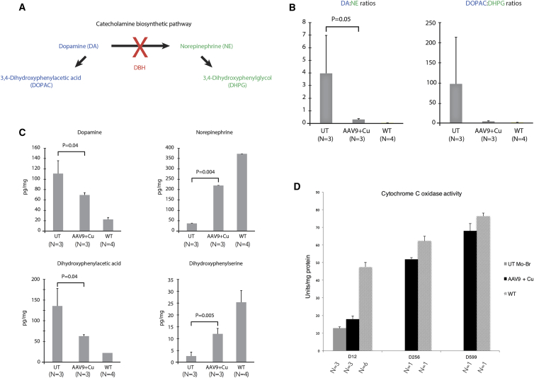
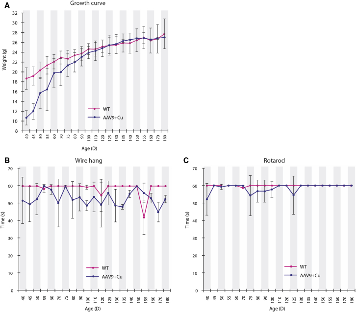
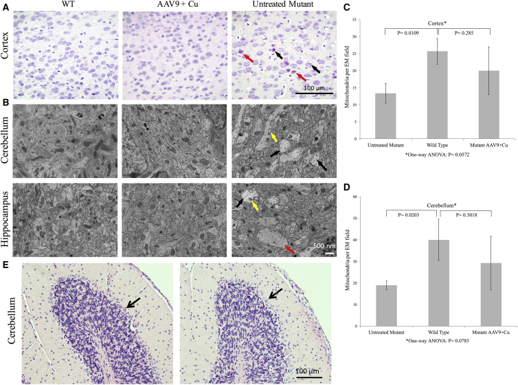
Menkes disease is a lethal neurodegenerative disorder of copper metabolism caused by mutations in an evolutionarily conserved copper transporter, ATP7A. Based on our prior clinical and animal studies, we seek to develop a therapeutic approach suitable for application in affected human subjects, using the mottled-brindled (mo-br) mouse model that closely mimics the Menkes disease biochemical and clinical phenotypes. Here, we evaluate the efficacy of low-, intermediate-, and high-dose recombinant adeno-associated virus serotype 9 (rAAV9)-ATP7A delivered to the cerebrospinal fluid (CSF), in combination with subcutaneous administration of clinical-grade copper histidinate (sc CuHis, IND #34,166). Mutant mice that received high-dose (1.6 × 10¹⁰ vg) cerebrospinal fluid-directed rAAV9-rsATP7A plus sc copper histidinate showed 53.3% long-term (≥300-day) survival compared to 0% without treatment or with either treatment alone. The high-dose rAAV9-rsATP7A plus sc copper histidinate-treated mutant mice showed increased brain copper levels, normalized brain neurochemical levels, improvement of brain mitochondrial abnormalities, and normal growth and neurobehavioral outcomes. This synergistic treatment effect represents the most successful rescue to date of the mo-br mouse model. Based on these findings, and the absence of a large animal model, we propose cerebrospinal fluid-directed rAAV9-rsATP7A gene therapy plus subcutaneous copper histidinate as a potential therapeutic approach to cure or ameliorate Menkes disease.

Keywords: ATP7A; Menkes disease; adeno-associated virus; choroid plexus epithelia; copper; dopamine-beta-hydroxylase.


Figures

None
Graphical abstract
Figure 1
Figure 1
Recombinant AAV9 Tropism in Mouse Brain Immunohistochemistry to illustrate reporter gene (GFP) expression after injection of rAAV9 into the cerebrospinal fluid of wild-type mice. (A) Low-power sagittal view of wild-type mouse brain on day 12 after cerebrospinal fluid administration of 1.6 × 1010 vg of rAAV9-GFP on day 2 (top). Staining with anti-GFP defines the transduction pattern of rAAV9 (brownish color) in these experiments since there is no reliable anti-ATP7A antibody for immunohistochemical applications. An uninjected mouse brain (bottom) shows no GFP expression. Scale bars, 1 mm. (B) Representative brain region images with GFP expression on day 12 after injection of 5 × 109 vg rAAV9-GFP into the cerebrospinal fluid of wild-type mice on day 2. CPLV, choroid plexus epithelia of lateral ventricle. Scale bars, 100 μm. (C–E) High-power views of cortex (C), cerebellum (D), and brainstem (E), respectively, from 12-day-old mice after cerebrospinal fluid injection of 5 × 109 vg of rAAV9-GFP on day 2, stained with anti-GFP on day 12. Closed arrows denote transduced neurons and open arrows, transduced glial cells. Scale bars, 25 μm.
Figure 2
Figure 2
Reduced Size ATP7A (rsATP7A) for rAAV9 Gene Therapy (A) Model of ATP7A including six copper (Cu)-binding domains in the N terminus and other functional domains, as indicated. The light blue-shaded area shows the segment removed to create rsATP7A, leaving only Cu-binding sites #5 and #6. Red asterisks indicate location of the two amino acids deleted in the mo-br mouse ortholog (Atp7a). (B) Diagram of the recombinant AAV9 construct used in this study. ITR, inverted terminal repeat of AAV serotype 2; CMV, cytomegalovirus (CMV) enhancer; CBA, chicken β-actin promoter; poly(A), rabbit β-globin polyadenylation signal. (C) Confocal microscopic images of HEK293T cells transfected with a Venus-tagged version of rsATP7A, showing normal trafficking (relocation from the perinuclear trans-Golgi network to plasma membrane) in response to increased concentration (from 0.5 μM to 200 μM) of copper. Scale bars, 10 μm. (D) Western blot of total protein from transfected HEK293T cells compared to non-transfected controls. Both rsATP7A protein (≈125 kDa) and endogenous full-length ATP7A (≈165 kDa) are detected in transfected cells. (E) Plasmid-mediated expression of rsATP7A in YSTT cells restores copper delivery to the trans-Golgi network and activates tyrosinase. This functional assay qualitatively assesses the physiological capacity of human copper-ATPases to transport copper into the trans-Golgi network (TGN) for biosynthetic incorporation into copper-dependent enzymes. YSTT cells lack endogenous ATP7A and are unable to transport copper into the TGN, thus rendering tyrosinase, a copper-dependent enzyme, inactive unless transfected with a functional version of ATP7A or ATP7B. Formation of black pigment [levo-3,4-dihydroxy-l-phenylalanine (L-DOPA) quinone] in the cells indicates activation of tyrosinase by both reduced size and full-length ATP7A. The assay was performed in triplicate, and representative images are shown. Scale bars, 50 μm.
Figure 3
Figure 3
Results of Cerebrospinal Fluid-Directed rAAV9-rsATP7A Plus sc Copper Histidinate versus Individual Treatments (A) Kaplan-Meier survival curve demonstrates superior survival with high-dose AAV9-rsATP7A plus sc copper histidinate (magenta) in comparison to untreated mutants (UT), rAAV9-rsATP7A alone (blue), and sc copper histidinate alone (brown) did not significantly enhance survival beyond that in untreated mutants (green) (p < 0.0001). (B) Viral genome quantification using real-time PCR to detect rsATP7A transgene in mo-br brain DNA after administration of 1.6 × 1010 viral particles of either AAV. Non-injected mutant (UT) and wild-type (WT) controls also shown. Error bars, SEM. (C) Western blot of total proteins from high-dose rAAV9-rsATP7A-injected wild-type mouse brain detects recombinant rsATP7A (≈125 kDa) as well as endogenous full-length murine ATP7A (≈165 kDa). Bottom, Beta-actin loading control. (D) Stable long-term expression of rsATP7A 597 days post-administration of 1.6 × 1010vg AAV9-rsATP7A, documented by RT-PCR from brain RNA extracted from a long-term surviving mutant male. −RT refers to a negative control for RT-PCR, with RNA but no reverse transcriptase included in the reaction. (E) Capillary DNA sequencing of the excised band verified that it represented the ATP7A transgene transcript (exon 5). Alignment to Homo sapiens ATP7A, sequence ID, gi|532691751|NM_001282224.1.
Figure 4
Figure 4
Cerebrospinal Fluid-Directed rAAV9-rsATP7A Gene Therapy Plus Subcutaneous Copper Histidinate Increases Brain Copper Levels in mo-br Mutant Mice (A) Brain copper levels, measured by ICP-MS, are significantly higher in 12-day-old mutant mice treated with rAAV9-rsATP7A (1.6 × 1010 vg) plus sc copper histidinate (AAV9 + Cu) compared to untreated mutants (UT). A two-tailed, paired Student’s t test was used to calculate p. (B) X-ray fluorescence microscopy (XFM) of representative 12-day-old mo-br mouse brain sections from untreated (top) and high-dose rAAV9-rsATP7A plus sc copper histidinate-treated (AAV9 + Cu, bottom) mice. Left, H&E-stained tissue sections. Boxed areas in white (left) correspond to the X-ray fluorescence microscopy elemental maps for Cu (middle) and phosphorus (P, right), respectively, the latter obtained as control. The elemental map for Cu shows diffusely increased signals within the choroid plexus of the lateral ventricle (box indicates region of focus) and subventricular zone of the rAAV9-rsATP7A plus sc copper histidinate-treated brain, whereas the phosphorus signals (right) are comparable. Red temperature scale represents copper concentration (0–53 μg/g) and phosphorus concentration 0–10 mg/g or 0–12 mg/g, as noted. Error bars, SEM.
Figure 5
Figure 5
Brain Neurochemical Measurements in rAAV9-rsATP7A Plus sc Copper Histidinate Combination-Treated mo-br Mice (A) Summarized version of catecholamine biosynthetic pathway. Dopamine (DA) is converted to norepinephrine (NE) by the copper-dependent enzyme dopamine-beta-hydroxylase (DBH). (B) Markedly increased DA:NE and DOPAC:DHPG ratios in 12-day-old untreated mo-br mutants (UT) compared to mutant mice that received high-dose rAAV9-rsATP7A plus sc copper histidinate. A two-tailed, paired Student’s t test was used to calculate p. Error bars, SEM. Elevated ratios of proximal:distal metabolites in this pathway are sensitive and specific for diagnosing Menkes disease., , (C) Individual brain neurochemical levels measured by high-performance liquid chromatography (HPLC). Proximal metabolites in the pathway (DA, DOPAC) are significantly lower in 12-day-old mutant mice treated with high-dose rAAV9-rsATP7A plus copper histidinate compared to untreated mutants (UT). Distal metabolites (NE, DHPG) are significantly higher. Two-tailed, paired Student’s t tests were used to calculate p values. Error bars, SEM. (D) Brain cytochrome c oxidase (CCO) activity in untreated mo-br (UT), high-dose rAAV9-rsATP7A plus copper histidinate-treated mo-br (AAV9 + Cu), and wild-type (WT) mice. Note progressive increase in cytochrome c oxidase activity with advancing age in long-term surviving rAAV9 plus sc copper histidinate-treated mutant mice (sacrificed at 256 days and 599 days). Results in the latter four animals reflect triplicate measurements in the four individual brains. Error bars, SEM.
Figure 6
Figure 6
Growth and Neurobehavioral Outcomes in rAAV9-rsATP7A Plus sc Copper Histidinate-Treated mo-br Mice (A) Weight gain in rAAV9 plus sc copper histidinate-treated mutant mice (blue line; n = 10 at start, n = 8 at end) was lower than in wild-type mice (red line; n = 21 at start, 18 at end), until 90–120 days of age, by which time “catch-up” growth was evident. Wire-hang (B) and rotarod (C) test results for mutant mice treated with rAAV9-rsATP7A plus sc copper histidinate combination treatment (blue; n = 10 at start, 8 at end) compared to wild-type (WT) male littermates (red). Error bars, SEM.
Figure 7
Figure 7
Brain Pathology at 12 Day of Age in mo-br Mice Treated with rAAV9-rsATP7A Plus sc Copper Histidinate Compared to Untreated Controls (A) H&E staining in 12-day-old wild-type (WT), high-dose rAAV9-rsATP7A plus sc copper histidinate-treated (AAV9 + Cu), and untreated mo-br (untreated mutant) mouse brains. The treated mutant brain (AAV9 + Cu) resembles the WT, whereas the untreated mutant manifests pathological abnormalities including pyknotic (black arrows) and dying (red arrow) neurons. (B) Electron micrographs of 12-day-old wild-type (WT), high-dose rAAV9-rsATP7A plus sc copper histidinate-treated, and untreated mo-br (untreated mutant) mouse brain. The untreated mo-br mouse brain shows palely stained, enlarged mitochondria (yellow arrow), swollen dendrites (red arrow), and reduced neurofilament density (black arrows); abnormalities not evident in the treated mutant or wild-type brains. (C and D) Brain mitochondria numbers in 12-day-old treated and untreated mo-br mutant mice brain, by treatment group. One-way ANOVA for the multigroup means was not statistically significant. There was no statistically significant difference between WT and mutant mice treated with AAV9-rsATP7A plus sc copper histidinate in either cerebral cortex (C) or cerebellum (D) on post-ANOVA Student’s t tests, suggesting modest mitochondrial treatment benefit. (E) H&E staining of cerebellar peduncles showing normal cerebellar Purkinje cell layers (arrows) in a 256-day-old rAAV9-rsATP7A plus sc Cu-treated mo-br mutant (left) and his wild-type littermate (right). Age-matched untreated mutant brain could not be shown since untreated mo-br mice typically die by 15 days of age. For comparison, the reader is referred to an abnormal cerebellum from a 300-day-old mo-br mouse treated with rAAV5 and cupric chloride depicted in an earlier publication (Figure 6C in Donsante et al.25).

References

    1. Menkes J.H., Alter M., Steigleder G.K., Weakley D.R., Sung J.H. A sex-linked recessive disorder with retardation of growth, peculiar hair, and focal cerebral and cerebellar degeneration. Pediatrics. 1962;29:764–779. - PubMed
    1. Danks D.M., Campbell P.E., Walker-Smith J., Stevens B.J., Gillespie J.M., Blomfield J., Turner B. Menkes’ kinky-hair syndrome. Lancet. 1972;1:1100–1102. - PubMed
    1. Vulpe C., Levinson B., Whitney S., Packman S., Gitschier J. Isolation of a candidate gene for Menkes disease and evidence that it encodes a copper-transporting ATPase. Nat. Genet. 1993;3:7–13. - PubMed
    1. Chelly J., Tümer Z., Tønnesen T., Petterson A., Ishikawa-Brush Y., Tommerup N., Horn N., Monaco A.P. Isolation of a candidate gene for Menkes disease that encodes a potential heavy metal binding protein. Nat. Genet. 1993;3:14–19. - PubMed
    1. Mercer J.F., Livingston J., Hall B., Paynter J.A., Begy C., Chandrasekharappa S., Lockhart P., Grimes A., Bhave M., Siemieniak D. Isolation of a partial candidate gene for Menkes disease by positional cloning. Nat. Genet. 1993;3:20–25. - PubMed