Review
. 2018 Oct;9(5):773-789.
doi: 10.1007/s13244-018-0638-x. Epub 2018 Aug 8.

Facet joint syndrome: from diagnosis to interventional management

Affiliations
Review

Facet joint syndrome: from diagnosis to interventional management

Romain Perolat et al. Insights Imaging. 2018 Oct.

Abstract

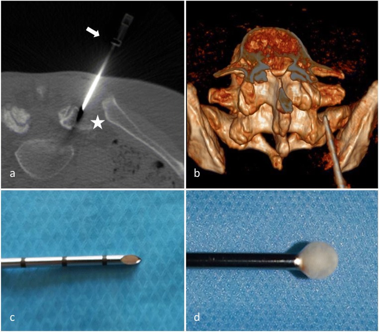
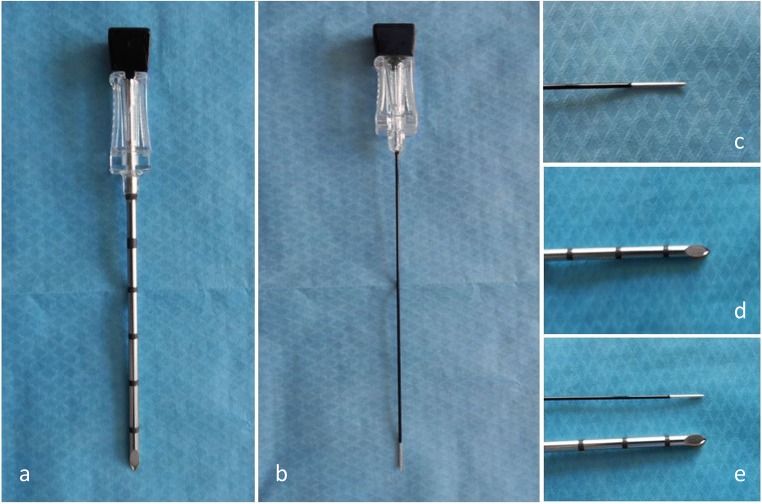
Low back pain (LBP) is the most common pain syndrome, and is an enormous burden and cost generator for society. Lumbar facet joints (FJ) constitute a common source of pain, accounting for 15-45% of LBP. Facet joint degenerative osteoarthritis is the most frequent form of facet joint pain. History and physical examination may suggest but not confirm facet joint syndrome. Although imaging (radiographs, MRI, CT, SPECT) for back pain syndrome is very commonly performed, there are no effective correlations between clinical symptoms and degenerative spinal changes. Diagnostic positive facet joint block can indicate facet joints as the source of chronic spinal pain. These patients may benefit from specific interventions to eliminate facet joint pain such as neurolysis, by radiofrequency or cryoablation. The purpose of this review is to describe the anatomy, epidemiology, clinical presentation, and radiologic findings of facet joint syndrome. Specific interventional facet joint management will also be described in detail. TEACHING POINTS: • Lumbar facet joints constitute a common source of pain accounting of 15-45%. • Facet arthrosis is the most frequent form of facet pathology. • There are no effective correlations between clinical symptoms, physical examination and degenerative spinal changes. • Diagnostic positive facet joint block can indicate facet joints as the source of pain. • After selection processing, patients may benefit from facet joint neurolysis, notably by radiofrequency or cryoablation.

Keywords: Block; Cryoablation; Facet joint; Low back pain; Neurolysis; Radiofrequency.


Conflict of interest statement

The authors of this manuscript declare no relationships with any companies whose products or services may be related to the subject matter of the article.

Figures

Fig. 1
Fig. 1
Innervation of facet joints (L3–4, L4–5 levels). Vr: ventral ramus. Dr: Dorsal ramus. m: medial branch. i: intermediate branch. l: lateral branch a: ascending branch. d: descending branch. Posterior (a) and posterolateral (b) view of the lumbar spine
Fig. 2
Fig. 2
Degenerative facet joint osteoarthritis (FJOA): Sagittal (a) and axial (b, c) CT views. Hypertrophy of the posterior articular process (black arrow). Joint space narrowing (thin white arrow). Joint capsule calcification (arrow head) and vacuum phenomenon (white arrow)
Fig. 3
Fig. 3
Isthmic lysis. a: Axial CT view at L4–5 level; b: axial CT view at L5-S1 level c: X-ray sagittal view at L5-S1 level; d: sagittal CT view L4–5 level
Fig. 4
Fig. 4
Septic facet joint arthritis. Axial (a) and coronal (b) T2 STIR views. Intra-articular effusion (white arrow) and articular process bone edema (white star). Unilateral signs should raise suspicion of a septic cause
Fig. 5
Fig. 5
Facet joint pain radiation. Posterior aspect of lower limb. Blue: from most frequent (dark blue), to less frequent (light blue) radiating pain areas. Dark blue: pain limited to lower back. Intermediate blue: radiating pain to the posterior aspect of the buttocks. Light blue: radiating pain to the posterior aspect of the lower limbs, may extend lower than the knee level. Green: anterior aspect of lower limb possible radiation areas. a anterior aspect of the lower limb (green). b posterior aspect of the lower limb etc
Fig. 6
Fig. 6
MRI imaging of facet joints. Active synovial inflammation and intra articular edema: axial and sagittal T2 STIR views (a, b) and T2 sagittal view (c). T2 STIR and T1 gado axial views (d, e): articular process bone edema
Fig. 7
Fig. 7
SPECT imaging of FJ. Hyperfixation on bone scintigraphy located on FJ capsule inflammation (white arrow)
Fig. 8
Fig. 8
Medial branch block under CT guidance. a: L4–5 level; b: L5-S1 level. c, d: Diffusion of contrast media prior to anesthetic injection confirming optimal needle tip placement (white arrows). Needle tip at the target point at injection should be placed at the middle of the base of the transverse process at its junction with the superior process at the L4–5 level. An analogous target point should be used at the L5-S1 level midway between the upper end and middle of the ala of the sacrum (white stars). Vr: ventral ramus. Dr: Dorsal ramus. m: medial branch. i: intermediate branch. l: lateral branch
Fig. 9
Fig. 9
Facet joint radiofrequency ablation. a: Radiofrequency ablation at the right L5-S1 level. Appropriate electrode placement (white arrow) parallel to the target nerve (white star) in order to achieve denervation along a substantial segment of the targeted nerve. b: 22G Radiofrequency needle showing uninsulated tip
Fig. 10
Fig. 10
Facet joint cryoneurolysis. CT-guided cryoablation at the right L5-S1 level. a: Anatomically accurate cryoprobe (white arrow) placement with the target midway between the upper end and middle of the ala of the sacrum (white star). b: 3D reconstruction showing the same cryoprobe placement. Cryoprobe needle tip (c): ice-ball formation at the cryoprobe tip (Joule–Thompson effect) (d)
Fig. 11
Fig. 11
Photographs of the coaxial needles: for cryodenervation (a, d) and radiofrequency (b, c), highlighting the difference in diameter 12G vs 22G (e)

References

    1. Breivik H, Collett B, Ventafridda V, Cohen R, Gallacher D. Survey of chronic pain in Europe: prevalence, impact on daily life, and treatment. Eur J Pain. 2006;10(4):287–333. - PubMed
    1. Manchikanti L, Hirsch JA, Falco FJ, Boswell MV. Management of lumbar zygapophysial (facet) joint pain. World J Orthop. 2016;7(5):315–337. - PMC - PubMed
    1. Kalichman L, Li L, Kim DH, et al. Facet joint osteoarthritis and low back pain in the community-based population. Spine (Phila Pa 1976) 2008;33(23):2560–2565. - PMC - PubMed
    1. Kalichman L, Kim DH, Li L, Guermazi A, Hunter DJ. Computed tomography-evaluated features of spinal degeneration: prevalence, intercorrelation, and association with self-reported low back pain. Spine J. 2010;10(3):200–208. - PMC - PubMed
    1. Manchikanti L, Singh V, Pampati V, et al. Evaluation of the relative contributions of various structures in chronic low back pain. Pain Physician. 2001;4(4):308–316. - PubMed