. 2018 Nov;61(11):2398-2411.
doi: 10.1007/s00125-018-4699-7. Epub 2018 Aug 8.

Human fetoplacental arterial and venous endothelial cells are differentially programmed by gestational diabetes mellitus, resulting in cell-specific barrier function changes

Affiliations

Human fetoplacental arterial and venous endothelial cells are differentially programmed by gestational diabetes mellitus, resulting in cell-specific barrier function changes

Silvija Cvitic et al. Diabetologia. 2018 Nov.

Abstract

Aims/hypothesis: An adverse intrauterine environment can result in permanent changes in the physiology of the offspring and predispose to diseases in adulthood. One such exposure, gestational diabetes mellitus (GDM), has been linked to development of metabolic disorders and cardiovascular disease in offspring. Epigenetic variation, including DNA methylation, is recognised as a leading mechanism underpinning fetal programming and we hypothesised that this plays a key role in fetoplacental endothelial dysfunction following exposure to GDM. Thus, we conducted a pilot epigenetic study to analyse concordant DNA methylation and gene expression changes in GDM-exposed fetoplacental endothelial cells.

Methods: Genome-wide methylation analysis of primary fetoplacental arterial endothelial cells (AEC) and venous endothelial cells (VEC) from healthy pregnancies and GDM-complicated pregnancies in parallel with transcriptome analysis identified methylation and expression changes. Most-affected pathways and functions were identified by Ingenuity Pathway Analysis and validated using functional assays.

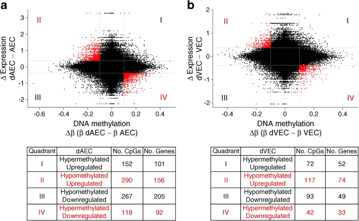
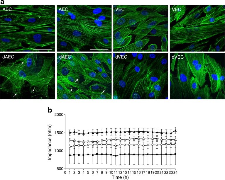
Results: Transcriptome and methylation analyses identified variation in gene expression linked to GDM-associated DNA methylation in 408 genes in AEC and 159 genes in VEC, implying a direct functional link. Pathway analysis found that genes altered by exposure to GDM clustered to functions associated with 'cell morphology' and 'cellular movement' in healthy AEC and VEC. Further functional analysis demonstrated that GDM-exposed cells had altered actin organisation and barrier function.

Conclusions/interpretation: Our data indicate that exposure to GDM programs atypical morphology and barrier function in fetoplacental endothelial cells by DNA methylation and gene expression change. The effects differ between AEC and VEC, indicating a stringent cell-specific sensitivity to adverse exposures associated with developmental programming in utero.

Data availability: DNA methylation and gene expression datasets generated and analysed during the current study are available at the National Center for Biotechnology Information (NCBI) Gene Expression Omnibus (GEO) database ( http://www.ncbi.nlm.nih.gov/geo ) under accession numbers GSE106099 and GSE103552, respectively.

Keywords: Actin organisation; DNA methylation; Fetoplacental endothelial cells; Gestational diabetes mellitus; Programming.


Conflict of interest statement

The authors declare that there is no duality of interest associated with this manuscript.

Figures

Fig. 1
Fig. 1
(a, c) PCA plot of DNA methylation (a) and gene expression (c) arrays. The first (PC1) and second principal components (PC2) are shown on the x- and y-axis, respectively. (b, d) Number of differentially methylated CpGs (b) and number of associated genes (d) in fetoplacental AEC and VEC exposed to GDM (dAEC and dVEC, respectively) vs control cells using a cut-off of p < 0.05, Δβ ≥ 0.2 for methylation and p < 0.05, FC ≥1.5 for gene expression. In (b), the grey section of the bars indicates the number of hypermethylated CpGs, the white section of the bars indicates the number of hypomethylated CpGs and the black bars indicate the number of associated genes. In (d), the grey section of the bars indicates the number of upregulated genes and the white section of the bars indicates the number of downregulated genes. (eg) Great distance analysis of altered CpG methylation relative to transcription start site (TSS). Associations for gene regions are shown for differentially methylated (hyper- and hypomethylated) CpGs in dAEC vs AEC (e) and dVEC vs VEC (f) (p < 0.05, Δβ ≥ 0.2) and for differentially methylated CpGs between AEC and VEC (g) (p < 0.05, Δβ ≥ 0.4). The absolute number of associated genes is indicated above each bar
Fig. 2
Fig. 2
Relationship between DNA methylation and gene expression changes associated with GDM in fetoplacental AEC and VEC. Scatterplot of DNA methylation (x-axis) and gene expression (y-axis) for HM450 probes with differences between control and GDM-exposed AEC (a) and VEC (b). Cut-offs are set to ≥10% (Δβ ≥ 0.1) methylation difference and to FC ≥1.3 in the gene expression. Points in red indicate genes likely to be under epigenetic regulation by DNA methylation, with increased methylation associated with decreased gene expression and vice versa. The tables below the scatterplots indicate the respective direction of methylation and gene expression changes as well as the number of affected CpGs and genes
Fig. 3
Fig. 3
Effect of GDM on F-actin organisation and barrier function. (a) F-actin fibres in primary fetoplacental AEC and VEC, after normal or GDM pregnancy were stained with phalloidin (green). Nuclei were stained with DAPI (blue). Note that AEC after normal pregnancy show a more organised actin fibre network, while the dAEC, exposed to GDM, reveal more cross-linked F-actin bundles. This difference was not found in the dVEC, exposed to GDM. Two representative stainings of n = 5 (AEC and dAEC) or n = 6 individual cell isolations (VEC and dVEC) per group are shown, each performed in quadruplicate. Original magnification ×400. Scale bar, 40 μm. White arrows indicate less organised actin fibres in dAEC. (b) Barrier function was assessed by real-time analysis of the electrical impedance of cell monolayers. After reaching maximum impedance, endothelial resistance of primary AEC and VEC, after normal (n = 10 and 8, respectively) or GDM pregnancy (n = 6 and 4, respectively), was followed over 24 h. Data are plotted as means ± SEM at 1 h intervals. Data were generated in three independent experiments for each cell type separately, in duplicate. For statistical comparison, a linear mixed-effects model was fitted, showing significantly increased average impedance in dAEC vs AEC (p < 0.01). Reduced average electrical impedance of dVEC was not significant (p = 0.24). White triangles, AEC; white squares, dAEC; black triangles, VEC; black squares, dVEC
Fig. 4
Fig. 4
Heat map of genes implicated in endothelial cell barrier function. The heat map depicts only genes that show significant expression difference (p < 0.05) in GDM-exposed endothelial cells vs control cells and that are related to focal adhesion, adherens and/or tight junctions, or cytoskeleton actin organisation. The colour bar indicates the expression of a particular gene from low (white) to high expression (black)

References

    1. Friedman JE. Obesity and gestational diabetes mellitus pathways for programming in mouse, monkey, and man-where do we go next? The 2014 Norbert Freinkel Award Lecture. Diabetes Care. 2015;38:1402–1411. doi: 10.2337/dc15-0628. - DOI - PMC - PubMed
    1. Padmanabhan V, Cardoso RC, Puttabyatappa M. Developmental programming, a pathway to disease. Endocrinology. 2016;157:1328–1340. doi: 10.1210/en.2016-1003. - DOI - PMC - PubMed
    1. Feil R, Fraga MF. Epigenetics and the environment: emerging patterns and implications. Nat Rev Genet. 2012;13:97–109. doi: 10.1038/nrg3142. - DOI - PubMed
    1. Novakovic B, Sibson M, Ng HK, et al. Placenta-specific methylation of the vitamin D 24-hydroxylase gene: implications for feedback autoregulation of active vitamin D levels at the fetomaternal interface. J Biol Chem. 2009;284:14838–14848. doi: 10.1074/jbc.M809542200. - DOI - PMC - PubMed
    1. Januar V, Desoye G, Novakovic B, Cvitic S, Saffery R. Epigenetic regulation of human placental function and pregnancy outcome: considerations for causal inference. Am J Obstet Gynecol. 2015;213:S182–S196. doi: 10.1016/j.ajog.2015.07.011. - DOI - PubMed

Publication types