Review
. 2018 Oct;132(5):416-429.
doi: 10.1037/bne0000260. Epub 2018 Aug 9.

The retrosplenial-parietal network and reference frame coordination for spatial navigation

Affiliations
Review

The retrosplenial-parietal network and reference frame coordination for spatial navigation

Benjamin J Clark et al. Behav Neurosci. 2018 Oct.

Abstract

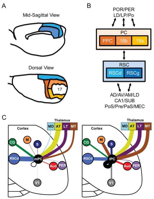
The retrosplenial cortex is anatomically positioned to integrate sensory, motor, and visual information and is thought to have an important role in processing spatial information and guiding behavior through complex environments. Anatomical and theoretical work has argued that the retrosplenial cortex participates in spatial behavior in concert with input from the parietal cortex. Although the nature of these interactions is unknown, a central position is that the functional connectivity is hierarchical with egocentric spatial information processed in the parietal cortex and higher-level allocentric mappings generated in the retrosplenial cortex. Here, we review the evidence supporting this proposal. We begin by summarizing the key anatomical features of the retrosplenial-parietal network, and then review studies investigating the neural correlates of these regions during spatial behavior. Our summary of this literature suggests that the retrosplenial-parietal circuitry does not represent a strict hierarchical parcellation of function between the two regions but instead a heterogeneous mixture of egocentric-allocentric coding and integration across frames of reference. We also suggest that this circuitry should be represented as a gradient of egocentric-to-allocentric information processing from parietal to retrosplenial cortices, with more specialized encoding of global allocentric frameworks within the retrosplenial cortex and more specialized egocentric and local allocentric representations in parietal cortex. We conclude by identifying the major gaps in this literature and suggest new avenues of research. (PsycINFO Database Record (c) 2018 APA, all rights reserved).


Figures

Figure 1
Figure 1
Cytoarchitectural organization of the retrosplenial (RSC) and parietal (PC) cortices. A, Panels adapted from Kolb and Walkey (1987) showing the cytoarchitectural organization of the retrosplenial and parietal cortex of the rat brain. B, Circuit diagram showing the primary differences in cortical and subcortical inputs to the retrosplenial and parietal cortex (PC). Note: anatomical and electrophysiological work have largely focused on PPC and the anterior regions of areas 18a and 18b (V2MM and V2ML in Paxinos & Watson, 2007). For clarity, we refer to the collection of these areas as PC. C, The density of cortical and thalamic inputs to PC have been precisely mapped in rats as illustrated here, more work is needed to map additional connections in the rat for this brain network (adapted from Wilber et al., 2015). For this study density of inputs to PC were shown to be similar in the anterior to posterior direction, but to vary considerably in the medial/lateral direction, therefore PC data was collapsed across the anterior/posterior direction and shown separately for medial (mPC) and lateral regions (lPC). Each brain region is indicated by a different color and the line thickness represents the strength of thalamic and cortical projections to the medial PC (left panel) and lateral PC (right panel). Only regions with significant medial versus lateral differences in projection strength are shown (i.e., non-overlapping error bars). Note that projections to lateral PC involve stronger projections from the somatosensory, motor, visual, auditory cortex, and motor thalamus. The medial PC receives stronger inputs from the dorsal retrosplenial cortex and cingulate region. Mediodorsal thalamus, anterior thalamus, lateral thalamus, perirhinal cortex. Key: AD, anterodorsal thalamus; AV, anteroventral thalamus; AM, anteromedial thalamus; Broadmann’s area 17, primary visual cortex; Broadmann’s areas 18a and 18b, secondary visual cortex; AT, anterior thalamus; AUD, auditory cortex; CG, cingulate region; lPC, lateral posterior parietal cortex and the rostral portion of area 18a; LD, laterodorsal thalamus; LP, lateroposterior thalamus; LT, lateral thalamus; mPC, medial posterior parietal cortex and the rostral portion of area 18b; MD, mediodorsal thalamus; M, motor; MT, motor thalamus; MEC, medial entorhinal cortex; PaS, parasubiculum; PER, perirhinal cortex; Po, posterior thalamic complex; POR, postrhinal cortex; PoS, postsubiculum; PPC, posterior parietal cortex; Pre, presubiculum; RSCg, retrosplenial cortex-granular; RSCd, retrosplenial cortex-dysgranular; S, somatosensory; SUB, subiculum; V1, visual.
Figure 2
Figure 2
Reference frames in PC and RSC. Top, Schematic of route-centered neuronal firing during maze traversal (adapted from Nitz, 2006). Shaded regions depict the locations of increased firing rate in cells tuned to the starting segment of the whole route (left) which corresponds to the peaks in firing rate on the line plot (right). The particular cell fires independently of both world-centered location (SE versus NW quadrant) and allocentric direction (inbound versus outbound). Note, this is an example of one of the “simplest” coding frames reported by Nitz and colleagues, see text for more complex forms of route centered coding in parietal cortex. Bottom, Schematic of allocentric modulation by an RSC neuron during locomotion across the W-shaped track when positioned in two locations (position A and position B) in the environment (adapted from Alexander & Nitz, 2015). Note that the line plot representing neural activity for an individual RSC neuron (right) fires robustly cell after the animal performs left-turns along the track when in maze position A, but not when in maze position B.
Figure 3
Figure 3
Diagram illustrating allocentric heading direction, allocentric landmark location, and egocentric translation of landmark location. The black dotted lines depict two examples of possible heading directions – NW and NE respectively. The red dotted line depicts the allocentric location of the building from where the person is standing. When facing NW, the person may think, “The building is slightly behind me and to the right.” When facing NE the person may think, “The building is slightly to my right.” However, for these two different egocentric representations of the building location, its allocentric representation is always the same from this point – “The building is east from where I am.”

References

    1. Agster KL, Burwell RD. Cortical efferents of the perirhinal, postrhinal, and entorhinal cortices of the rat. Hippocampus. 2009;19(12):1159–1186. - PMC - PubMed
    1. Aguirre GK, D’Esposito M. Topographical disorientation: a synthesis and taxonomy. Brain. 1999;122(9):1613–1628. doi: 10.1093/brain/122.9.1613. - DOI - PubMed
    1. Alexander AS, Conner AM, Tung JC, Nitz DA. Hippocampal and Posterior Parietal Cortex Spatial Encoding During Pursuit. Paper presented at the Society for Neuroscience; San Diego, CA. 2016.
    1. Alexander AS, Nitz DA. Retrosplenial cortex maps the conjunction of internal and external spaces. Nat Neurosci. 2015;18(8):1143–1151. doi: 10.1038/nn.4058. http://www.nature.com/neuro/journal/v18/n8/abs/nn.4058.html#supplementar.... - DOI - PubMed
    1. Alexander AS, Nitz DA. Spatially Periodic Activation Patterns of Retrosplenial Cortex Encode Route Sub-spaces and Distance Traveled. Current Biology. 2017;27(11):1551–1560. e1554. doi: 10.1016/j.cub.2017.04.036. - DOI - PubMed

MeSH terms