. 2018 Oct 1;29(20):2470-2480.
doi: 10.1091/mbc.E17-08-0504. Epub 2018 Aug 9.

WDR62 mediates TNFα-dependent JNK activation via TRAF2-MLK3 axis

Affiliations

WDR62 mediates TNFα-dependent JNK activation via TRAF2-MLK3 axis

Elad Prinz et al. Mol Biol Cell. .

Abstract

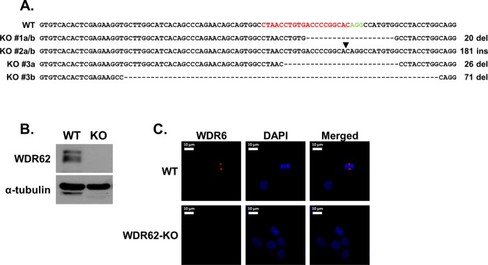
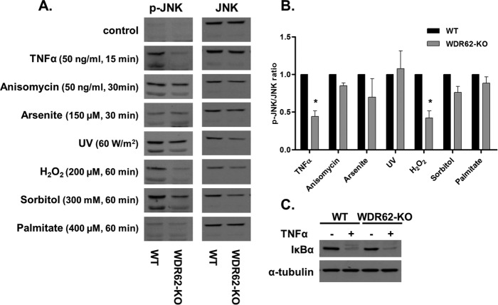
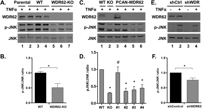
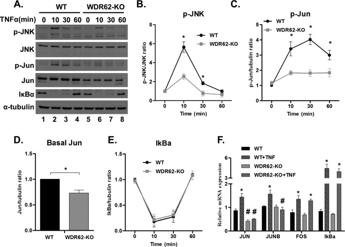
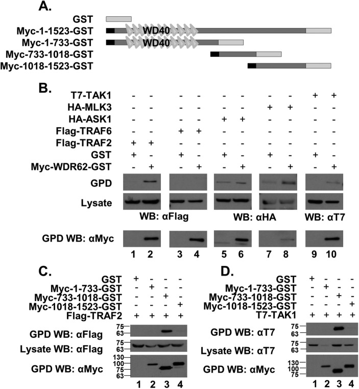
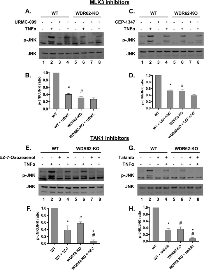
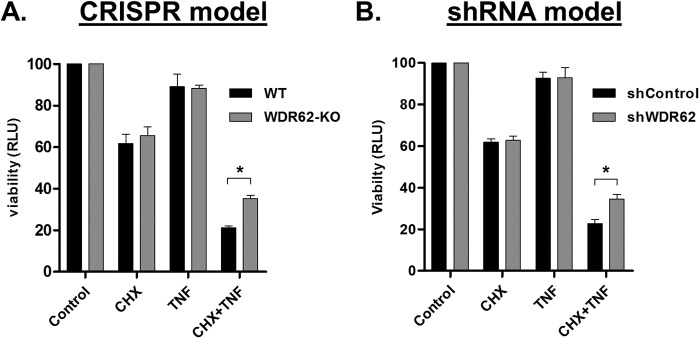
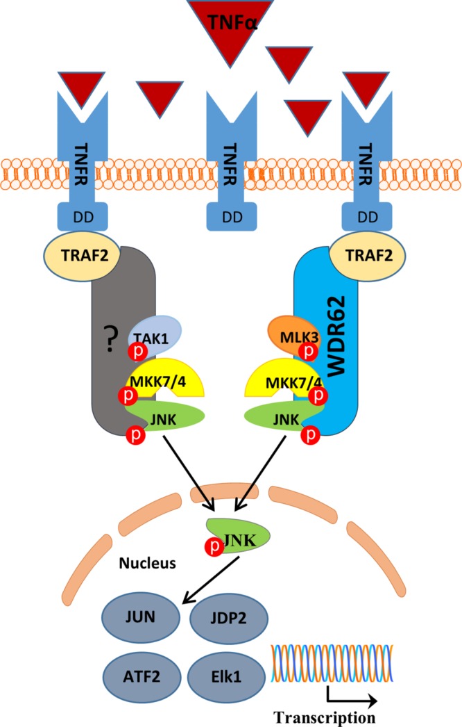
The mitogen-activated protein kinases (MAPKs) regulate a variety of cellular processes. The three main MAPK cascades are the extracellular signal-regulated kinases (ERK), c-Jun N-terminal kinase (JNK), and p38 kinases. A typical MAPK cascade is composed of MAP3K-MAP2K-MAPK kinases that are held by scaffold proteins. Scaffolds function to assemble the protein tier and contribute to the specificity and efficacy of signal transmission. WD repeat domain 62 (WDR62) is a JNK scaffold protein, interacting with JNK, MKK7, and several MAP3Ks. The loss of WDR62 in human leads to microcephaly and pachygyria. Yet the role of WDR62 in cellular function is not fully studied. We used the CRISPR/Cas9 and short hairpin RNA approaches to establish a human breast cancer cell line MDA-MB-231 with WDR62 loss of function and studied the consequence to JNK signaling. In growing cells, WDR62 is responsible for the basal expression of c-Jun. In stressed cells, WDR62 specifically mediates TNFα-dependent JNK activation through the association with both the adaptor protein, TNF receptor-associated factor 2 (TRAF2), and the MAP3K protein, mixed lineage kinase 3. TNFα-dependent JNK activation is mediated by WDR62 in HCT116 and HeLa cell lines as well. MDA-MB-231 WDR62-knockout cells display increased resistance to TNFα-induced cell death. Collectively, WDR62 coordinates the TNFα receptor signaling pathway to JNK activation through association with multiple kinases and the adaptor protein TRAF2.


Figures

FIGURE 1:
FIGURE 1:
Generation of MDA-MB-231 WDR62-KO cells. (A) Sequence of exon 2 of WT WDR62 and three clones with WDR62-KO. The sequences a and b in each clone represents the two mutated alleles. The gRNA sequence that was used is shown in red and the protospacer adjacent motif (PAM) is shown in green. Hyphens indicate deleted nucleotides; arrowhead indicates site of insertion. Clone #1 was selected for further characterization. (B) Western blot analysis of WDR62-KO clone #1 cells. WT and WDR62-KO MDA-MB-231 were immunoblotted using WDR62 antibody. The expression level of α-tubulin served as loading control. (C) WDR62 expression in MDA-MB-231 WT and WDR62-KO dividing cells: Cells were fixed and stained with anti-WDR62 polyclonal antibody (red) and DAPI (blue). Representative images of dividing cells are shown.
FIGURE 2:
FIGURE 2:
JNK activation in WT and WDR62-KO cells. (A) WT and WDR62-KO MDA-MB-231 cells were exposed to various stimuli as indicated. Following stimulation, cells were harvested and lysates were subjected to Western blot analysis with anti-phospho-JNK antibody. Total JNK antibody served as reference. (B) Densitometric quantification of the Western blot analysis shown in A. The ratio of p-JNK/JNK obtained with WT cells was determined as 1 while the ratio obtained with WDR62-KO was determined relatively. Data are expressed as mean ratio ± SEM from three independent experiments. *p ≤ 0.05. (C) WT and WDR62-KO cells were treated with TNFα (50 ng/ml) for 15 min. IκBα expression level was examined by Western blot. The expression level of α-tubulin served as loading control.
FIGURE 3:
FIGURE 3:
Validation of WDR62 role in TNFα signaling. (A, B) Parental MDA-MB-231 cells, three WT clones, and three WDR62-KO clones were treated with TNFα (50 ng/ml) for 15 min. Following stimulation, cells were harvested and subjected to Western blot and densitometric analysis with p-JNK and JNK antibodies. Results are expressed as the mean ratio ± SEM of the three clones from each group. *p ≤ 0.05 compared with WT cells. (C, D) WDR62-KO cells were stably transfected with empty vector or WDR62 expression plasmids. One WDR62-positive clone and three WDR62-negative clones were treated with TNFα (50 ng/ml) for 15 min and subjected to Western blot and densitometric analysis. WT and WDR62-KO cells were used as controls. *p ≤ 0.05 compared with WT, #p ≤ 0.05 compared with WDR62-KO cells. (E,F) MDA-MB-231 cells were infected with shControl or shWDR62 lentiviruses. Cells were treated with TNFα (50 ng/ml) for 15 min and subjected to Western blot and densitometric analysis. *p ≤ 0.05 compared with shControl cells. The ratio of p-JNK/JNK obtained with TNFα-treated WT cells was determined as 1 while the ratio obtained with other TNFα-treated cells was determined relatively to their appropriate WT control. Results are expressed as the mean ratio ± SEM of three to five independent experiments.
FIGURE 4:
FIGURE 4:
Time course of TNFα-induced JNK activation. (A–E) WT and WDR62-KO cells were treated with TNFα (50 ng/ml) for the indicated time points. Following stimulation, cells were harvested and subjected to Western blot analysis and densitometric analysis with the indicated antibodies. p-JNK level is expressed relative to total JNK (B); p-Jun, c-Jun, and IκBα levels are expressed relative to α-tubulin (C–E). The ratio of p-JNK/JNK, p-Jun/α-tubulin, c-Jun/α-tubulin, and IκBα/α-tubulin obtained with untreated WT cells was determined as 1, while the ratio obtained with all other samples was determined relatively. Results are expressed as the mean ± SEM of three independent experiments. *p ≤ 0.05 comparing between WT and WDR62-KO counterparts. (F) WT and WDR62-KO cells were treated with TNFα (50 ng/ml) for 30 min. Then, mRNA was extracted and the indicated genes were analyzed by qRT-PCR. The expression level of GAPDH gene was used to normalization. Results are expressed as the mean ratio ± SEM of three independent experiments. *p ≤ 0.05 comparing treated to untreated cells of the same genotype; #p ≤ 0.05 comparing between WT and WDR62-KO counterparts.
FIGURE 5:
FIGURE 5:
Interaction of WDR62 with key component of TNFα signaling. (A) Schematic representation of the WDR62 fragments used in this experiment. The black square represents the Myc epitope-tag and the light-gray rectangle represents the GST tag. (B–D) HEK-293T cells were cotransfected with expression plasmids as indicated. Cell lysates were pulled down with glutathione beads, and eluted proteins were separated by SDS–PAGE, followed by Western blot with the appropriate antibodies as indicated. The expression level of transfected plasmids was determined by blotting the total cell lysate with the appropriate antibodies.
FIGURE 6:
FIGURE 6:
WDR62 mediates TNFα-induced JNK activation through MLK3. (A–D) WT and WDR62-KO cells were preincubated with the selective MLK3 inhibitors URMC-099 (100 nM) or CEP-1347 (50 nM) for 1 h and then treated with TNFα (50 ng/ml) for 15 min. (E–H) WT and WDR62-KO cells were preincubated with the selective TAK1 inhibitors 5Z-7-Oxozeaenol (20 nM) or takinib (2 µM) for 1 h then treated with TNFα (50 ng/ml) for 15 min. JNK activation was determined by Western blotting and densitometric analysis with p-JNK and JNK antibodies. The ratio of p-JNK/JNK obtained with TNFα-only treated WT cells was determined as 1 while the ratio obtained with all other treatments was determined relatively. Results are expressed as the mean ratio ± SEM of three independent experiments. *p ≤ 0.05, comparing pretreated to not pretreated cells of the same genotype; #p ≤ 0.05, comparing between WT and WDR62-KO counterparts.
FIGURE 7:
FIGURE 7:
WDR62-deficient cells are less sensitive to TNFα-induced apoptosis. (A) MDA-MB-231 WT and WDR62-KO cells; (B) MDA-MB-231 shRNA control and WDR62-KD cells were treated with CHX (10 µg/ml) and TNFα (50 ng/ml) for 24 h. Viability was assayed with CellTiter-Glo reagent. Each experiment was performed in triplicate. The viability obtained with control untreated cells was determined as 100, while the viability obtained with all other treatments was determined relatively. Results are expressed as the mean relative light unit (RLU) ± SEM of three independent experiments. *p ≤ 0.05.
FIGURE 8:
FIGURE 8:
Schematic model for WDR62-mediated TNFα-dependent JNK activation. Following TNFα binding to TNF receptor, WDR62 recruits MLK3, MKK7, and JNK to TRAF2, allowing efficient activation of JNK by TNFα. Another scaffold protein mediating the TRAF2/TAK1/JNK axis is yet to be determined. Red circles represent phosphorylation.

References

    1. Aggarwal BB. (2003). Signalling pathways of the TNF superfamily: a double-edged sword. Nat Rev Immunol , 745–756. - PubMed
    1. Aguirre V, Uchida T, Yenush L, Davis R, White MF. (2000). The c-Jun NH(2)-terminal kinase promotes insulin resistance during association with insulin receptor substrate-1 and phosphorylation of Ser(307). J Biol Chem , 9047–9054. - PubMed
    1. Balkwill F. (2009). Tumour necrosis factor and cancer. Nat Rev Cancer , 361–371. - PubMed
    1. Batard P, Jordan M, Wurm F. (2001). Transfer of high copy number plasmid into mammalian cells by calcium phosphate transfection. Gene , 61–68. - PubMed
    1. Bilguvar K, Ozturk AK, Louvi A, Kwan KY, Choi M, Tatli B, Yalnizoglu D, Tuysuz B, Caglayan AO, Gokben S, et al. (2010). Whole-exome sequencing identifies recessive WDR62 mutations in severe brain malformations. Nature , 207–210. - PMC - PubMed

Publication types

MeSH terms

Substances