Review
. 2018 Jul;24(4):203-208.
doi: 10.5152/dir.2018.17462.

Imaging features of leadless cardiovascular devices

Affiliations
Review

Imaging features of leadless cardiovascular devices

Jesse M Conyers et al. Diagn Interv Radiol. 2018 Jul.

Abstract

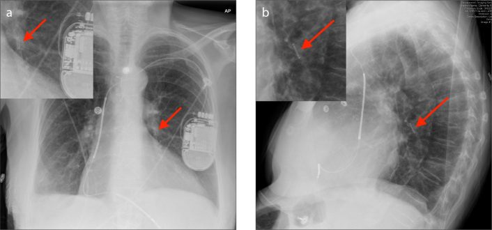
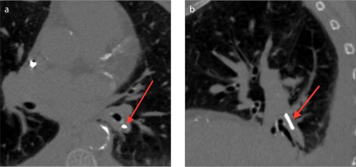
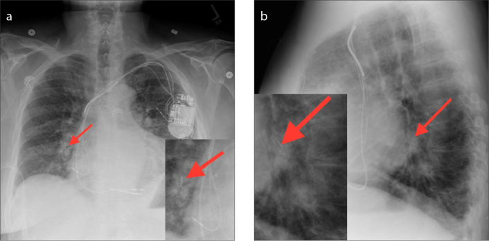
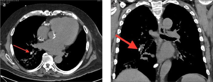
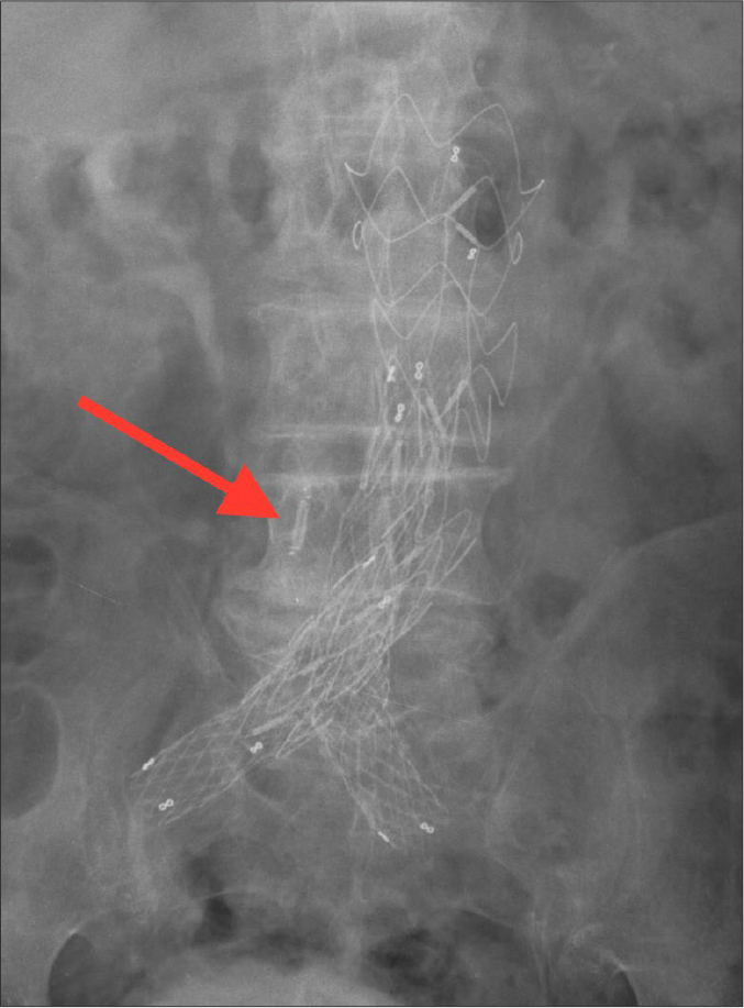
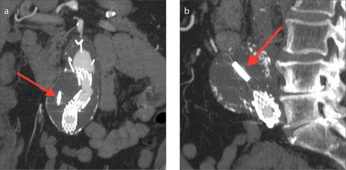
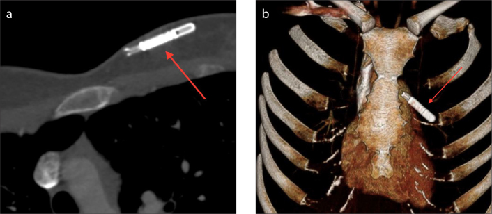
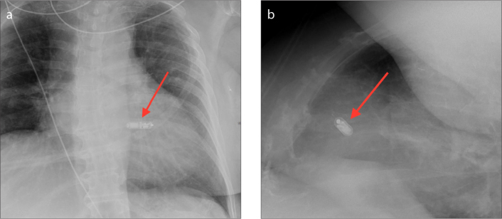
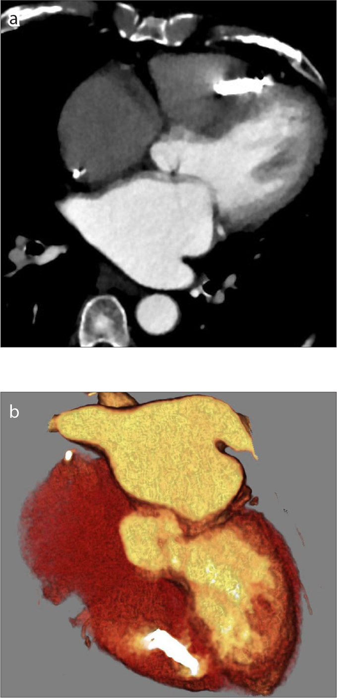
Cardiovascular devices and hemodynamic monitoring systems continue to evolve with the goal of allowing for rapid clinical intervention and management. Cardiovascular devices including the CardioMicroelectromechanical (CardioMEMS) device, implantable loop recorder, and right ventricular (RV) leadless pacemaker are now widely used for treatment and monitoring of advanced cardiac conditions, as many of these devices have been shown to significantly improve patient outcomes. Additionally, hemodynamic monitoring devices have shown utility in monitoring patients with aortic aneurysms after endovascular aortic repair (EVAR) for early detection of Type I and Type II endoleaks. There is limited published data regarding the imaging features of these devices. As these devices become more widely used, it is important for radiologists to become familiar with the normal imaging features and potential complications. The goal of this review is to summarize the data regarding the use of leadless cardiovascular devices including the CardioMEMS device, implantable loop recorder, and RV leadless pacemaker, and to present cases demonstrating their utility and normal imaging features.


Conflict of interest statement

Conflict of interest disclosure

The authors declared no conflicts of interest.

Figures

Figure 1
Figure 1
Illustration of the CardioMEMS device positioned in the left lower lobe segmental pulmonary artery.
Figure 2 a, b
Figure 2 a, b
AP with zoomed image (a) and lateral with zoomed image (b) radiographs of the CardioMEMS device inserted in the left lower lobe segmental pulmonary artery (arrows).
Figure 3 a, b
Figure 3 a, b
Axial (a) and sagittal (b) CT images of the CardioMEMS device inserted in the left lower lobe segmental pulmonary artery (arrows).
Figure 4 a, b
Figure 4 a, b
AP with zoomed image (a) and lateral with zoomed image (b) chest radiographs showing the CardioMEMS device inserted in the right lower lobe lobar pulmonary artery (arrows).
Figure 5 a, b
Figure 5 a, b
Axial (a) and coronal (b) CT images showing the CardioMEMS device inserted in the right lower lobe segmental pulmonary artery (arrows).
Figure 6
Figure 6
Illustration of the CardioMEMS device inserted in an excluded aortic aneurysm status post endovascular aortic repair.
Figure 7
Figure 7
AP abdominal radiograph of the CardioMEMS device inserted in an excluded aortic aneurysm status post endovascular aortic repair.
Figure 8 a, b
Figure 8 a, b
Coronal (a) and sagittal (b) MIP CT images of CardioMEMS device inserted in an excluded aortic aneurysm status post endovascular aortic repair (arrows).
Figure 9 a, b
Figure 9 a, b
Axial CT (a) and 3D volume rendering (b) images of an implantable loop recorder (Medtronic LINQ) inserted in the left chest wall (arrows).
Figure 10 a, b
Figure 10 a, b
AP (a) and lateral (b) chest radiographs showing an RV leadless pacemaker (Medtronic Micra) inserted in the right ventricle.
Figure 11 a, b
Figure 11 a, b
Axial CT image (a) and oblique coronal 3D volume rendering image (b) showing an RV wireless pacemaker (Medtronic Micra) inserted in the right ventricle.

Similar articles

Cited by

References

    1. Sperzel J, Burri H, Gras D, et al. State of the art of leadless pacing. Europace. 2015;17:1508–1513. doi: 10.1093/europace/euv096. - DOI - PMC - PubMed
    1. Yandrapalli S, Raza A, Tariq S, Aronow WS. Ambulatory pulmonary artery pressure monitoring in advanced heart failure patients. World J Cardiol. 2017;9:21–26. doi: 10.4330/wjc.v9.i1.21. - DOI - PMC - PubMed
    1. Ricci RP, Morichelli L, Quarta L, et al. Long-term patient acceptance of and satisfaction with implanted device remote monitoring. EP Europace. 2010;12:674–679. doi: 10.1093/europace/euq046. - DOI - PubMed
    1. Benjamin EJ, Blaha MJ, Chiuve SE, et al. Heart Disease and Stroke Statistics—2017 Update: A Report From the American Heart Association. Circulation. 2017;136:e1–e23. doi: 10.1161/CIR.0000000000000485. - DOI - PMC - PubMed
    1. Tang WH, Girod JP, Lee MJ, et al. Plasma B-type natriuretic peptide levels in ambulatory patients with established chronic symptomatic systolic heart failure. Circulation. 2003;108:2964–2966. doi: 10.1161/01.CIR.0000106903.98196.B6. - DOI - PubMed