. 2018 Aug 9;13(8):e0201578.
doi: 10.1371/journal.pone.0201578. eCollection 2018.

The neurofilament derived-peptide NFL-TBS.40-63 enters in-vitro in human neural stem cells and increases their differentiation

Affiliations

The neurofilament derived-peptide NFL-TBS.40-63 enters in-vitro in human neural stem cells and increases their differentiation

Kristell Barreau et al. PLoS One. .

Abstract

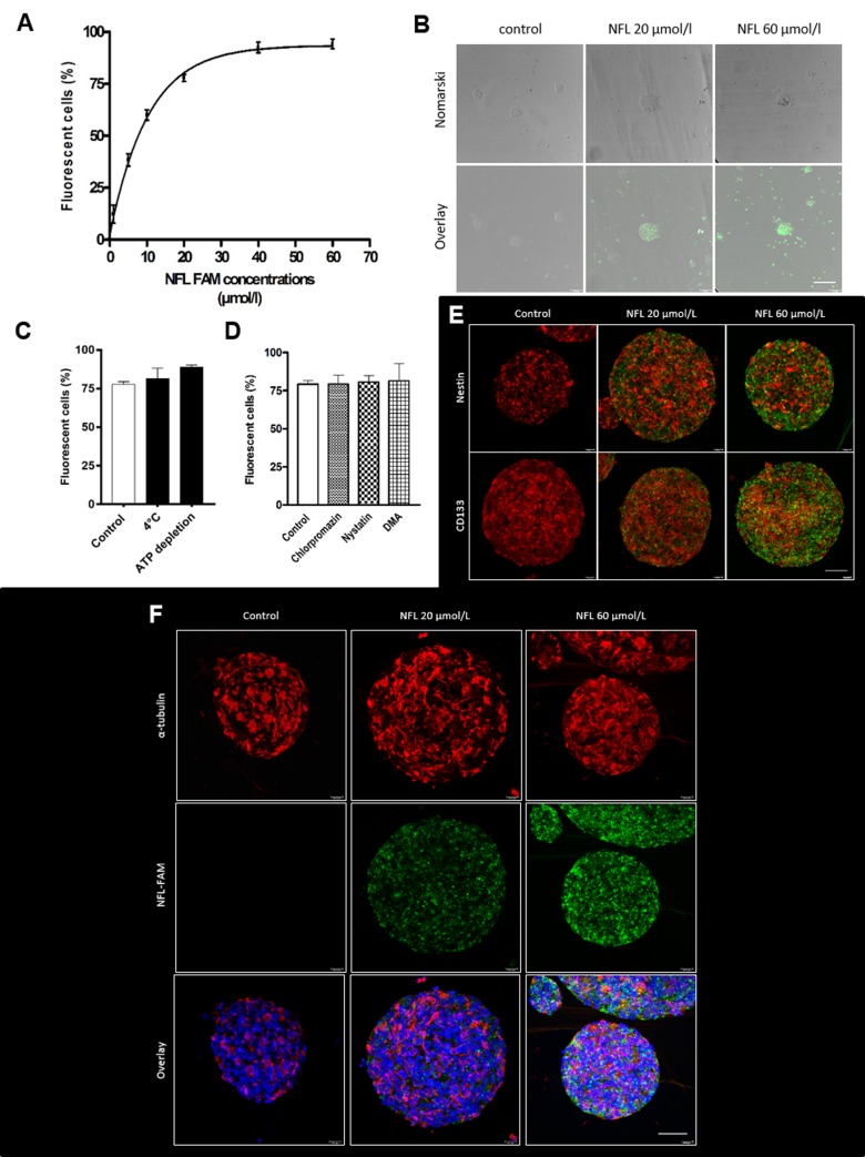
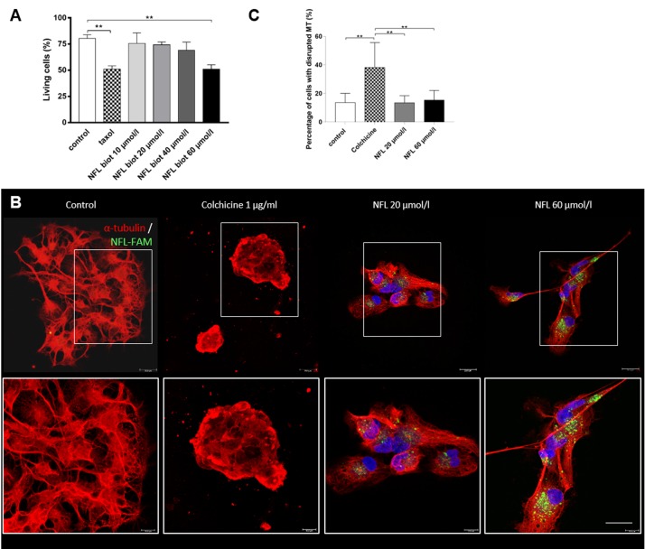
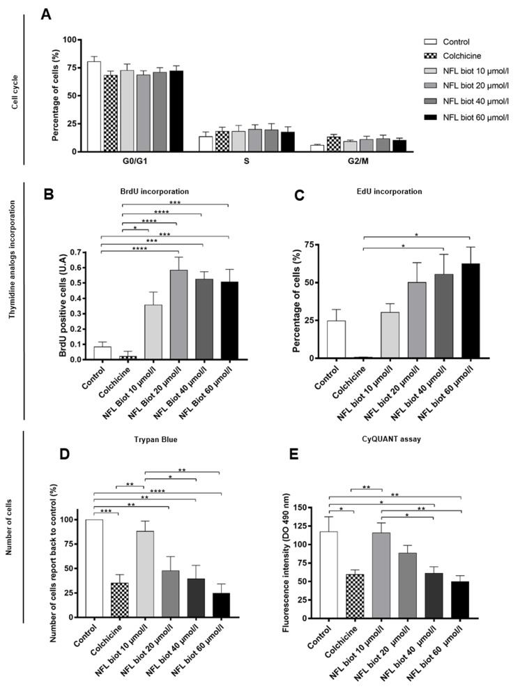
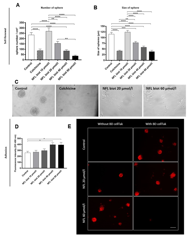
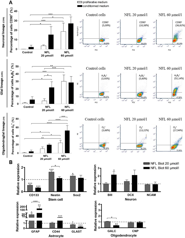
Regenerative medicine is a promising approach to treat neurodegenerative diseases by replacing degenerating cells like neurons or oligodendrocytes. Targeting human neural stem cells directly in the brain is a big challenge in such a strategy. The neurofilament derived NFL-TBS.40-63 peptide has recently been introduced as a novel tool to target neural stem cells. Previous studies showed that this peptide can be internalized by rat neural stem cells in vitro and in vivo, which coincided with lower proliferation and self-renewal capacity and increase of differentiation. In this study, we analyzed the uptake and potential effects of the NFL-TBS.40-63 peptide on human neural stem cells isolated from human fetuses. We showed that the peptide inhibits proliferation and the ability to produce neurospheres in vitro, which is consistent with an increase in cell adhesion and differentiation. These results confirm that the peptide could be a promising molecule to target and manipulate human neural stem cells and thus could serve as a strategic tool for regenerative medicine.


Conflict of interest statement

The authors have declared that no competing interests exist.

Figures

Fig 1
Fig 1. Uptake of the FAM-labeled NFL-TBS.40-63 peptide in hNSCs.
(A) Percentage of FAM-labeled NFL-TBS.40-63 internalized by hNSCs with increasing concentrations of peptide after 1 hour incubation at 37°C. (B) Visualization of the FAM-labeled peptide after 1 hour incubation with 20 or 60 µmol/L of peptide. Scale bar: 50 µm. Green: FAM-labeled peptide (C) Internalization of 20 µmol/L of peptide after pre-treatment at 4°C or ATP depletion or (D) in the presence of inhibitors of the endocytosis pathways. Data are presented as means ± SEM. *P<0.05. (E-F) Immunofluorescence after incubation of cells for 1 hour with 20 or 60 µmol/L FAM-labeled peptide. Cells were stained with specific neural stem cells markers: Nestin and CD133 (E) or with α-tubulin (F). Images were taken with the confocal microscope. Red: Nestin, CD133 or α-tubulin; Green: FAM-labeled peptide; Blue: DAPI. Scale bar: 50 µm.
Fig 2
Fig 2. The NFL-TBS.40-63 peptide has no toxic effect on cell viability and the microtubule network.
(A) Percentage of viable cells was evaluated after incubation for 72 hours with increasing concentrations of NFL-TBS.40-63 peptide or with Taxol (0.1 µmol/L). Cell viability was measured by FACS (Annexin-V-FITC / PI assay kit). (B) Effect of 20 or 60 µmol/L of FAM peptide (green) on the microtubule network (α-tubulin: red; NFL FAM peptide: green) after 24 hours. Images were taken with the confocal microscope. Scale bar: 20 µm. (C) Percentage of cells with a disrupted microtubule network in control condition, or with different treatments (colchicine, NFL 20 or 60 µmol/l). Data presented as means ± SEM. **P<0.01.
Fig 3
Fig 3. The NFL-TBS.40-63 peptide affects the proliferation of hNSCs.
(A) Percentage of hNSCs at G0/G1, S or G2/M of the cell cycle after treatment with colchicine (1µg/ml) or increasing concentrations of biotinylated-NFL-TBS.40-63 peptide. Data are presented as means ± SEM. (B-C) Thymidine analogue incorporation: The number of BrdU (B) or EdU positive cells (C) was counted after 72 hours incubation with 1 µg/ml of colchicine or with increasing concentrations of peptide, and compared to the control condition. Thymidine analogues were incubated for 5 hours before the end of the test. Data presented as means ± SEM. (D-E) Cell proliferation: Number of cells after 72 hours incubation with the NFL peptide was quantified using Trypan blue exclusion (D) or using the CyQUANT assay (E). *P<0.05, **P<0.01, ***P<0.001, ****P<0,0001.
Fig 4
Fig 4. The NFL-TBS.40-63 peptide affects the capability to form neurospheres and increase adhesion of hNSCs.
(A-C) Sphere formation from hNSCs: quantification of the number of neurospheres per square centimeter (A) and their size (B) after 7 days incubation with increasing concentrations of the peptide, or with 1 µg/ml of colchicine. Scale bar: 150 µm. (C) Morphology of neurospheres observed after 7 days incubation in normal conditions, or with colchicine, or with the NFL peptide at 20 or 60 µmol/L. (D-E) Quantification of adherent cells after 72 hours with increasing concentrations of NFL peptide using the CyQUANT assay (D) or by immunostaining of cells after 72 hours with increasing concentrations of biotinylated peptide (E). Adherent cells were observed on coverslips without coating (top pictures), while non-adherent (floating) cells were recovered following centrifugation and 1 hour incubation on BD Cell-Tak coated coverslips (Bottom pictures). Cells were visualized by Phalloïdin staining (red) and DAPI staining the nucleus (blue). Scale bar: 100 µm. Data presented as means ± SEM. *P<0.05, **P<0.01, ***P<0.001, ****P<0,0001.
Fig 5
Fig 5. The NFL-TBS.40-63 peptide increases differentiation of hNSCs.
(A-B) Differentiation of cells induced after 5 days by increasing concentrations of NFL peptide in a proliferative or conditioned medium. (A) Percentage of cells which express the neuronal marker CD90 (top graph), the glial marker A2B5 (medium graph) or the oligodendroglial marker O4 (bottom graph) after 5 days in the presence of the NFL peptide at 20 or 60 µmol/L in the proliferative medium (white bars) or the conditioned medium (dark bar). Markers were analyzed by FACS technique and some examples are given in the left part of each graph. (B) The relative expression level of CD133, Nestin, Sox2, βIII tubulin, DCX, NCAM, GFAP, CD44, GLAST, GALC, CNP genes was quantified by reverse transcription polymerase chain reaction after 5 days with 0 (control condition), 20 or 60 µmol/L of peptide. The relative gene expression was compared with control conditions after normalization with the GAPDH gene (value of the control condition = 1) with the 2-ΔΔCt method. All data were presented as means ± SEM. *P<0.05, **P<0.01, ***P<0.001, ****P<0,0001. Stars above bars represent significant data compared to control.

References

    1. Hsu YC, Chen SL, Wang DY, Chiu IM. Stem cell-based therapy in neural repair. Biomed J. 2013;36(3):98–105. 10.4103/2319-4170.113226 . - DOI - PubMed
    1. Muller FJ, Snyder EY, Loring JF. Gene therapy: can neural stem cells deliver? Nat Rev Neurosci. 2006;7(1):75–84. 10.1038/nrn1829 . - DOI - PubMed
    1. Mazzini L, Gelati M, Profico DC, Sgaravizzi G, Projetti Pensi M, Muzi G, et al. Human neural stem cell transplantation in ALS: initial results from a phase I trial. J Transl Med. 2015;13:17 10.1186/s12967-014-0371-2 ; PubMed Central PMCID: PMCPMC4359401. - DOI - PMC - PubMed
    1. Abokrysha NKaN. Stem cells in neurological disorders In: Gholamrezanezhad DA, editor. Stem Cells in clinic and research: Intech; 2011.
    1. Pino A, Fumagalli G, Bifari F, Decimo I. New neurons in adult brain: distribution, molecular mechanisms and therapies. Biochemical pharmacology. 2017;141:4–22. 10.1016/j.bcp.2017.07.003 . - DOI - PubMed

Publication types

LinkOut - more resources