. 2019 Apr:77:23-40.
doi: 10.1016/j.matbio.2018.08.003. Epub 2018 Aug 7.

RNA binding protein HuR regulates extracellular matrix gene expression and pH homeostasis independent of controlling HIF-1α signaling in nucleus pulposus cells

Affiliations

RNA binding protein HuR regulates extracellular matrix gene expression and pH homeostasis independent of controlling HIF-1α signaling in nucleus pulposus cells

Hehai Pan et al. Matrix Biol. 2019 Apr.

Abstract

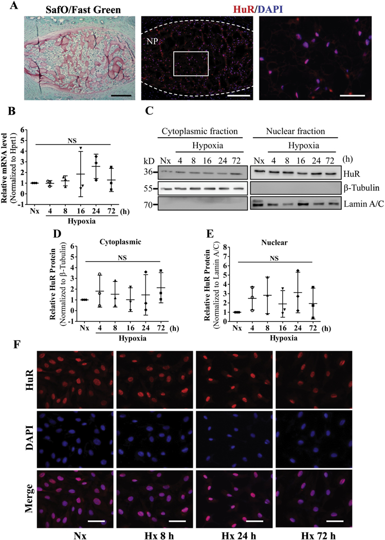
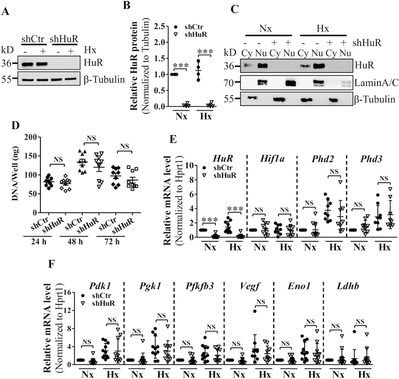
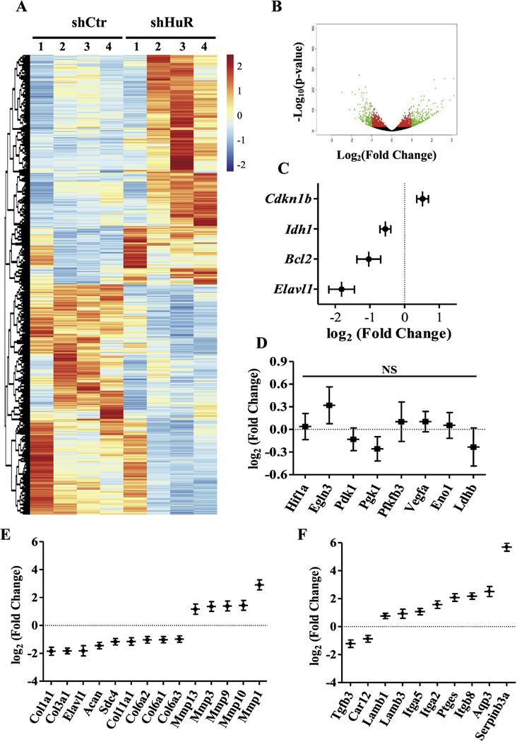
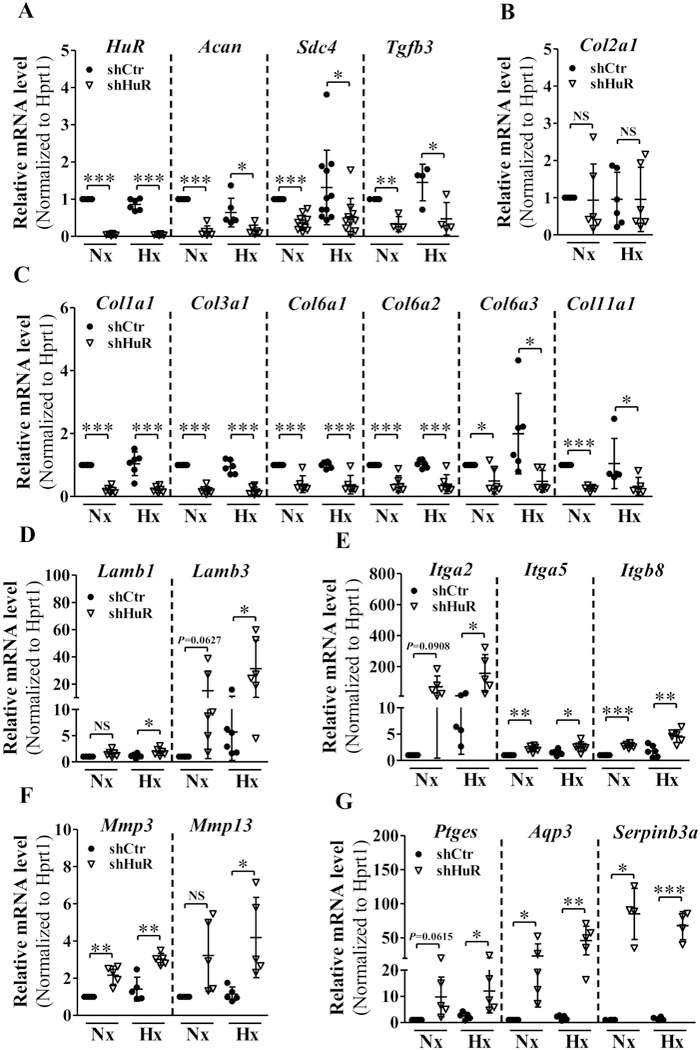
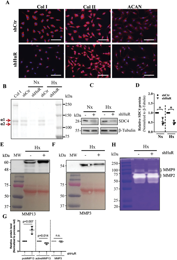
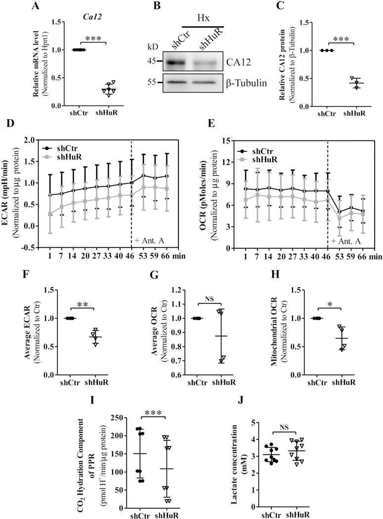
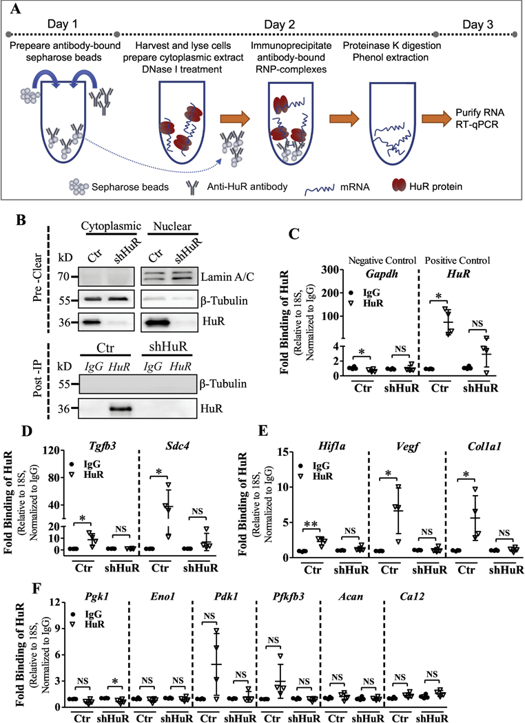
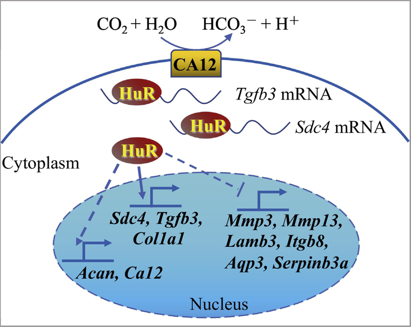
Nucleus pulposus (NP) cells reside in the hypoxic niche of the intervertebral disc. Studies have demonstrated that RNA-binding protein HuR modulates hypoxic signaling in several cancers, however, its function in the disc is unknown. HuR did not show cytoplasmic translocation in hypoxia and its silencing did not alter levels of Hif-1α or HIF-targets in NP cells. RNA-Sequencing data revealed that important extracellular matrix-related genes including several collagens, MMPs, aggrecan, Tgf-β3 and Sdc4 were regulated by HuR. Further analysis of HuR-silenced NP cells confirmed that HuR maintained expression of these matrix genes. We confirmed decreased levels of secreted collagen I and Sdc4 and increased pro-MMP13 in HuR-knockdown cells. In addition, messenger ribonucleoprotein immunoprecipitation demonstrated HuR binding to Tgf-β3 and Sdc4 mRNAs. Interestingly, while HuR bound to Hif-1α and Vegf mRNAs, it was clear that compensatory mechanisms sustained their expression when HuR was silenced. Noteworthy, despite the presence of multiple HuR-binding sites and reported interaction in other cell types, HuR showed no binding to Pgk1, Eno1, Pdk1 and Pfkfb3 in NP cells. Metabolic studies showed a significant decrease in the extracellular acidification rate (ECAR) and mitochondrial oxygen consumption rate (OCR) and acidic pH in HuR-silenced NP cells, without appreciable change in total OCR. These changes were likely due to decreased Ca12 expression in HuR silenced cells. Taken together, our study demonstrates for the first time that HuR regulates extracellular matrix (ECM) and pH homeostasis of NP cells and has important implications in the maintenance of intervertebral disc health.

Keywords: Extracellular matrix (ECM); HIF-1α; Human antigen R (HuR); Intervertebral disc; Nucleus pulposus.


Conflict of interest statement

Conflict of Interest: The authors all declare no conflicts of interest.

Figures

Figure 1.
Figure 1.
A) Safranin O/Fast Green staining of mouse intervertebral disc. Scale bar: 100 μm. A’, A’’) Immunofluorescence staining of HuR in NP. Scale bar: 100 μm (A’); 50 μm (A’’). B) HuR mRNA level in NP cells cultured under hypoxia up to 72 h. C) Cytoplasmic and nuclear levels of HuR protein in NP cells culture under hypoxia upto 72 h. D, E) Corresponding densitometric quantification of HuR cytoplasmic and nuclear protein fractions. F) Immunofluorescence staining of HuR in NP cells cultured in hypoxia. Western blot images shown in C are from one representative experiment that was performed independently at least thrice. Quantitative data are presented as mean ± SD of at least three independent experiments. NS, not significant; Nx, Normoxia; Hx, Hypoxia.
Figure 2.
Figure 2.
A, B) Measurement of HuR protein by Western blot (A) and corresponding densitometric analysis (B) confirmed successful knockdown of HuR after transduction with HuR-directed shRNA under normoxia and hypoxia. C) Both cytoplasmic levels of HuR significantly decreased following NP cell transduction with shHuR. D) Proliferation potential of HuR-silenced and control NP cells under hypoxia up to 72 h. E, F) HuR silencing exhibited no effects on select HuR/HIF-1α transcriptional targets - Hif-1α, Phd2, Phd3, Pdk1, Pgk1, Pfkfb3, Vegf, Eno1, Ldhb under both normoxia and hypoxia. Western blot images shown in A and C are from one representative experiment performed in ≥ 3 biological replicates. Quantitative data are represented as mean ± SD of at least three independent experiments. ***, p ≤ 0.001; NS, not significant; Cy, Cytoplasmic; Nu, Nuclear; Nx, Normoxia; Hx, Hypoxia.
Figure 3.
Figure 3.
A, B) Heat map and volcano plot depicting differentially expressed genes between control and HuR knockdown NP cells. C) RNA sequencing of HuR-silenced NP cells confirmed decreased expression of HuR (Elavl1), along with significant changes in positively regulated (Bcl2, Idh1) and negatively regulated (Cdkn1b) known HuR target genes. Log2(fold change) values are shown. D) RNA sequencing of HuR-silenced NP cells showed no significant changes of select HuR/HIF-1α transcriptional targets - Hif-1α, Phd3, Pdk1, Pgk1, Pfkfb3, Vegf, Eno1, Ldhb. E, F) Log2(fold change) values for matrix-related transcripts that were differentially expressed between control and HuR knockdown NP cells. NS, not significant.
Figure 4.
Figure 4.
A-C) qRT-PCR analysis of mRNA levels of Acan, Sdc4, Tgfb3 (A) and C Col1a1, Col3a1, Col6a1, Col6a2, Col6a3, Col11a1 (C) were significantly decreased under both normoxia and hypoxia following HuR silencing. Col2a1 (B) mRNA level did not change with HuR silenced NP cells. D-G) Levels of Lamb1, Lamb3 (D), Itga2 (E), Mmp13 (F) and Ptges (G) mRNA levels increased only under hypoxia in HuR silenced cells. Whereas, Itga5, Itgb8, Mmp3 (F), Aqp3 and Serpinb3a (G) mRNA levels increased under both normoxia and hypoxia following HuR knockdown. PCR data is represented as Mean ± SD of at least three independent experiments performed in duplicate. *, p ≤ 0.05; **, p ≤ 0.01; ***, p ≤ 0.001; NS, not significant; Nx, Normoxia; Hx, Hypoxia.
Figure 5.
Figure 5.
A) Immunofluorescence staining of NP cells demonstrated a dramatic decrease in Collagen I and a slight reduction in Aggrecan expression after HuR knockdown, while no change was observed in Collagen II levels. B) Electrophoresis of precipitated collagens from NP cell conditioned medium and Coomassie blue staining of precipitated fraction showed both α1 and α2 chains of Collagen I were diminished in HuR-silenced cells. C, D) Western blot and corresponding densitometric quantification showed decreased SDC4 protein levels after stable HuR knockdown under both normoxia and hypoxia. E) Western blot showed increased in levels of pro-MMP13 (60 kDa) but a slight decrease in active form (48 kDa) after stable HuR knockdown under hypoxia. F) Levels of MMP3 were not appreciably different between shcontrol and shHuR groups. G) Densitometry of Western blots from 3 independent experiments in E and F. H) Gelatin zymography showed decreased enzyme activity of MMP9 after HuR knockdown under hypoxia but no changes in MMP2 activity were seen. Images shown are from a representative experiment, which was performed at least at three independent times. *, p ≤ 0.05; **, p ≤ 0.01; NS, not significant; Nx, Normoxia; Hx, Hypoxia.
Figure 6.
Figure 6.
A-C) qRT-PCR and Western blot analysis showed carbonic anhydrase 12 (Ca12) mRNA and protein levels were attenuated after silencing HuR in hypoxia. D, E) Extracellular acidification rate (ECAR) of control and HuR-silenced NP cells subjected to hypoxia for 24 h, measured by Seahorse XF Analyzer, exhibited a significant decrease in the ECAR of HuR-silenced NP cells. F, G and H) HuR silencing did not affect total Oxygen consumption rate (OCR) but resulted in a slight reduction in Mitochondrial OCR of NP cells. I) CO2 hydration component of proton production rate (PPR) was reduced after HuR silencing. J) Extracellular lactate concentrations were unaffected in HuR silenced NP cells. Western blot images shown in (B) are from one representative experiment, which was performed at three independent times. Quantitative data are represented as mean ± SD of at least three independent experiments. *, p ≤ 0.05; **, p ≤ 0.01; ***, p ≤ 0.001; NS, not significant; Ant. A, Antimycin A; Hx, Hypoxia.
Figure 7.
Figure 7.
A) Schematic showing the workflow of the Messenger ribonucleoprotein immunoprecipitation (mRNP-IP or RIP) assay. B) Validation of cell lysates and post-IP samples from HuR RIP assay. HuR was immunoprecipitated (IP) using a rabbit polyclonal antibody, and IP was validated by a mouse monoclonal antibody by Western blot analysis. β-Tubulin was used as a loading control for the input and a negative control for the post-IP samples. Lamin A/C was used as a control to detect nuclear contamination in the input. C-E) The relative abundance of Tgfb3, Sdc4, Hif-1α, Vegf, Col1a1, Pgk1, Pdk1, Eno1, Pfkfb3, Acan and Ca12 mRNA bound to HuR, normalized to IgG isotype controls, was determined by qRT-PCR using 18S rRNA as a loading control. Gapdh was used as a negative control and HuR as a positive control. Western blot images shown in B are from one representative experiment out of 4 independent experiments. Quantitative data are represented as Mean ± SD. *, p ≤ 0.05; **, p ≤ 0.01; NS, not significant.
Figure 8.
Figure 8.
Schematic of proposed HuR regulation of extracellular matrix (ECM) and pH homeostasis in NP cells. HuR positively regulates Tgfb3, Sdc4, Col1a1, Acan and Ca12 expression through direct binding of Tgfb3, Sdc4 and Col1a1 mRNAs, and negatively regulates levels of Mmp3, Mmp13, Lamb3, Itgb8, Ptges, and Serpinb3a.

References

    1. Hoy D, March L, Brooks P, Blyth F, Woolf A, Bain C, Williams G, Smith E, Vos T, Barendregt J, Murray C, Burstein R, Buchbinder R, The global burden of low back pain: estimates from the Global Burden of Disease 2010 study, Ann. Rheum. Dis 73 (2014) 968–974. - PubMed
    1. Balague F, Mannion AF, Pellise F, Cedraschi C, Non-specific low back pain, Lancet 379 (2012) 482–91. - PubMed
    1. Murray CJ, Atkinson C, Bhalla K, Birbeck G, Burstein R, Chou D, Dellavalle R, Danaei G, Ezzati M, Fahimi A, et al., The state of US health, 1990–2010: burden of diseases, injuries, and risk factors, JAMA 310 (2013) 591–608. - PMC - PubMed
    1. Yang G, Wang Y, Zeng Y, Gao GF, Liang X, Zhou M, Wan X, Yu S, Jiang Y, Naghavi M, Vos T, Wang H, Lopez AD, Murray CJ, Rapid health transition in China, 1990–2010: findings from the Global Burden of Disease Study 2010, Lancet 381 (2013) 1987–2015. - PMC - PubMed
    1. Vos T, Barber RM, Bell B, Bertozzi-Villa A, Biryukov S, Bolliger I, Charlson F, Davis A, Degenhardt L, Dicker D, et al., Global, regional, and national incidence, prevalence, and years lived with disability for 301 acute and chronic diseases and injuries in 188 countries, 1990–2013: a systematic analysis for the Global Burden of Disease Study 2013, Lancet 386 (2015) 743–800. - PMC - PubMed

Publication types

MeSH terms