Review
. 2019 Mar:133:88-100.
doi: 10.1016/j.freeradbiomed.2018.08.005. Epub 2018 Aug 6.

Handling heme: The mechanisms underlying the movement of heme within and between cells

Affiliations
Review

Handling heme: The mechanisms underlying the movement of heme within and between cells

Rebecca K Donegan et al. Free Radic Biol Med. 2019 Mar.

Abstract

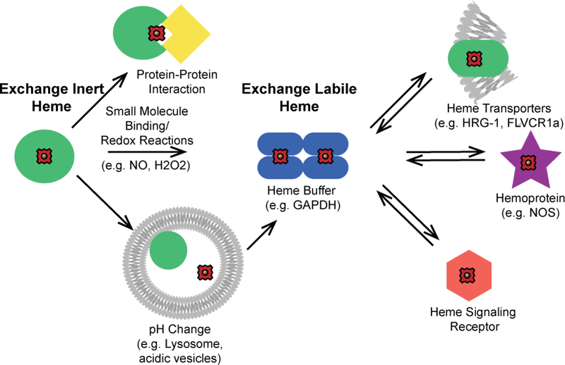
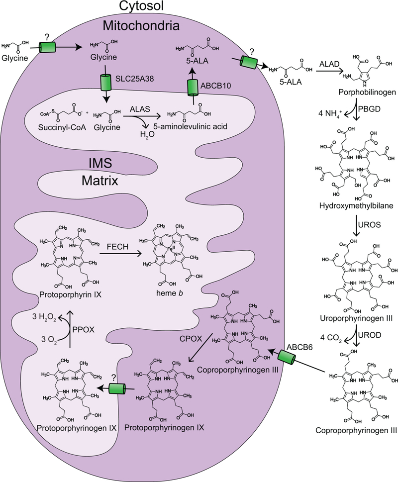
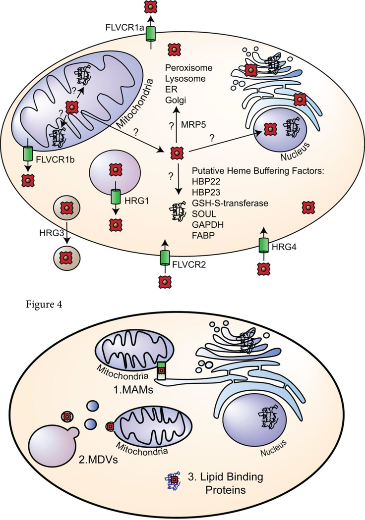
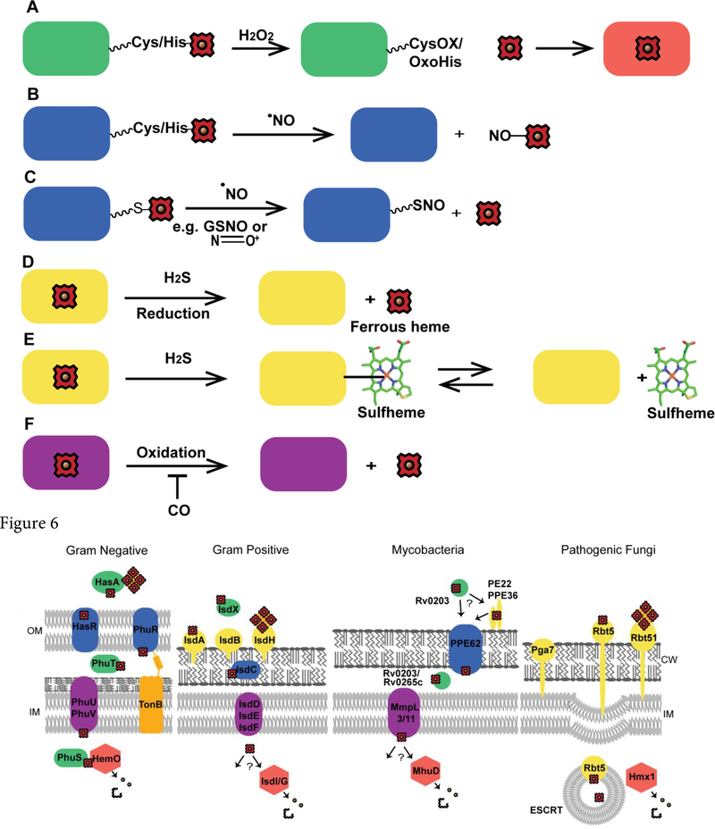
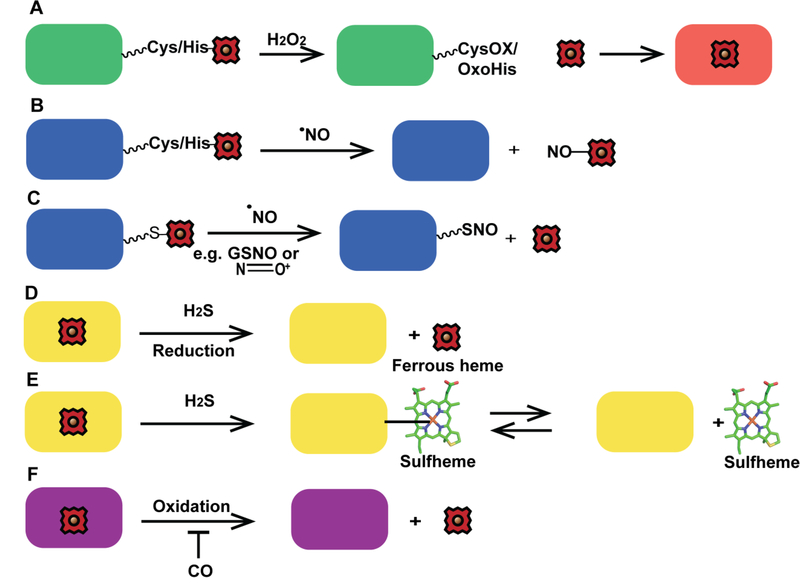
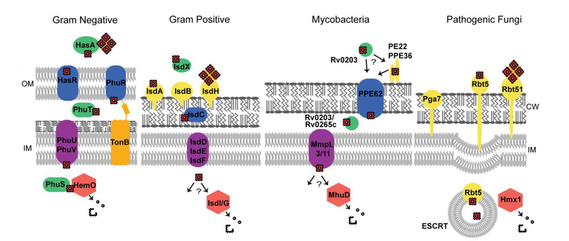
Heme is an essential cofactor and signaling molecule required for virtually all aerobic life. However, excess heme is cytotoxic. Therefore, heme must be safely transported and trafficked from the site of synthesis in the mitochondria or uptake at the cell surface, to hemoproteins in most subcellular compartments. While heme synthesis and degradation are relatively well characterized, little is known about how heme is trafficked and transported throughout the cell. Herein, we review eukaryotic heme transport, trafficking, and mobilization, with a focus on factors that regulate bioavailable heme. We also highlight the role of gasotransmitters and small molecules in heme mobilization and bioavailability, and heme trafficking at the host-pathogen interface.

Keywords: Heme; Heme trafficking; Heme transport; Host-pathogen interactions; Hydrogen peroxide; Iron; Nitric oxide.


Conflict of interest statement

Disclosure:

The authors declare no conflict of interest.

Figures

Figure 1.
Figure 1.
Conceptual model for the mobilization of exchange inert and labile heme. Heme is bound by biomolecules that are either exchange inert (green circles) or labile (blue ellipses). Exchange inert heme can be mobilized by specific protein-protein interactions that drive the transfer of heme or by post-translational modifications that can induce the release of heme, e.g. protonation of heme coordinating residues, allosteric regulation, or redox reactions at the heme iron center or amino acid side chains. Labile heme may exchange with downstream clients based on thermodynamic gradients, including with transporters (green ellipse), heme enzymes (purple star), and proteins that are targets of heme-based signaling (orange hexagon).
Figure 2.
Figure 2.
Heme synthesis in eukaryotes. Heme is synthesized in eukaryotes via eight highly conserved enzymes, four in the mitochondria and four in the cytoplasm. The final step of heme biosynthesis is the ferrochelatase catalyzed insertion of ferrous iron into protoporphyrin IX at the matrix side of the mitochondrial inner-membrane. ALAS = 5-aminolevulinic acid (5-ALA) synthase; ALAD = 5-ALA dehydratase; PBGD = porphobilinogen deaminase; UROS = uroporphyrinogen synthase; UROD = uroporphyrinogen decarboxylase; CPOX = coproporphyrinogen oxidase; PPOX = protoporphyrinogen oxidase; FECH = ferrochelatase.
Figure 3.
Figure 3.
Consensus model of heme transport and trafficking. Heme trafficking from the matrix of the mitochondria to various subcellular locations involve transporters and chaperones. Putative transporters are shown at their predicted location in the cell and denoted via green cylinders. Their direction of transport is noted with arrows. Putative heme buffering factors are listed as well.
Figure 4.
Figure 4.
Trafficking of heme via membranes and vesicles. Heme may be trafficked from the mitochondria via (1) direct inter-organelle contact points such as MAMs, (2) via vesicular mediated trafficking in MDVs, or (3) chaperoned by lipid binding proteins.
Figure 5.
Figure 5.
Schemes for heme mobilization via small molecules. Heme mobilization may be regulated via small molecules. (A) H2O2 can oxidize hemoproteins, including hemecoordinating residues, e.g. Cys or His, and potentially release heme for use by other hemoproteins. CysOX is cysteine sulfenic acid and OxoHis is 2-oxo-histidine. (B) Nitrosylation of heme by NO may cause heme dissociation. (C) S-nitrosation may also initiate the release of heme. (D) H2S can reduce ferric heme, potentially causing the dissociation of ferrous heme. (E) H2S can bind to heme yielding sulfheme, where sulfur is incorporated into one of the porphyrin rings of heme, (structure modified from PDB ID 1YMC) which could cause heme dissociation. (F) CO could potentially inhibit heme mobilization by preventing oxidation of ferrous heme.
Figure 6.
Figure 6.
Heme uptake in pathogenic microbes. Representative heme uptake systems for gram negative bacteria (P. aeruginosa), gram positive bacteria (S. aureus), mycobacteria (M. tuberculosis), and fungal pathogens (C. albicans). OM is the outer membrane, IM is inner membrane and CW is the cell wall.

References

    1. Hooda J, Shah A, Zhang L, Heme, an essential nutrient from dietary proteins, critically impacts diverse physiological and pathological processes, Nutrients 6(3) (2014) 1080–1102. - PMC - PubMed
    1. Aich A, Freundlich M, Vekilov PG, The free heme concentration in healthy human erythrocytes, Blood Cells, Molecules, and Diseases 55(4) (2015) 402–409. - PMC - PubMed
    1. Hanna DA, Martinez-Guzman O, Reddi AR, Heme Gazing: Illuminating Eukaryotic Heme Trafficking, Dynamics, and Signaling with Fluorescent Heme Sensors, Biochemistry 56(13) (2017) 1815–1823. - PMC - PubMed
    1. Reddi AR, Hamza I, Heme mobilization in animals: a Metallolipid’s journey, Accounts of chemical research 49(6) (2016) 1104–1110. - PMC - PubMed
    1. Atamna H, Boyle K, Amyloid-β peptide binds with heme to form a peroxidase: Relationship to the cytopathologies of Alzheimer’s disease, Proceedings of the National Academy of Sciences of the United States of America 103(9) (2006) 3381–3386 doi10.1073/pnas.0600134103. - DOI - PMC - PubMed

Publication types