Review
. 2018 Dec:53:139-145.
doi: 10.1016/j.conb.2018.07.007. Epub 2018 Aug 6.

Dendrite morphogenesis from birth to adulthood

Affiliations
Review

Dendrite morphogenesis from birth to adulthood

Cameron L Prigge et al. Curr Opin Neurobiol. 2018 Dec.

Abstract

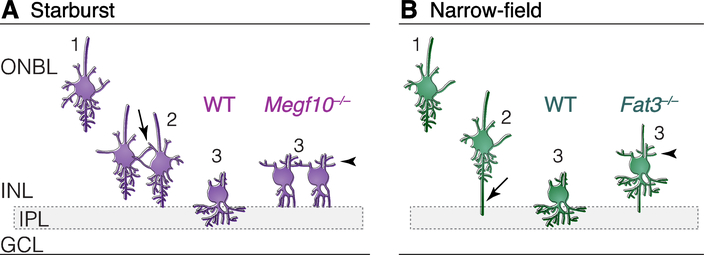
Dendrites are the conduits for receiving (and in some cases transmitting) neural signals; their ability to do these jobs is a direct result of their morphology. Developmental patterning mechanisms are critical to ensuring concordance between dendritic form and function. This article reviews recent studies in vertebrate retina and brain that elucidate key strategies for dendrite functional maturation. Specific cellular and molecular signals control the initiation and elaboration of dendritic arbors, and facilitate integration of young neurons into particular circuits. In some cells, dendrite growth and remodeling continues into adulthood. Once formed, dendrites subdivide into compartments with distinct physiological properties that enable dendritic computations. Understanding these key stages of dendrite patterning will help reveal how circuit functional properties arise during development.


Figures

Fig. 1:
Fig. 1:. Organization of cells and synapses in mammalian retina.
Cellular layers (shaded) include outer and inner nuclear layers (ONL, INL); ganglion cell layer (GCL). Outer and inner plexiform layers (OPL, IPL) contain synapses. In outer retina, horizontal cells (purple) receive synapses from cone photoreceptors (red) on their dendrites, and from rods (dark blue) on their axons (a postsynaptic arbor with many dendrite-like properties). Rods and cones also connect to dendrites of specific bipolar cell types; cone bipolar cells (light blue) are illustrated here. In IPL, the direction-selective circuit is illustrated as an example of how inner retinal circuits are organized. Circuit partners converge upon particular IPL sublayers, where direction-selective retinal ganglion cells (RGCs, pink) receive glutamatergic excitation from bipolar cells (light blue) and inhibition from starburst amacrine cells (green). Direction-selective circuit occupies two sublayers (OFF, ON); starburst and bipolar cells project to one or the other, depending on the polarity of their light response. ON-OFF direction-selective RGCs project to both sublayers.
Fig. 2:
Fig. 2:. Initiation of dendrite formation in migrating amacrine cells.
Two types of amacrine cells, starburst (A) and late-born narrow-field cells (B), highlight distinct dendrite initiation strategies. Each cell type is illustrated at three time points: 1) Early radial migration through the outer neuroblast layer (ONBL); 2) nearing their final position adjacent to the IPL; and 3) initiating dendrite projections into the IPL. Starburst cells use homotypic contacts (A, arrow) to detect that they have arrived in the right place for IPL innervation. Narrow-field cells send out long basal processes (B, arrow) to obtain IPL-derived polarity signals that control timing and location of dendrite initiation. Dendrite initiation is impaired when recognition receptors MEGF10 and Fat3 are eliminated. Megf10–/– starburst cells (A) do not receive contact-mediated signals initiating IPL innervation, and therefore remain multipolar (A, time 3, right). Fat3–/– narrow-field cells (B) lose cytoskeletal polarity, which slows migration and permits dendrite initiation on the wrong side of the cell (B, time 3, right). WT, wild-type cells. Arrowheads, mislocalized dendrites in mutant cells. Layer abbreviations as in Fig. 1.
Fig. 3:
Fig. 3:. Functional compartmentalization of starburst amacrine cell dendrites.
A: Starburst dendrites are radially symmetric. GABAergic presynaptic boutons (magenta) are restricted to the distal third of their dendrites. B: Detail of part of a starburst dendritic arbor. Circles illustrate restricted distribution of inhibitory (orange) and excitatory (blue) synaptic inputs. C: Individual wild-type starburst boutons have preferred directions (PDs, magenta arrows) aligned to the cell’s centrifugal axis (gray arrow). Local dendrite angle determines bouton PD, as shown by Sema6a–/– dendrites (illustrated at right). Dendrite mispatterning causes uncoupling of bouton PD and centrifugal axis (arrows, right panel).

References

    1. Cline H, Haas K: The regulation of dendritic arbor development and plasticity by glutamatergic synaptic input: a review of the synaptotrophic hypothesis. J Physiol 2008, 586:1509–1517. - PMC - PubMed
    1. Hoshiba Y, Toda T, Ebisu H, Wakimoto M, Yanagi S, Kawasaki H: Sox11 Balances Dendritic Morphogenesis with Neuronal Migration in the Developing Cerebral Cortex. J Neurosci 2016, 36:5775–5784. - PMC - PubMed
    1. Guerrier S, Coutinho-Budd J, Sassa T, Gresset A, Jordan NV, Chen K, Jin WL, Frost A,Polleux F: The F-BAR domain of srGAP2 induces membrane protrusions required for neuronal migration and morphogenesis. Cell 2009, 138:990–1004. - PMC - PubMed
    1. Icha J, Kunath C, Rocha-Martins M, Norden C: Independent modes of ganglion cell translocation ensure correct lamination of the zebrafish retina. J Cell Biol 2016, 215:259–275. - PMC - PubMed
    1. Sawada M, Ohno N, Kawaguchi M, Huang SH, Hikita T, Sakurai Y, Bang Nguyen H, Quynh Thai T, Ishido Y, Yoshida Y, et al.: PlexinD1 signaling controls morphological changes and migration termination in newborn neurons. EMBO J 2018, 37. - PMC - PubMed

Publication types

LinkOut - more resources