. 2018 Aug 9;13(1):43.
doi: 10.1186/s13024-018-0276-2.

Immunotherapy targeting toll-like receptor 2 alleviates neurodegeneration in models of synucleinopathy by modulating α-synuclein transmission and neuroinflammation

Affiliations

Immunotherapy targeting toll-like receptor 2 alleviates neurodegeneration in models of synucleinopathy by modulating α-synuclein transmission and neuroinflammation

Changyoun Kim et al. Mol Neurodegener. .

Abstract

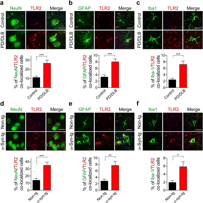
Background: Synucleinopathies of the aging population are an heterogeneous group of neurological disorders that includes Parkinson's disease (PD) and dementia with Lewy bodies (DLB) and are characterized by the progressive accumulation of α-synuclein in neuronal and glial cells. Toll-like receptor 2 (TLR2), a pattern recognition immune receptor, has been implicated in the pathogenesis of synucleinopathies because TLR2 is elevated in the brains of patients with PD and TLR2 is a mediator of the neurotoxic and pro-inflammatory effects of extracellular α-synuclein aggregates. Therefore, blocking TLR2 might alleviate α-synuclein pathological and functional effects. For this purpose, herein, we targeted TLR2 using a functional inhibitory antibody (anti-TLR2).

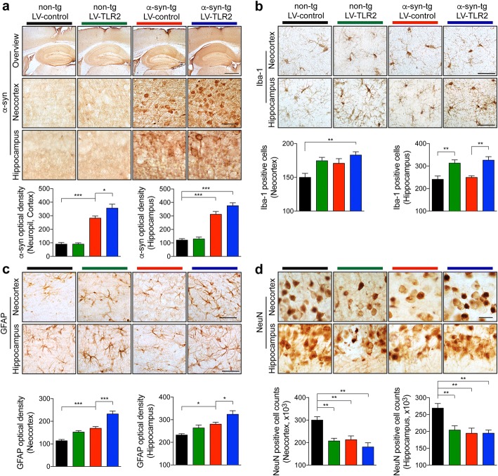
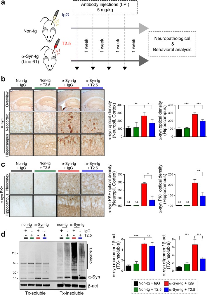
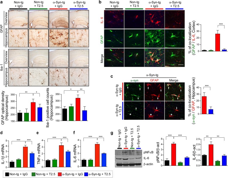
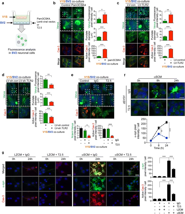
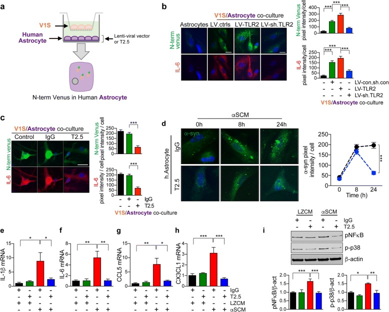
Methods: Two different human α-synuclein overexpressing transgenic mice were used in this study. α-synuclein low expresser mouse (α-syn-tg, under the PDGFβ promoter, D line) was stereotaxically injected with TLR2 overexpressing lentivirus to demonstrate that increment of TLR2 expression triggers neurotoxicity and neuroinflammation. α-synuclein high expresser mouse (α-Syn-tg; under mThy1 promoter, Line 61) was administrated with anti-TLR2 to examine that functional inhibition of TLR2 ameliorates neuropathology and behavioral defect in the synucleinopathy animal model. In vitro α-synuclein transmission live cell monitoring system was used to evaluate the role of TLR2 in α-synuclein cell-to-cell transmission.

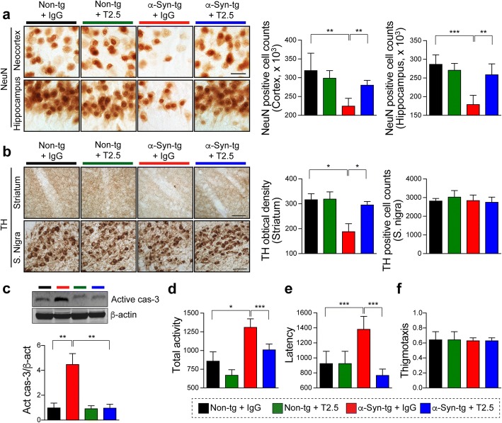
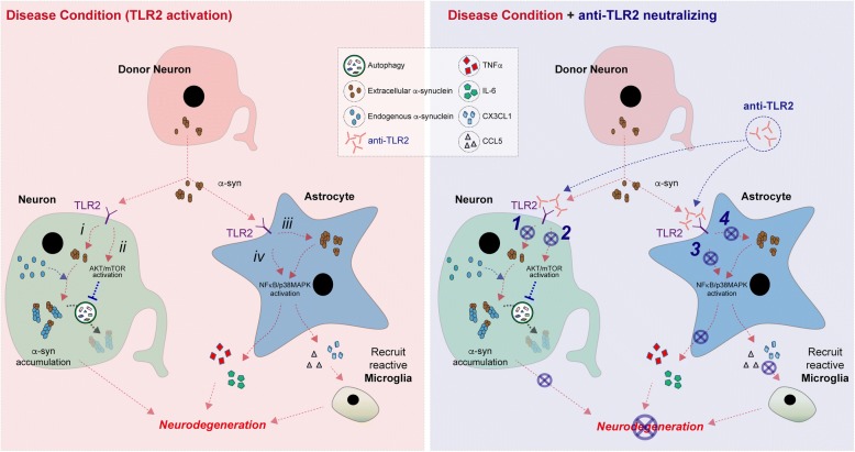
Results: We demonstrated that administration of anti-TLR2 alleviated α-synuclein accumulation in neuronal and astroglial cells, neuroinflammation, neurodegeneration, and behavioral deficits in an α-synuclein tg mouse model of PD/DLB. Moreover, in vitro studies with neuronal and astroglial cells showed that the neuroprotective effects of anti-TLR2 antibody were mediated by blocking the neuron-to-neuron and neuron-to-astrocyte α-synuclein transmission which otherwise promotes NFκB dependent pro-inflammatory responses.

Conclusion: This study proposes TLR2 immunotherapy as a novel therapeutic strategy for synucleinopathies of the aging population.

Keywords: Immunotherapy; Neurodegeneration; Neuroinflammation; Parkinson’s disease; Synucleinopathy; Toll-like receptor 2; Transmission; α-synuclein.


Conflict of interest statement

All procedures for animal use were approved by the institutional Animal Care and Use Committee at University of California, San Diego under protocol S02221.

All authors have approved of the consents of this manuscript and provided consent for publication.

The authors declare that they have no competing interests.

Springer Nature remains neutral with regard to jurisdictional claims in published maps and institutional affiliations.

Figures

Fig. 1
Fig. 1
Expression of TLR2 in the neocortex of synucleinopathy patients and an animal model. a–c Representative images from double-immunolabeling for TLR2 with cellular markers in the neocortex of normal and PD/DLB patients. The percentages of TLR2-positive neurons (NeuN) (a), astrocytes (GFAP) (b), and microglia (Iba1) (c) in the neocortex (n = 8 per group). df Representative images from double-immunolabeling for TLR2 with cellular markers in the neocortex of non-tg and α-Syn-tg (Line 61) mice (9–10 month olds). The percentages of TLR2-positive neuron (NeuN) (d), astrocyte (GFAP) (e), and microglia (Iba1) (f) in the neocortex (n = 6 per group). Data are mean ± SEM. *p < 0.05, **p < 0.01, and ***p < 0.001; unpaired t test. Scale bar, 20 μm
Fig. 2
Fig. 2
Neuropathology analysis of LV-TLR2-delivered non-tg and α-syn-tg mice. Either LV-control or LV-TLR2 was injected into the hippocampus of non-tg or α-syn-tg mice (D line). ad Representative images from immunohistochemical staining of α-synuclein (a), Iba-1 (b), GFAP (c), and NeuN (d) in the neocortex and hippocampus of lentiviral vector-delivered mice. The level of α-synuclein (a) or GFAP (c) was analyzed in the neocortex and hippocampus of the mice by optical density quantification. The number of Iba-1 (b) or NeuN (d) positive cell was counted in neocortex and hippocampus of the mice. Data are mean ± SEM (n = 6 per group). *p < 0.05, **p < 0.01, and ***p < 0.001; one way ANOVA. Scale bars, 250 μm (low magnification) and 25 μm (high magnification)
Fig. 3
Fig. 3
Administration of anti-TLR2 (T2.5) decreases α-synuclein pathology in a synucleinopathy mouse model. a Experimental scheme. Non-tg and α-Syn-tg (Line 61) mice were administrated with either IgG (5 mg/kg) or T2.5 (5 mg/kg) weekly for 4 weeks. The levels of α-synuclein pathology, glial cell reactivity, neurodegeneration, and behavioral deficits were analyzed after a 5-weeks post injection. b Representative images from immunohistochemical staining of α-synuclein in the neocortex and hippocampus of mice. The level of α-synuclein was analyzed by optical density quantification (n = 6 per group). c Representative images from immunohistochemical staining of PK-resistant α-synuclein in the neocortex and hippocampus of mice. The levels of PK-resistant α-synuclein were analyzed by optical density quantification (n = 6 per group). d Immunoblot analysis of mice brain lysates. Triton-soluble and -insoluble brain lysates were probed for α-synuclein and β-actin. The levels of triton-insoluble α-synuclein monomer and high molecular weight oligomers were determined by densitometric quantification (n = 4 per group). Data are mean ± SEM. *p < 0.05, **p < 0.01, and ***p < 0.001, n.d. not detected; one way ANOVA for (b, d) and unpaired t test for (c). Scale bars, 250 μm (low magnification) and 25 μm (high magnification)
Fig. 4
Fig. 4
Administration of anti-TLR2 (T2.5) decreases neuroinflammation in synucleinopathy mouse model. Non-tg or α-Syn-tg (Line 61) mice were administrated with either IgG (5 mg/kg) or T2.5 (5 mg/kg) weekly for 4 weeks. a Representative images from immunohistochemical staining of GFAP and Iba-1 in the hippocampus of mice. The level of GFAP was analyzed by optical density quantification and the number of Iba-1 positive cell was counted in the hippocampus of mice (n = 6 per group). b Representative images from co-localization of GFAP (green) and IL-6 (red) in the antibody-administrated mice. The percentages of GFAP/IL-6 double positive cells were analyzed in the hippocampus of mice. (n = 6 per group). c Double immunolabeling analysis for human α-synuclein (green) and GFAP (red) in tg mice. The percentages of α-synuclein and GFAP positive cells were analyzed in the hippocampus of tg mice (n = 6 per group). df Quantitative analysis of the cytokine gene expressions in the cortex of mice. The expressions of IL-1β (d), TNFα (e), and IL-6 (f) were normalized to the levels of β-actin (n = 4 per group). g Immunoblot analysis of the whole brain lysates probed for NFκB, IL-6, and β-actin. The levels of NFκB and IL-6 were determined by densitometric quantification (n = 3 per group). Data are mean ± SEM. *p < 0.05, **p < 0.01, and ***p < 0.001; one way ANOVA for (a, dg) and unpaired t test for (b and c). Scale bars, 250 μm (low magnification) and 25 μm (high magnification)
Fig. 5
Fig. 5
Neuroprotective effect of anti-TLR2 (T2.5) treatment in synucleinopathy mouse model. Non-tg or α-Syn-tg (Line 61) mice were administrated with either IgG (5 mg/kg) or T2.5 (5 mg/kg) weekly for 4 weeks. a Representative images from immunohistochemical staining of NeuN in the neocortex and hippocampus of mice. The numbers of NeuN positive cells were counted in the neocortex and hippocampus of mice (n = 6 per group). b Representative images from immunohistochemical staining of Thyroxine hydroxylase (TH) in the antibody-administrated mice. The level of TH was analyzed by optical density quantification and the numbers of TH positive cell were counted in the striatum and substantia nigra of mice, respectively (n = 6 per group). c Immunoblotting analysis of whole-brain lysates. The lysates were probed for active form of casepase-3 and β-actin. The level of active caspase 3 was determined by densitometric quantification (n = 3 per group). df Behavioral analysis of the mice. The total activity (d), latency (e), and thigmotaxis (f) were analyzed by open field test (n = 6 per group). Data are mean ± SEM. *p < 0.05, **p < 0.01, and ***p < 0.001; one way ANOVA. Scale bar, 25 μm
Fig. 6
Fig. 6
TLR2 mediates neurotoxic neuron-to-neuron α-synuclein transmission. a Overview diagram. Donor neuronal cells (V1S), expressing α-synuclein-conjugated with N-terminus of venus were plated in trans-well insert and the recipient neuronal cells (SV2), expressing α-synuclein conjugated with C-terminus of venus were seeded onto cover slips in the bottom well. Only SV2 cells were treated with pam3CSK4 (10 μg/ml), lentiviral vectors, or antibodies. Images were taken from SV2 cells after a 3-days co-culture. be Representative confocal images for BiFC fluorescence and caspase-3 activity in SV2 cells. Middle panels are enlargements of cropped regions outlined with dashed lines from upper panels. Lower panels are double-immunolabeling assay with active casepase-3. The average numbers of venus fluorescence intensity in each cell, the average size of the venus punctum diameters, and caspase-3 fluorescence intensity were analyzed. b V1S and SV2 cells were co-cultured in the presence or absence of pam3CSK4 (10 μg/ml) (n = 3). c V1S and SV2 cells were co-cultured with either LV-control or LV-TLR2 (n = 3). d V1S and SV2 cells were co-cultured with either LV-sh.control or LV-shTLR2 (n = 3). e V1S and SV2 cells were co-cultured with either IgG (5 μg/ml) or T2.5 (5 μg/ml) (n = 3). f The kinetics of α-synuclein internalization in the presence of antibodies. dSY5Y cells were incubated with αSCM for indicated hours in the presence of either IgG (5 μg/ml) or T2.5 (5 μg/ml). The kinetics was analyzed by immunolabeling assay (n = 3). g Neuronal internalization of α-synuclein in the presence of antibodies. Mouse primary cortical neurons were incubated with αSCM or LZCM for indicated hours in the presence of either IgG (5 μg/ml) or T2.5 (5 μg/ml). Neurons were double immunolabelled with human α-synuclein (Middle panels) and active form of caspase-3 (low panels) (n = 3). Data are mean ± SEM (n = 3 per group). *p < 0.05, **p < 0.01, and ***p < 0.001; unpaired t test for all analysis except (g) (one way ANOVA). Scale bar, 20 μm
Fig. 7
Fig. 7
Astrocyte responses by TLR2 mediated neuron-to-astrocyte α-synuclein transmission. a Overview diagram. Donor neuronal cells (V1S), expressing α-synuclein-conjugated with N-terminus of venus were plated in trans-well insert and the recipient human primary astrocytes were plated onto the cover slips in the bottom well. Only astrocytes were treated with either lentiviral vectors or antibodies. Images were taken from astrocytes after a 3-days co-culture. b, c Representative confocal images for N-terminus of venus (Upper panel) and IL-6 (Lower panel) in recipient astrocytes. The fluorescence intensity of N-term venus and IL-6 were analyzed in randomly chosen area. b V1S and astrocytes were co-cultured in the presence of either LV-control/sh.control, LV-TLR2, or LV-sh.TLR2 (n = 3). c V1S and astrocytes were co-cultured in the presence of either IgG (5 μg/ml) or T2.5 (5 μg/ml) (n = 3). d The kinetics of astroglial α-synuclein internalization in the presence of antibodies. Human primary astrocytes were incubated with αSCM for indicated hours in the presence of either IgG (5 μg/ml) or T2.5 (5 μg/ml). The kinetics was analyzed by immunolabeling assay (n = 3). e–h Quantitative analysis of the cytokine/chemokine gene expressions in astrocytes. The cells were incubated with either LZCN or αSCM for 24 h in the presence of indicated antibodies. The expressions of IL-1β (e), IL-6 (f), CCL5 (g), and CX3CL1 (h) were normalized to the levels of β-actin (n = 4). Data are mean ± SEM. *p < 0.05, **p < 0.01, and ***p < 0.001; one way ANOVA for all analysis except (d) (unpaired t test). Scale bar, 20 μm
Fig. 8
Fig. 8
Model for TLR2 immunotherapy ameliorates neurodegeneration in synucleinopathy. In disease condition, TLR2 mediates neurotoxicity. In neuron, i) TLR2 induces pathological internalization of extracellular α-synuclein into neuron and ii) extracellular α-synuclein activates neuronal TLR2 which results in mTOR-mediated autophagy inhibition. Thus, neuronal TLR2 induces neurotoxic α-synuclein accumulation. In astrocyte, iii) TLR2 increased abnormal α-synuclein accumulation which leads astroglial activation and iv) extracellular α-synuclein activates astroglial TLR2 signaling cascade through NFκB/p38 MAPK which results in neurotoxic astroglial responses such as pro-inflammatory cytokine expression and induction of reactive microglia recruiting chemokines. Therefore, TLR2 immunotherapy ameliorates α-synuclein-mediated neurotoxicity via inhibition of 1) TLR2-mediated neuronal α-synuclein internalization, 2) activation of neuronal autophagy via TLR2-mTOR signaling cascade, 3) inhibition of TLR2-mediated astroglial responses, and 4) reduction of astroglial α-synuclein accumulation. Thereby, TLR2 immunotherapy might be a novel therapeutic strategy for synucleinopathy

References

    1. Lee HJ, Bae EJ, Lee SJ. Extracellular alpha--synuclein-a novel and crucial factor in Lewy body diseases. Nat Rev Neurol. 2014;10:92–98. doi: 10.1038/nrneurol.2013.275. - DOI - PubMed
    1. Fellner L, Jellinger KA, Wenning GK, Stefanova N. Glial dysfunction in the pathogenesis of alpha-synucleinopathies: emerging concepts. Acta Neuropathol. 2011;121:675–693. doi: 10.1007/s00401-011-0833-z. - DOI - PMC - PubMed
    1. Halliday GM, Stevens CH. Glia: initiators and progressors of pathology in Parkinson's disease. Movement disorders : official journal of the Movement Disorder Society. 2011;26:6–17. doi: 10.1002/mds.23455. - DOI - PubMed
    1. Kim C, Lee SJ. Controlling the mass action of alpha-synuclein in Parkinson's disease. J Neurochem. 2008;107:303–316. doi: 10.1111/j.1471-4159.2008.05612.x. - DOI - PubMed
    1. Ingelsson M. Alpha-Synuclein oligomers-neurotoxic molecules in Parkinson's disease and other Lewy body disorders. Front Neurosci. 2016;10:408. doi: 10.3389/fnins.2016.00408. - DOI - PMC - PubMed

Publication types