Review
. 2018 Aug;11(8):1150-1172.
doi: 10.1016/j.jcmg.2018.06.004.

Cardiovascular Magnetic Resonance in the Oncology Patient

Affiliations
Review

Cardiovascular Magnetic Resonance in the Oncology Patient

Jennifer H Jordan et al. JACC Cardiovasc Imaging. 2018 Aug.

Abstract

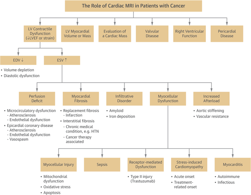
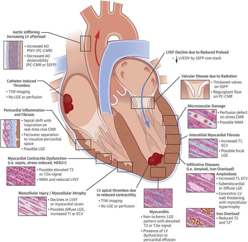
Patients with or receiving potentially cardiotoxic treatment for cancer are susceptible to developing decrements in left ventricular mass, diastolic function, or systolic function. They may also experience valvular heart disease, pericardial disease, or intracardiac masses. Cardiovascular magnetic resonance may be used to assess cardiac anatomy, structure, and function and to characterize myocardial tissue. This combination of features facilitates the diagnosis and management of disease processes in patients with or those who have survived cancer. This report outlines and describes prior research involving cardiovascular magnetic resonance for assessing cardiovascular disease in patients with or previously having received treatment for cancer.

Keywords: cardio-oncology; cardiovascular magnetic resonance; tissue characterization.


Figures

FIGURE 1
FIGURE 1
The Role of Cardiovascular Magnetic Resonance in Patients With Cancer
FIGURE 2
FIGURE 2
Early Changes in Left Ventricular Systolic Function After Anthracycline-Based Chemotherapy
FIGURE 3
FIGURE 3
Cardiovascular Magnetic Resonance Case Examples of Left Ventricular Ejection Fraction Due to Either a Decline in Left Ventricular End-Diastolic Volume or an Increase in Left Ventricular End-Systolic Volume After Chemotherapy
FIGURE 4
FIGURE 4
Cardiovascular Magnetic Resonance Case Example of Transient Left Ventricular Dysfunction Syndrome in Patient With Cancer
FIGURE 5
FIGURE 5
Case Example of Amyloidosis in Patient With Cancer
FIGURE 6
FIGURE 6
Myocardial Fibrosis Imaging in Anthracycline-Treated Cancer Survivors Using Extracellular Volume Mapping
FIGURE 7
FIGURE 7
Cardiovascular Magnetic Resonance Case of Pericardial Constriction in a Patient with Cancer
FIGURE 8
FIGURE 8
Cardiovascular Magnetic Resonance Case of Right Atrial Thrombus
FIGURE 9
FIGURE 9
Cardiovascular Magnetic Resonance Features of Cardiac Masses
CENTRAL ILLUSTRATION
Adverse Cardiovascular Effects Related to Cancer Treatment and Key Cardiovascular Magnetic Resonance Features

References

    1. Barac A, Murtagh G, Carver JR, et al. Cardio-vascular health of patients with cancer and cancer survivors: a roadmap to the next level. J Am Coll Cardiol 2015;65:2739–46. - PMC - PubMed
    1. Mulrooney DA, Yeazel MW, Kawashima T, et al. Cardiac outcomes in a cohort of adult survivors of childhood and adolescent cancer: retrospective analysis of the Childhood Cancer Survivor Study cohort. BMJ 2009;339: b4606. - PMC - PubMed
    1. Vasu S, Hundley WG. Understanding cardio-vascular injury after treatment for cancer: an overview of current uses and future directions of cardiovascular magnetic resonance. J Cardiovasc Magn Reson 2013;15:66–83. - PMC - PubMed
    1. Kongbundansuk S, Hundley WG. Noninvasive imaging of cardiovascular injury related to the treatment of cancer. J Am Coll Cardiol Img 2014;7: 824–38. - PMC - PubMed
    1. Thavendiranathan P, Grant AD, Negishi T, Plana JC, Popovic ZB, Marwick TH. Reproducibility of echocardiographic techniques for sequential assessment of left ventricular ejection fraction and volumes: application to patients undergoing cancer chemotherapy. J Am Coll Cardiol 2013;61: 77–84. - PubMed

Publication types

MeSH terms

Substances