

Current Advances in COPD Imaging
- PMID: 30093217
- PMCID: PMC11247962
- DOI: 10.1016/j.acra.2018.05.023
Current Advances in COPD Imaging
Abstract
Objective: To review the recent advances in available technologies for imaging COPD and present the novel optical coherence tomography (OCT) airway imaging technology.
Materials and methods: This is an unstructured review of published evidence of available pulmonary imaging technologies along with a demonstration of state-of-the-art OCT imaging technology of in vivo human and animal airways.
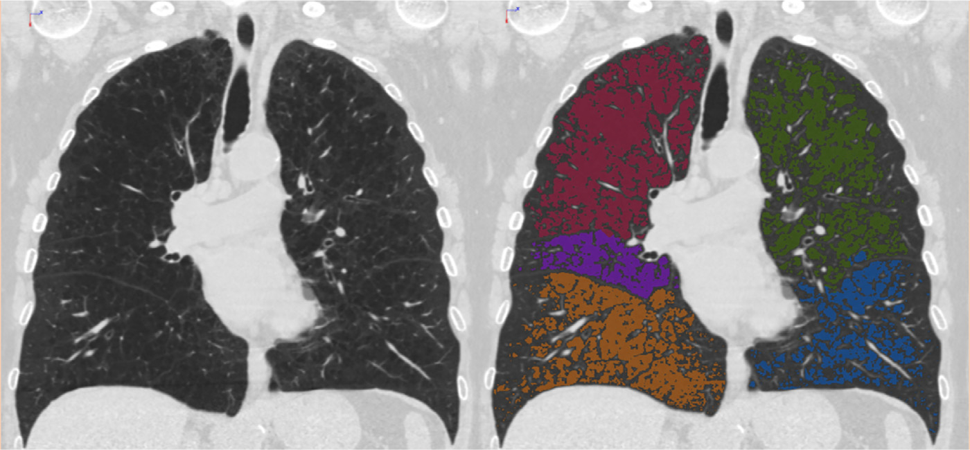
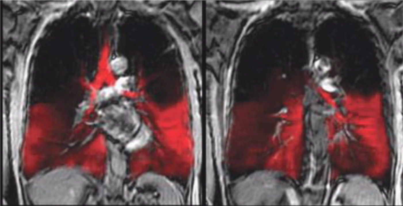
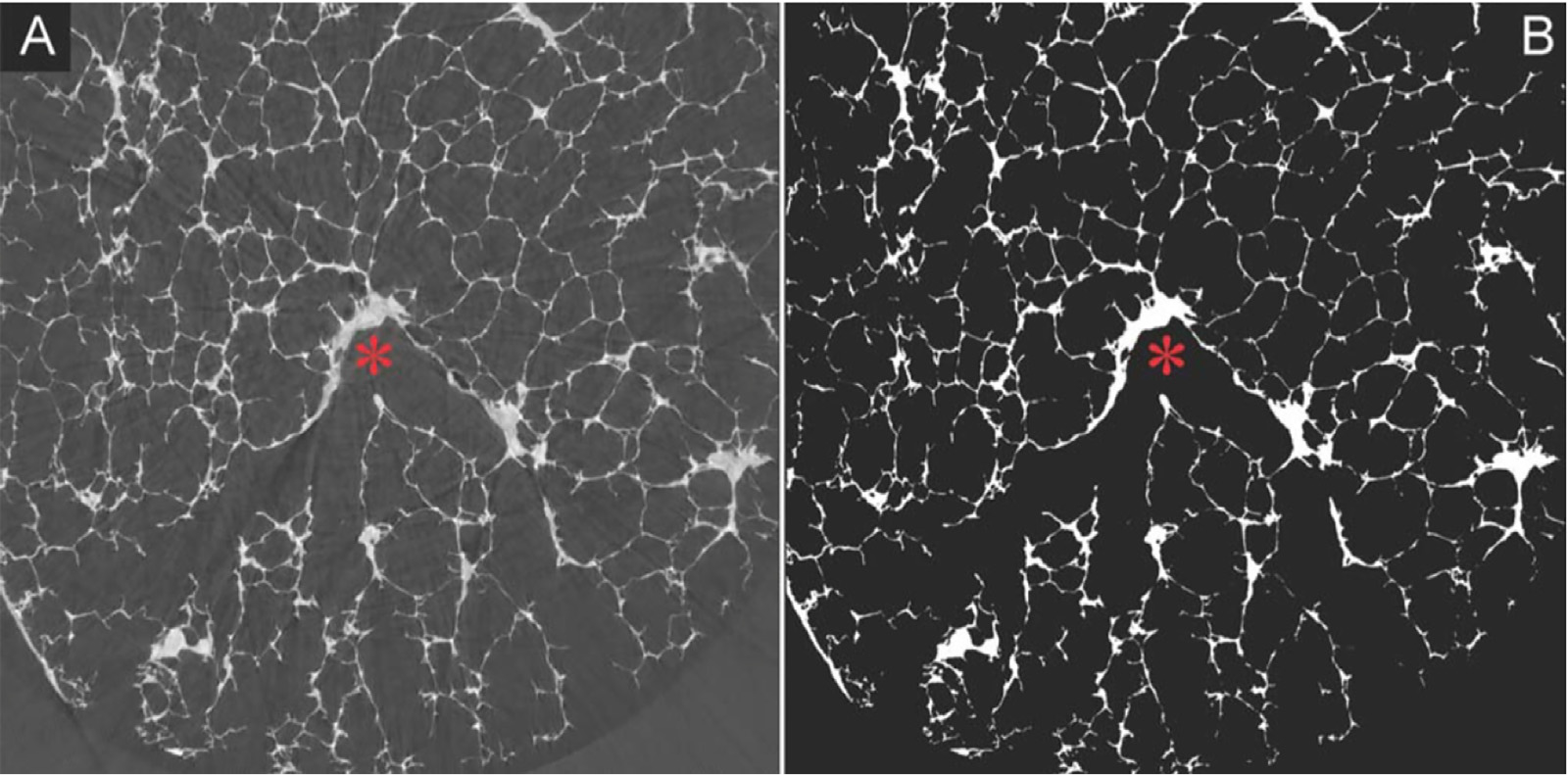
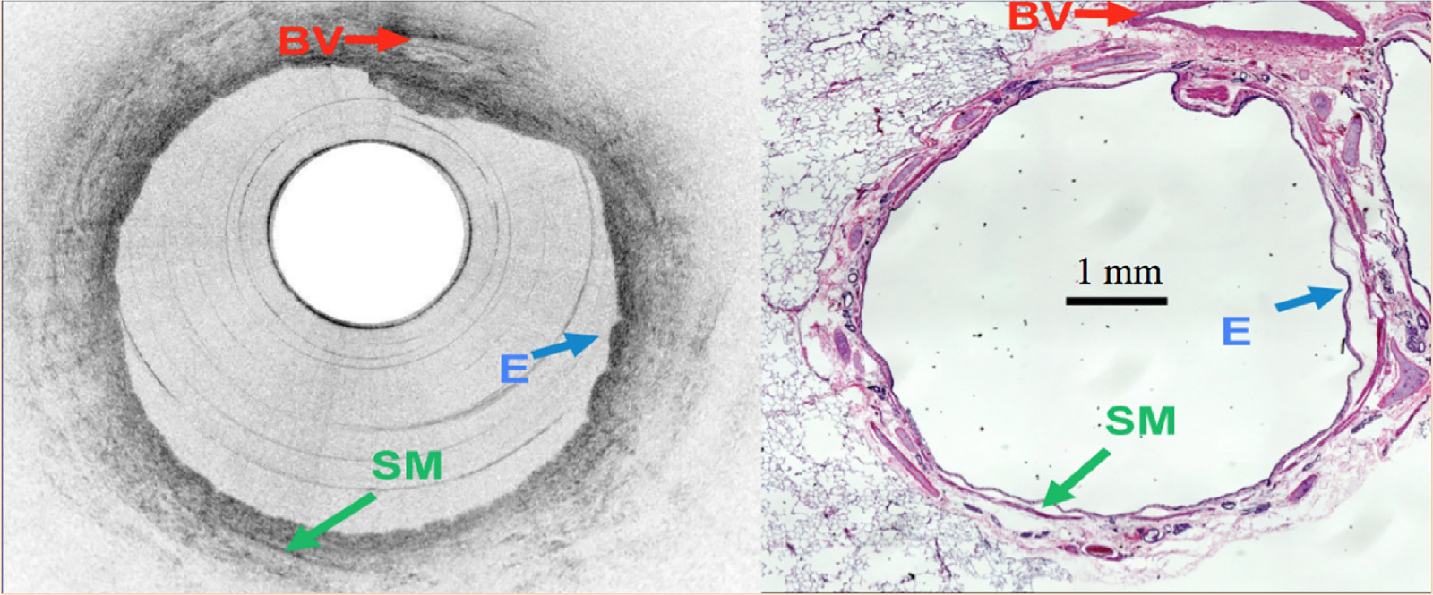
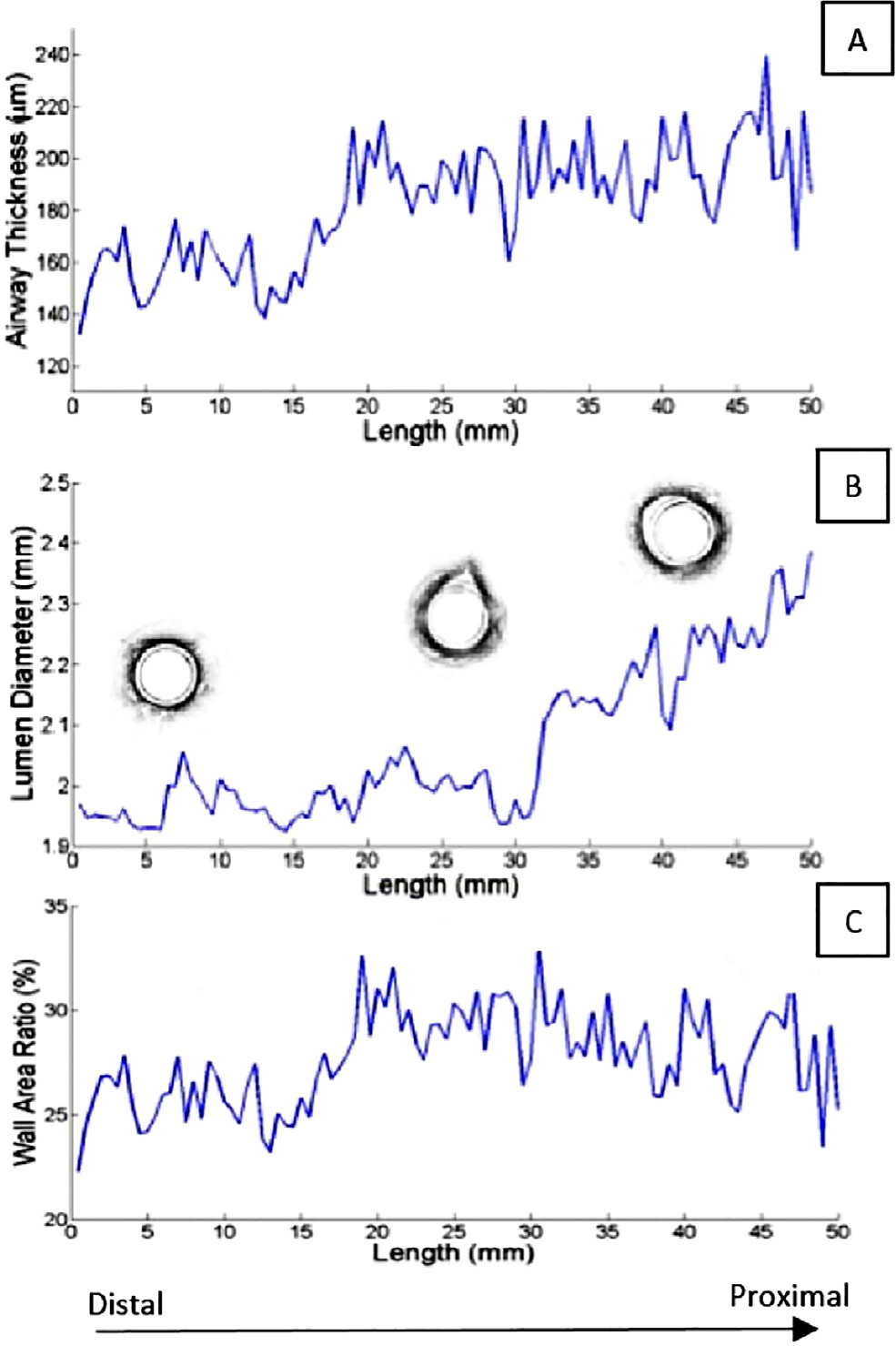
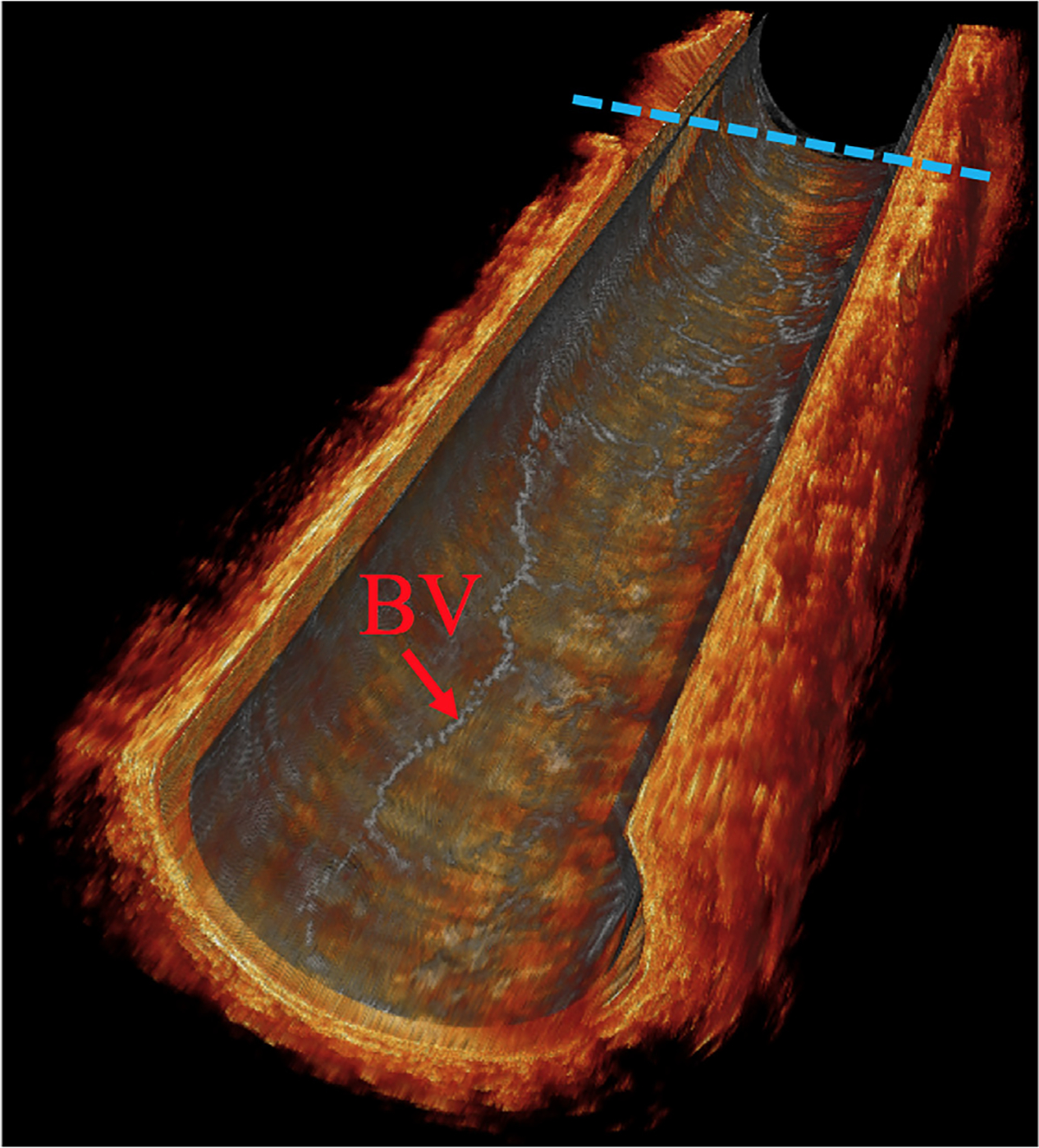
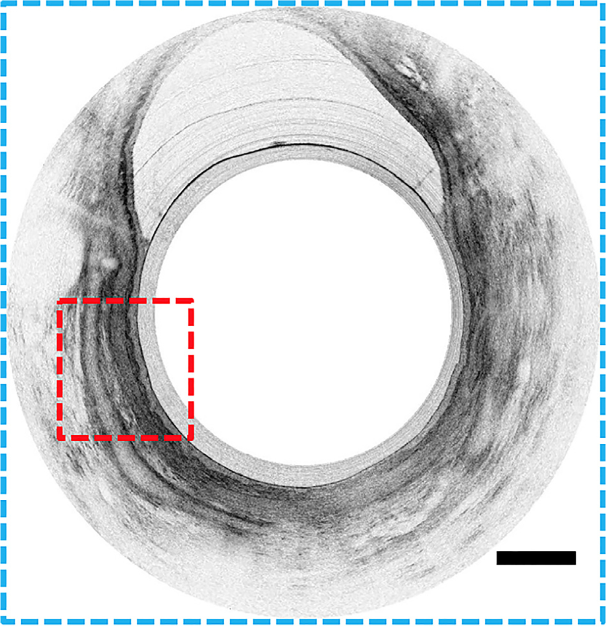
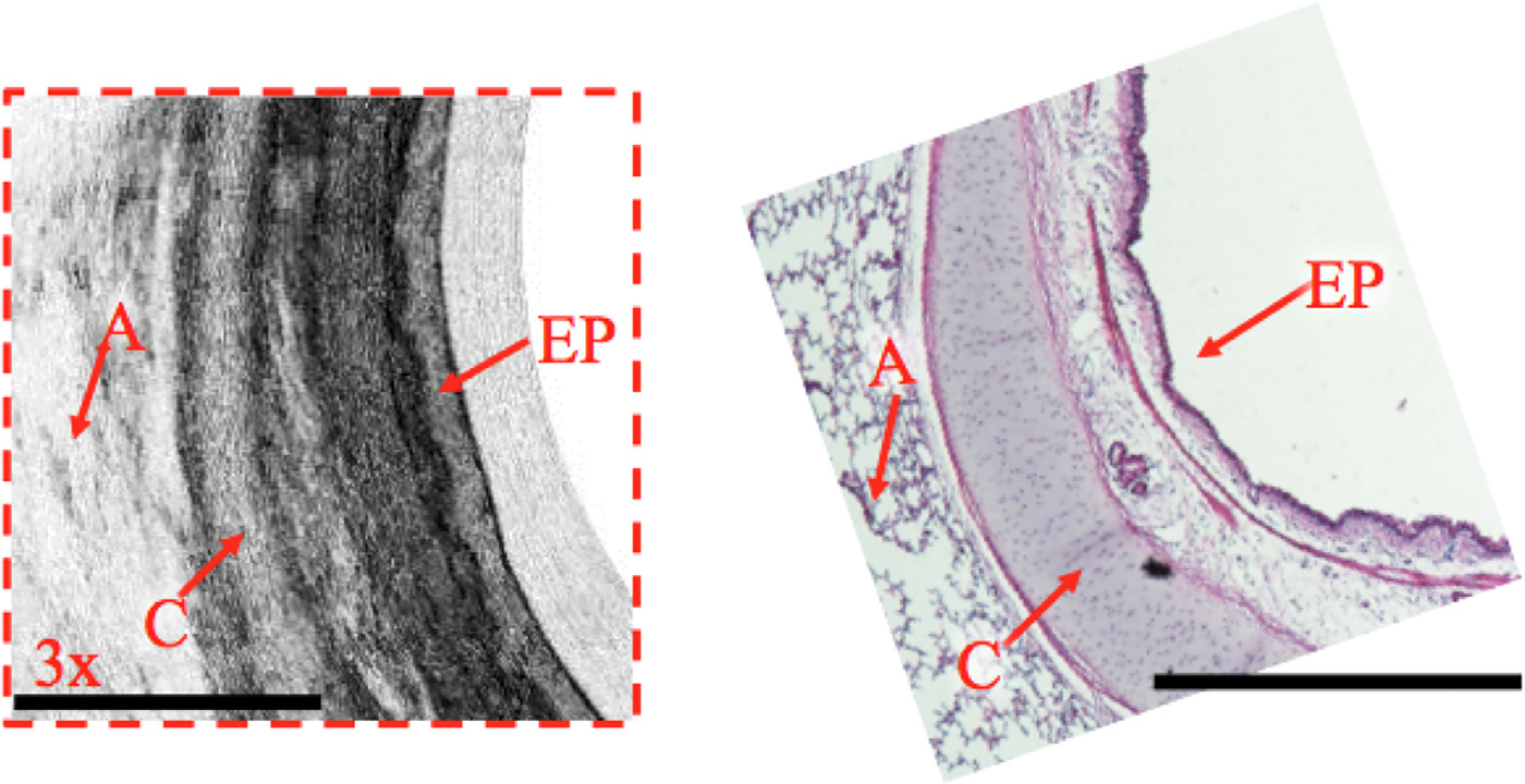
Results: Advanced imaging techniques such as Magnetic Resonance (MR) imaging using hyperoloarized noble gases, micro-Computed Tomography (micro-CT), and OCT aim to further our understanding of COPD. Lung densitometry can aid in identifying an exacerbation prone phenotype which may have implications for targeting specific therapies to these individuals. MR ventilation scans have the ability to provide a functional and regional distribution of airflow obstruction offering insight into the airway and parenchymal changes induced by COPD. Micro-CT gives a near microscopic view of the terminal bronchioles and alveoli permitting study of the microarchitecture of the lung ex vivo. Optical coherence tomography can visualize the microstructure of the airway walls (epithelium, smooth muscle, blood vessels, cartilage) permitting real time in vivo as well as longitudinal evaluation of airway changes in patients with COPD.
Conclusion: Advanced imaging techniques play a vital role in expanding our current understanding of COPD.
Keywords: COPD; Optical coherence tomography.
Copyright © 2018 The Association of University Radiologists. Published by Elsevier Inc. All rights reserved.
Figures
Similar articles
-
Analysis of airway pathology in COPD using a combination of computed tomography, micro-computed tomography and histology.Eur Respir J. 2018 Feb 14;51(2):1701245. doi: 10.1183/13993003.01245-2017. Print 2018 Feb. Eur Respir J. 2018. PMID: 29444912 Free PMC article.
-
Significances of spirometry and impulse oscillometry for detecting small airway disorders assessed with endobronchial optical coherence tomography in COPD.Int J Chron Obstruct Pulmon Dis. 2018 Oct 1;13:3031-3044. doi: 10.2147/COPD.S172639. eCollection 2018. Int J Chron Obstruct Pulmon Dis. 2018. PMID: 30319251 Free PMC article.
-
Recent advances in airway imaging using micro-computed tomography and computed tomography for chronic obstructive pulmonary disease.Korean J Intern Med. 2021 Nov;36(6):1294-1304. doi: 10.3904/kjim.2021.124. Epub 2021 Oct 6. Korean J Intern Med. 2021. PMID: 34607419 Free PMC article. Review.
-
Small airways disease in mild and moderate chronic obstructive pulmonary disease: a cross-sectional study.Lancet Respir Med. 2018 Aug;6(8):591-602. doi: 10.1016/S2213-2600(18)30196-6. Epub 2018 Jul 4. Lancet Respir Med. 2018. PMID: 30072106
-
Small airway obstruction in COPD: new insights based on micro-CT imaging and MRI imaging.Chest. 2013 May;143(5):1436-1443. doi: 10.1378/chest.12-1766. Chest. 2013. PMID: 23648907 Free PMC article. Review.
Cited by
-
A high-resolution 3D atlas of the spectrum of tuberculous and COVID-19 lung lesions.EMBO Mol Med. 2022 Nov 8;14(11):e16283. doi: 10.15252/emmm.202216283. Epub 2022 Oct 26. EMBO Mol Med. 2022. PMID: 36285507 Free PMC article.
-
Imaging in animal models: bridging experimental findings and human pathophysiology.Crit Care. 2025 Jul 26;29(1):327. doi: 10.1186/s13054-025-05574-6. Crit Care. 2025. PMID: 40713694 Free PMC article. Review.
-
Advances in Chronic Obstructive Pulmonary Disease Imaging.Barc Respir Netw Rev. 2020 May-Dec;6(2):128-143. doi: 10.23866/brnrev:2019-0023. Barc Respir Netw Rev. 2020. PMID: 33758787 Free PMC article.
-
Optimizing the Yield of Abnormal Preoperative Chest Radiographs in Elective Non-cardiothoracic Surgery: Development of a Risk Prediction Score and External Validation.World J Surg. 2023 Nov;47(11):2698-2707. doi: 10.1007/s00268-023-07146-7. Epub 2023 Sep 6. World J Surg. 2023. PMID: 37674044
-
Current Perspectives of Pharmacotherapies for COPD.Respir Care. 2023 Jul;68(7):927-938. doi: 10.4187/respcare.10952. Respir Care. 2023. PMID: 37353337 Free PMC article.
References
-
- Kurmi OP, Semple S, Simkhada P, et al. COPD and chronic bronchitis risk of indoor air pollution from solid fuel: a systematic review and meta-analysis. Thorax 2010; 65:221–228. - PubMed
-
- Global Strategy for the Diagnosis MaPoC, Global Initiative for Chronic Obstructive Lung Disease (GOLD) 2017. Available from: http://goldcopd.org/. Accessed July 19, 2017.
Publication types
MeSH terms
Grants and funding
LinkOut - more resources
Full Text Sources
Other Literature Sources
Medical
