. 2018 Sep 28;293(39):15095-15106.
doi: 10.1074/jbc.RA118.003579. Epub 2018 Aug 9.

Structural insight into proline cis/ trans isomerization of unfolded proteins catalyzed by the trigger factor chaperone

Affiliations

Structural insight into proline cis/ trans isomerization of unfolded proteins catalyzed by the trigger factor chaperone

Soichiro Kawagoe et al. J Biol Chem. .

Abstract

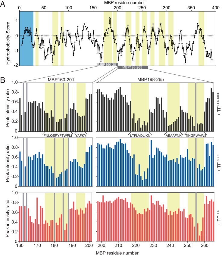
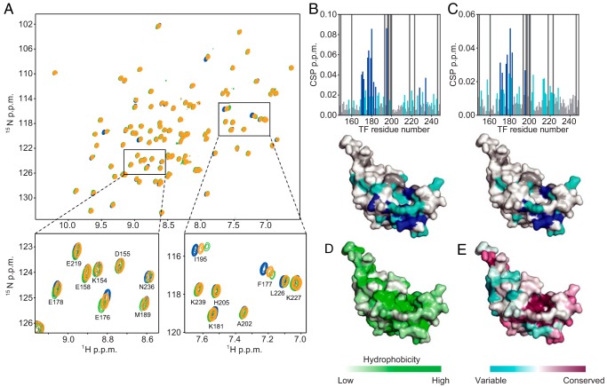
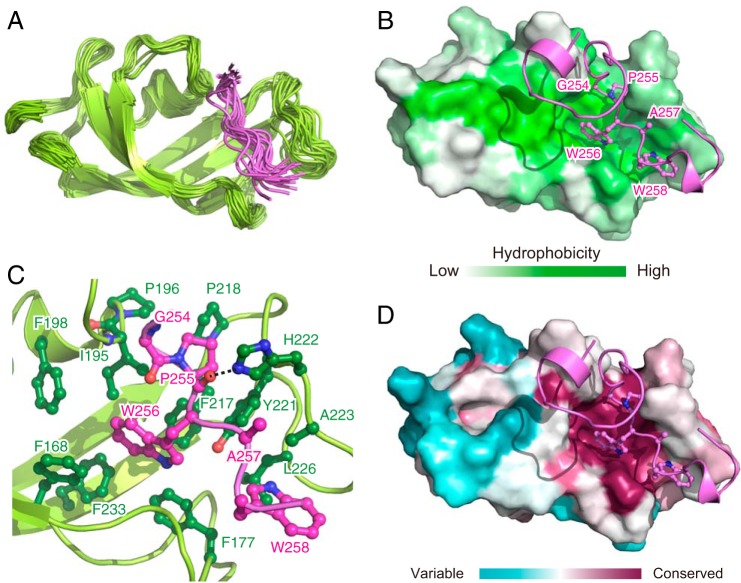
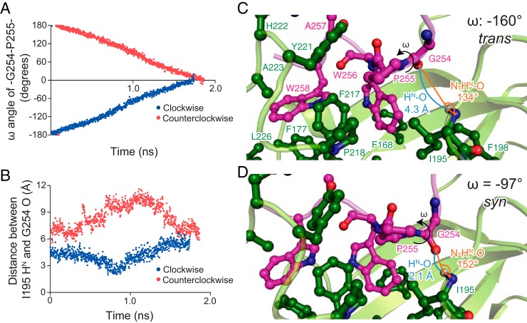
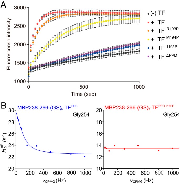
Molecular chaperones often possess functional modules that are specialized in assisting the formation of specific structural elements, such as a disulfide bridges and peptidyl-prolyl bonds in cis form, in the client protein. A ribosome-associated molecular chaperone trigger factor (TF), which has a peptidyl-prolyl cis/trans isomerase (PPIase) domain, acts as a highly efficient catalyst in the folding process limited by peptidyl-prolyl isomerization. Herein we report a study on the mechanism through which TF recognizes the proline residue in the unfolded client protein during the cis/trans isomerization process. The solution structure of TF in complex with the client protein showed that TF recognizes the proline-aromatic motif located in the hydrophobic stretch of the unfolded client protein through its conserved hydrophobic cleft, which suggests that TF preferentially accelerates the isomerization of the peptidyl-prolyl bond that is eventually folded into the core of the protein in its native fold. Molecular dynamics simulation revealed that TF exploits the backbone amide group of Ile195 to form an intermolecular hydrogen bond with the carbonyl oxygen of the amino acid residue preceding the proline residue at the transition state, which presumably stabilizes the transition state and thus accelerates the isomerization. The importance of such intermolecular hydrogen-bond formation during the catalysis was further corroborated by the activity assay and NMR relaxation analysis.

Keywords: hydrophobic interaction; molecular chaperone; molecular dynamics; nuclear magnetic resonance (NMR); peptidyl-prolyl isomerase domain; prolyl isomerase; protein folding; structure-function; trigger factor.


Conflict of interest statement

The authors declare that they have no conflicts of interest with the contents of this article

Figures

Figure 1.
Figure 1.
Recognition of unfolded MBP by TF. A, plot of the hydrophobicity score (Roseman algorithm, window = 9) of MBP as a function of its primary sequence. A hydrophobicity score higher than 0 denotes increased hydrophobicity. The regions recognized by TF as identified by NMR titration experiments are highlighted in yellow. The signal sequence is highlighted in blue. The MBP segments depicted in the B are indicated as a gray bar. B, plots of peak intensity change of MBP160–201 (left panels) and MBP198–265 (right panels) by the addition of TFPPD-SBD at a ratio of MBP:TFPPD-SBD 1:0.1 (top panels), TFSBD at a ratio of MBP:TFSBD 1:0.2 (middle panels), and TFPPD at a ratio of MBP:TFPPD 1:0.5 (bottom panels), as a function of the primary sequences of the MBP peptides. The regions recognized by TF are highlighted in yellow and labeled with the primary sequences. The proline residue and unassigned residue are indicated by gray bar.
Figure 2.
Figure 2.
The substrate-binding sites on TFPPD. A, 1H-15N HSQC spectra of TFPPD in the absence (blue) and presence of MBP160–201 (green) or MBP198–265 (orange). The spectra were recorded with several titration points, TFPPD:MBP 1:0.1, 1:0.2, 1:0.5, 1:1, and 1:2, but only the spectra at 1:2 ratio are shown for clarity. The regions indicated by boxes in the top panel are expanded in the bottom panels with resonance assignments. B and C, chemical shift perturbations of amide moieties of TFPPD upon binding to MBP160–201 (B) or MBP198–265 (C), plotted as a function of the residue number of TFPPD. Proline and unassigned residues are indicated by bars colored dark gray. The lower panels show chemical shift perturbation mapping on the structure of TFPPD (PDB code 1W26). The residues with perturbations larger than the threshold values of 0.015 and 0.025 ppm are colored light blue and dark blue, respectively. Proline and unassigned residues are colored dark gray. D, mapping of the hydrophobicity on the structure of TFPPD. E, TFPPD sequence conservation mapped on the structure of TFPPD.
Figure 3.
Figure 3.
NMR structure of TFPPD in complex with MBP. A, superimposition of 20 structures of TFPPD in complex with MBP 238–266. For MBP, only the converged region, Asn253–Ser259, is shown for clarity. TFPPD and MBP are shown in green and pink ribbons, respectively. B and D, the lowest-energy structure of MBP–TFPPD complex. TFPPD is shown as a solvent-exposed surface to which the hydrophobicity (B) or the conservation of the amino acid residues (D) is mapped. MBP is shown as pink ribbons. The MBP residues that directly interact with TFPPD are drawn in a ball-and-stick representation. C, close-up view of the lowest-energy structure of MBP–TFPPD complex. TFPPD and MBP are shown as green and pink ribbons, respectively. The residues of TFPPD and MBP involved in the interaction are drawn in a ball-and-stick representation and colored green and pink, respectively.
Figure 4.
Figure 4.
Molecular dynamics simulation of cis/trans isomerization by TFPPD. A, plots of the ω dihedral angle for the peptidyl–prolyl bond between MBP Gly254 and Pro255. The simulation was started from the NMR structure of MBP–TFPPD complex in which the ω angle for the peptidyl–prolyl bond between MBP Gly254 and Pro255 is −174° (trans state). Because of the ω angle constraint at a rate of 0.2° per 2 ps, the ω angle changed linearly as a function of time. B, plots of the distance between the HN atom of TF Ile195 and the carbonyl oxygen of MBP Gly254 as a function of time when the ω angle was rotated clockwise (blue) or counterclockwise (red). When the ω angle was rotated in clockwise direction, the two atoms approached each other, and the distance became shorter than 2.5 Å at ∼0.8 ns. As seen in A, the ω dihedral angle at 0.8 ns was at approximately −90° (syn state). C and D, snap shots of the cis/trans isomerization at the trans state (0.200 ns) (C) and the syn state (0.794 ns) (D). The intermolecular hydrogen bond between backbone amide group of TFPPD Ile195 and the backbone carbonyl oxygen of MBP Gly254 is formed at the syn state, which tethers the peptidyl–prolyl bond closer onto the hydrophobic cleft formed by TFPPD Ile195, Phe217, and Pro218.
Figure 5.
Figure 5.
PPIase activity of TF and TF mutants. A, evaluation of PPIase activity of TF and TF variants by refolding assay of RCM-RNase T1. Refolding of RCM-RNase T1 in the absence and presence of TF or TF mutants was monitored by increase of intrinsic tryptophan fluorescence at 320 nm after excitation at 268 nm. The experiments were performed at 15 °C. Because of the complex process of the refolding of RCM-RNase T1 (37), refolding rates were not extracted. B, evaluation of PPIase activity of TF and TF variants by NMR relaxation dispersion experiments. The chemical exchange in MBP Gly254 coupled with cis/trans isomerization of peptidyl–prolyl bond between MBP Gly254 and Pro255 in complex with TFPPD (left panel) or TFPPD, I195P (right panel) was monitored.
Figure 6.
Figure 6.
Schematic representation of peptidyl–prolyl cis/trans isomerization by TFPPD. TFPPD and the substrate protein are show in green and magenta, respectively. TFPPD captures the proline-aromatic motif in the trans form located in the hydrophobic stretches of the substrate protein. The interaction is mainly mediated by hydrophobic interactions with the conserved hydrophobic cleft of TFPPD that is decollated by TF His222 forming a hydrogen bond with the backbone carbonyl oxygen of the proline residue in the substrate. When the peptidyl–prolyl bond rotates to syn form, the carbonyl oxygen of the amino acid residue preceding the proline residue forms an intermolecular hydrogen bond with backbone amide group of TFPPD Ile195, which tethers the peptidyl–prolyl bond onto the hydrophobic surface of the TFPPD. Both the intermolecular hydrogen bond and the hydrophobic environment are important for efficient cis/trans isomerization. When the peptidyl–prolyl bond rotates to cis form, the intermolecular hydrogen bond, and consequently the close hydrophobic contact are released.

References

    1. Dobson C. M. (2003) Protein folding and misfolding. Nature 426, 884–890 10.1038/nature02261 - DOI - PubMed
    1. Hartl F. U., Bracher A., and Hayer-Hartl M. (2011) Molecular chaperones in protein folding and proteostasis. Nature 475, 324–332 10.1038/nature10317 - DOI - PubMed
    1. Baneyx F., and Mujacic M. (2004) Recombinant protein folding and misfolding in Escherichia coli. Nat. Biotechnol. 22, 1399–1408 10.1038/nbt1029 - DOI - PubMed
    1. Wedemeyer W. J., Welker E., and Scheraga H. A. (2002) Proline cis-trans isomerization and protein folding. Biochemistry 41, 14637–14644 10.1021/bi020574b - DOI - PubMed
    1. Wedemeyer W. J., Welker E., Narayan M., and Scheraga H. A. (2000) Disulfide bonds and protein folding. Biochemistry 39, 4207–4216 10.1021/bi992922o - DOI - PubMed

Publication types

LinkOut - more resources