. 2018 Aug 10;361(6402):594-599.
doi: 10.1126/science.aat1699.

Single-cell transcriptomes from human kidneys reveal the cellular identity of renal tumors

Affiliations

Single-cell transcriptomes from human kidneys reveal the cellular identity of renal tumors

Matthew D Young et al. Science. .

Abstract

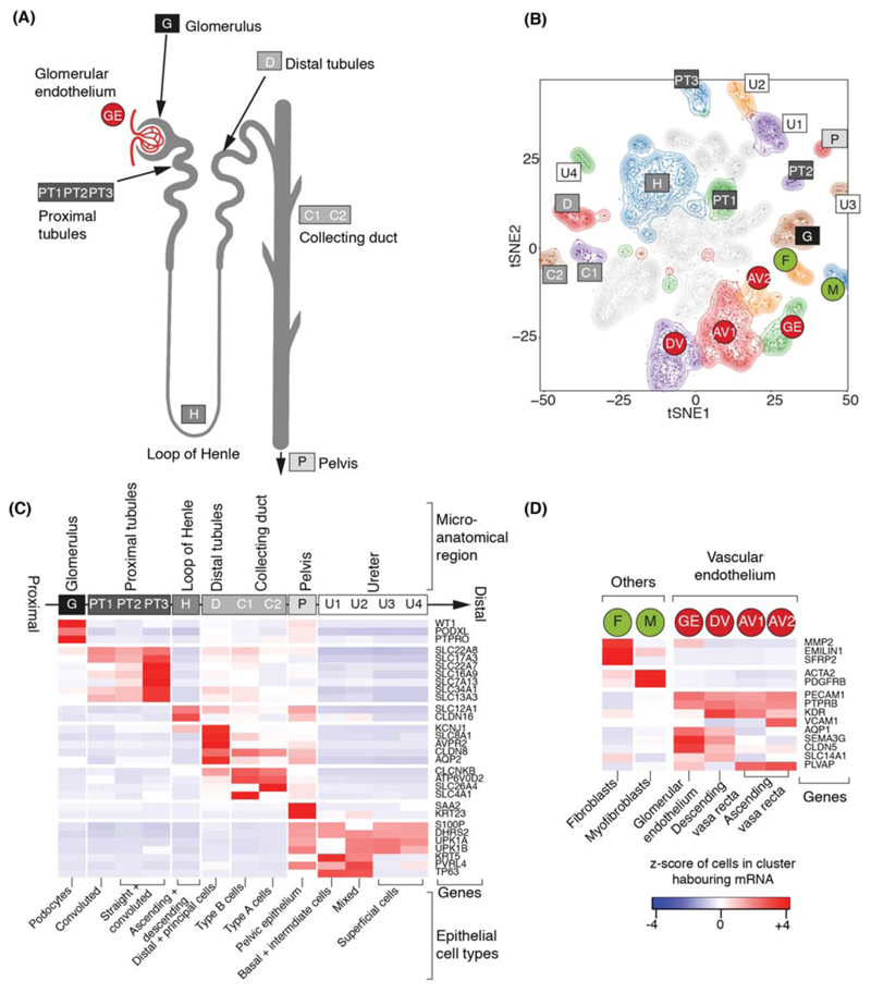
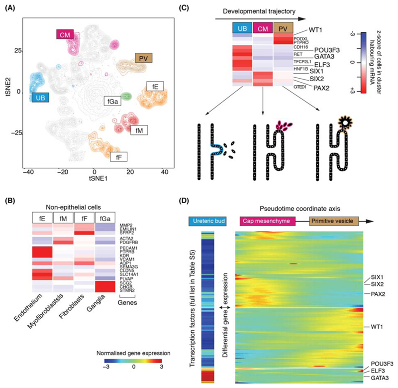
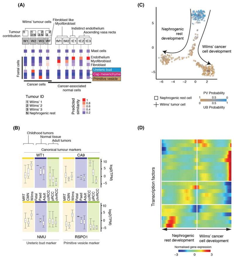
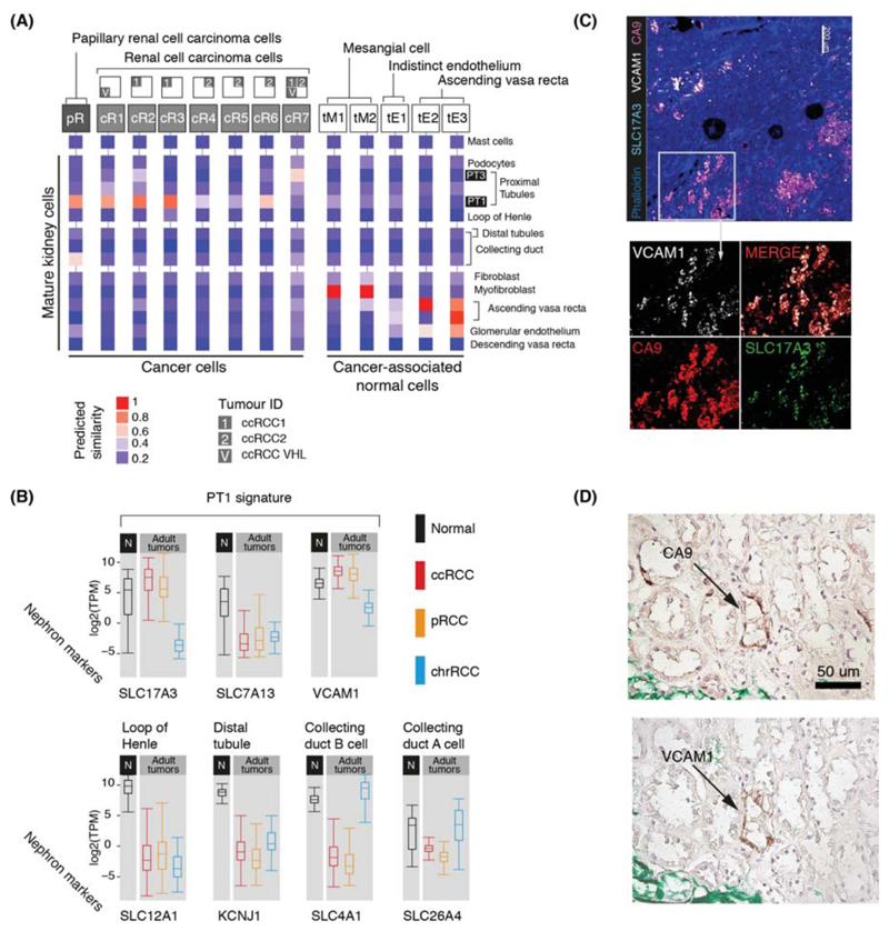
Messenger RNA encodes cellular function and phenotype. In the context of human cancer, it defines the identities of malignant cells and the diversity of tumor tissue. We studied 72,501 single-cell transcriptomes of human renal tumors and normal tissue from fetal, pediatric, and adult kidneys. We matched childhood Wilms tumor with specific fetal cell types, thus providing evidence for the hypothesis that Wilms tumor cells are aberrant fetal cells. In adult renal cell carcinoma, we identified a canonical cancer transcriptome that matched a little-known subtype of proximal convoluted tubular cell. Analyses of the tumor composition defined cancer-associated normal cells and delineated a complex vascular endothelial growth factor (VEGF) signaling circuit. Our findings reveal the precise cellular identities and compositions of human kidney tumors.


Conflict of interest statement

Competing interests: None.

Figures

Figure 1
Figure 1. Canonical cell types in normal human kidneys.
(A) Illustration of nephron anatomy with cell clusters marked. (B) tSNE representation of 8,707 normal epithelial and vascular cells. Clusters are colored, uniquely labelled and emphasized with density contours. Ambiguous clusters are de-emphasized and fully shown in Fig. S2. (C) Expression of canonical nephron specific genes (Table S3) in clusters from (A). Colors give the fraction of cells expressing each gene in a cluster, scaled to have mean 0 and standard deviation 1 across all clusters. (D) Expression of clusters in (A) not shown in (C) and their canonical genes.
Figure 2
Figure 2. Fetal cell types and nephrogenesis.
(A) tSNE representation of 4,858 fetal epithelial and vascular cells, colored and labelled as in Fig. 1A. (B) Expression of markers of clusters in (A), colored as in Fig. 1C. (C) Expression of nephrogenesis markers from clusters in (A) with illustration of nephron development. Formation of nephrons emanates from the ureteric bud, which induces condensation of the overlying mesenchyme into the cap mesenchyme. The cap mesenchyme then forms the primitive vesicle, the precursor of the glomerulus. The tubular system grows out from both ends of the fetal nephron: ureteric bud and primitive vesicle. (D) The expression of transcription factor which vary significantly (p<0.01; likelihood ratio test) along the pseudo-time trajectory defined using the CM and PV cells from (C), or differentially expressed between UB versus CM and PV. UB expression is shown in a separated block on the left. Within the right block, pseudo-time increases from left to right and rows are clustered and grouped by hierarchical clustering with canonical transcription factors of nephrogenesis highlighted (see Table S6).
Figure 3
Figure 3. Matching childhood tumors with normal fetal cells.
(A) Similarity of Wilms’ tumor and cancer-associated normal cells to the reference fetal kidney map (Fig. 2A), with mast cells added as a negative control. Square boxes indicate sample contribution. Colors represent the probability that the cluster identified in the column header is “similar” to the fetal cluster identified by the row label (2). (B) Expression of canonical tumor markers and representative UB and PV specific genes (Table S8) in RNA-seq from childhood cancers (yellow), normal tissue (blue) or adult cancers (green). MRT: malignant rhabdoid tumor; CMN: congenital mesoblastic nephroma. As positive controls, canonical tumor markers are shown: WT1 (Wilms’); CA9 (ccRCC). (C) Pseudo-time trajectory of all Wilms tumor and nephrogenic rest cell. Color indicates similarity of each cell to the PV or UB fetal population. Jitter has been added to each point’s position with the original position plotted underneath in black (2). (D) Transcription factors identified as varying significantly along the pseudo-time trajectory in (C). The center of the heatmap corresponds to the cells at the top of (C) and then proceeding left/right along the arrows shown in (C).
Figure 4
Figure 4. Matching adult tumors with normal mature kidney cells.
(A) Similarity of adult cancer and cancer-associated normal cells to the mature kidney reference map (Fig. 1B), with mast cells added as a negative control. Square boxes indicate sample contribution. Colors represent the probability that the cluster identified in the column header is “similar” to the normal cluster identified by the row label (2). (B) Expression of nephron specific genes in bulk RNA-seq as in Fig. 3B. pRCC samples are both type 1 and 2. (C) Confocal microscopy showing co-localization of PT1 markers (VCAM1, SLC17A3) in ccRCC cells (CA9). (D) Staining of a proximal tubular ccRCC precursor lesion (CA9) for the PT1 marker, VCAM1.

References

    1. Ziegenhain C, et al. Comparative Analysis of Single-Cell RNA Sequencing Methods. Molecular cell. 2017;65:631–643.e634. - PubMed
    1. Supplementary Methods

    1. Pritchard-Jones K, et al. Omission of doxorubicin from the treatment of stage II-III, intermediate-risk Wilms' tumour (SIOP WT 2001): an open-label, non-inferiority, randomised controlled trial. Lancet (London, England) 2015;386:1156–1164. - PubMed
    1. Perlman EJ, et al. MLLT1 YEATS domain mutations in clinically distinctive Favourable Histology Wilms tumours. Nature communications. 2015;6 10013. - PMC - PubMed
    1. Gadd S, et al. A Children's Oncology Group and TARGET initiative exploring the genetic landscape of Wilms tumor. Nature genetics. 2017;49:1487–1494. - PMC - PubMed

Publication types