. 2018 Aug 10;361(6402):607-612.
doi: 10.1126/science.aah4065.

Apoptosis propagates through the cytoplasm as trigger waves

Affiliations

Apoptosis propagates through the cytoplasm as trigger waves

Xianrui Cheng et al. Science. .

Abstract

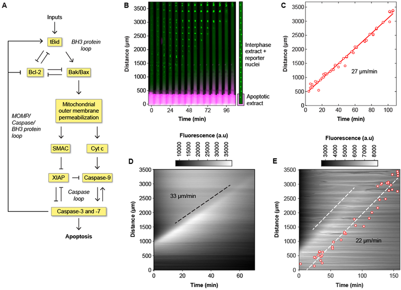
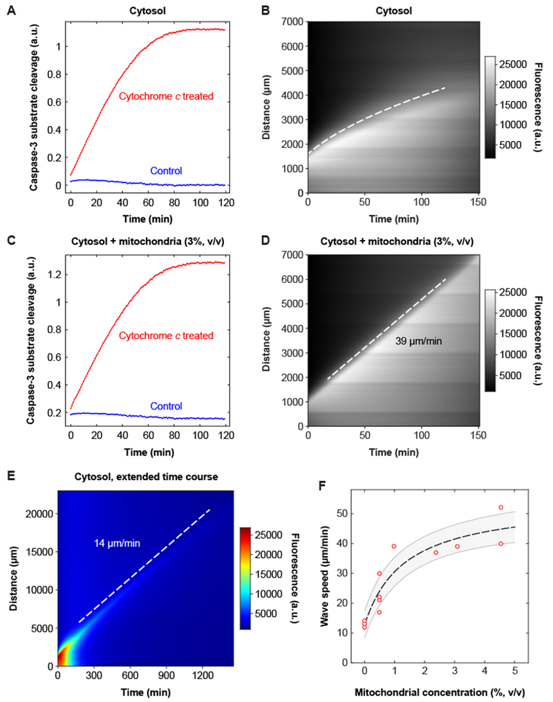
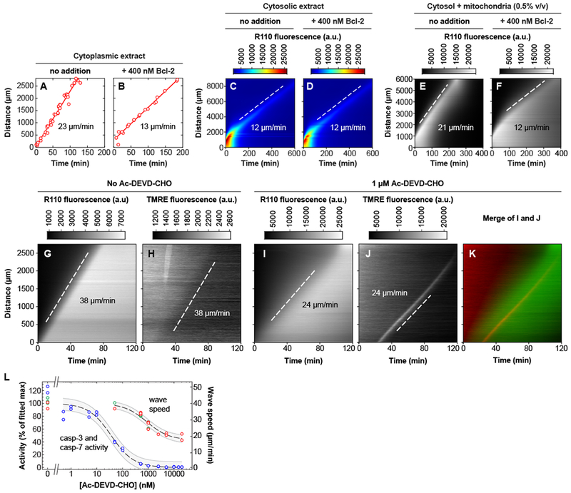
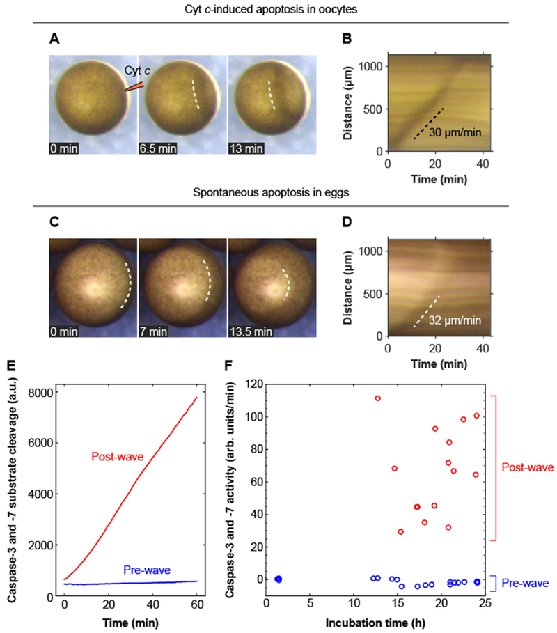
Apoptosis is an evolutionarily conserved form of programmed cell death critical for development and tissue homeostasis in animals. The apoptotic control network includes several positive feedback loops that may allow apoptosis to spread through the cytoplasm in self-regenerating trigger waves. We tested this possibility in cell-free Xenopus laevis egg extracts and observed apoptotic trigger waves with speeds of ~30 micrometers per minute. Fractionation and inhibitor studies implicated multiple feedback loops in generating the waves. Apoptotic oocytes and eggs exhibited surface waves with speeds of ~30 micrometers per minute, which were tightly correlated with caspase activation. Thus, apoptosis spreads through trigger waves in both extracts and intact cells. Our findings show how apoptosis can spread over large distances within a cell and emphasize the general importance of trigger waves in cell signaling.


Conflict of interest statement

Competing interests:None declared.

Figures

Fig. 1.
Fig. 1.. Apoptosis propagates in interphase-arrested cytoplasmic Xenopus laevis egg extracts by trigger waves.
(A) The control circuit for apoptosis, adapted from previously published work (–28). (B) Time-lapse montage of GST-GFP-NLS filled nuclei (green) in a cytoplasmic extract in a Teflon tube with its lower end in contact with an apoptotic extract reservoir. The extract in the reservoir is marked with 10-kD dextran-Texas Red dye, shown in magenta. A time-lapse video of this experiment can be found in movie S1. (C) Correlation between timing and position of nuclear disappearance for the experiment shown in panel (B). The solid red line is a linear least squares fit to the data. The propagation speed (the slope of the fitted line) is 27 μm/min. (D) Kymograph showing the spatial propagation of caspase-3 and/or caspase-7 activity (indicated by R110 fluorescence) in a crude cytoplasmic extract. The black dashed line was manually fitted to the fluorescence front, and it yielded a propagation speed of 33 μm/min. (E) R110 fluorescence and nuclear disappearance, using GST-mCherry-NLS as a nuclear marker, measured in the same tube. Note that the presence of the nuclei makes the R110 fluorescence less diffuse than it is in panel D. One dashed line is manually fitted to the fluorescence front, and the other is a least-squares fit to the nuclear data. The propagation speed was 22 μm/min for both waves.
Fig. 2
Fig. 2. The speed of the apoptotic trigger wave depends upon the concentration of mitochondria.
(A, B) Cytosolic extract. Panel A shows activation of caspase-3 and/or caspase-7, by a chromogenic assay, and panel B is a kymograph showing diffusive spread of caspase-3/7 activation, as read out by the Z-DEVD-R110 probe, over this time scale and distance scale. The dashed curve was obtained by defining an equal-fluorescence isocline, replotting the isocline on a distance squared vs. time plot, carrying out a linear least squares fit, and then transforming the fitted line for plotting on the original distance vs. time axes. Further details are provided in figure S4. (C, D) Reconstituted extract from the same experiment. Panel C shows activation of caspase-3 and/or −7 and panel D is a kymograph, here showing trigger wave propagation of caspase activation. (E) Slow apoptotic trigger waves detected in cytosolic extracts. This kymograph shows a cytosolic extract incubated for 24 h in a 3 cm tube. R110 fluorescence is displayed here on a heat map scale to allow the shape of the wave front to be appreciated both early and late in the time course. (F) Wave speed as a function of mitochondrial concentration. Data are from 18 tubes and 8 independent experiments (there are 9 overlapping data points with 0% mitochondria). The dashed line is a Michaelian dose-response curve given by the equation y=y0+ymaxxK+x; the fitted parameters are y0 = 13.0±1.4 μm/min, ymax = 41.3±6.1 μm/min, and K = 1.3±0.6% (means ± S.E.), and r2 = 0.98. The grey region is the standard error (68.2% confidence interval) single prediction confidence band calculated with Mathematica 11.1.1.
Fig. 3.
Fig. 3.. Both GST-Bcl-2 addition and inhibition of caspase-3 and caspase-7 affect the speed of the trigger waves.
(A, B) GST-Bcl-2 reduces the speed of trigger waves in cytoplasmic extracts. Apoptosis was monitored by the disappearance of reporter nuclei containing GST-mCherry-NLS. (C, D) GST-Bcl-2 has no effect on the speed of trigger waves in cytosolic extracts. Apoptosis was detected with Z-DEVD-R110, whose fluorescence can be activated by caspase-3 or caspase-7. The pseudocolor heat map scale allows both the initial and final shapes of the wave front to be discerned. (E, F) GST-Bcl-2 reduces the speed of trigger waves in extracts reconstituted with cytosol and mitochondria (0.5% v/v). (G-K) Inhibition of caspase-3 and −7 slows trigger waves. The reporters were Z-DEVD-R110 and the mitochondrial probe TMRE. (G, H) Cytosolic extract reconstituted with mitochondria (2.4% v/v). (I-K) A reconstituted extract treated with the caspase inhibitor Ac-DEVD-CHO (1 μM). In experiments with higher concentrations of Ac-DEVD-CHO, a brief (~10 min) period when the TMRE wave appeared to be parabolic rather than linear was seen (figure S9). (L) Inhibition of caspase-3 and −7 activity, and slowing of trigger waves, as a function of Ac-DEVD-CHO concentration in cytosolic extracts reconstituted with mitochondria (2.4% v/v). Blue data points show caspase activities, measured in extracts diluted 1:20. Green data points indicate wave speeds estimated from Z-DEVD-R110 fluorescence, and red points from TMRE fluorescence. The curves are fits to a Michaelian inhibition function, y=y0+(ymaxy0)KK+x, where y is the caspase activity or trigger wave speed, x is the Ac-DEVD-CHO concentration, and y0, ymax, and K are parameters determined by fitting the data. For the caspase activity curve, the fitted parameter values were: y0 = 0.1 ± 2.5; ymax = 100 ± 3; and K = 36 ± 8 nM (mean ± S.E.). For the wave speed curve, the fitted parameter values were y0 = 17 ± 1 μm/min; ymax = 40 ± 1 μm/min; and K = 856 ± 160 nM (mean ± S.E.). The shaded regions are the standard error (68.2% confidence interval) single prediction confidence bands calculated with Mathematica 11.1.1.
Fig. 4.
Fig. 4.. Apoptotic trigger waves in intact oocytes and eggs.
(A, B) Injection of immature Stage VI oocytes with cytochrome c (10 nl of 1 mg/ml cytochrome c) causes a wave of pigmentation changes to spread from the injection site to the opposite side of the oocyte. Panel A shows one example of this surface wave in montage form; panel B shows the kymograph. Two other examples of these waves are shown in movie S9. (C, D) Surface waves occur in spontaneously dying eggs. Panel C shows an example of this wave in montage form, and panel D shows the kymograph. (E) Caspase-3 and/or caspase-7 assays for one pre-wave and one post-wave egg. (F) Caspase-3 and/or −7 activities for eggs pre- and post-wave. The data are from 19 pre-wave eggs and 15 post-wave eggs.

References

    1. Iguchi S, Iwasaki T, Fukami Y, Tokmakov AA, Unlaid Xenopus eggs degrade by apoptosis in the genital tract. BMC Cell Biol. 14, 11 (March 03, 2013). - PMC - PubMed
    1. Newmeyer DD, Farschon DM, Reed JC, Cell-free apoptosis in Xenopus egg extracts: inhibition by Bcl-2 and requirement for an organelle fraction enriched in mitochondria. Cell 79, 353–364 (1994). - PubMed
    1. Deming P, Kornbluth S, Study of apoptosis in vitro using the Xenopus egg extract reconstitution system. Meth. Mol. Biol 322, 379–393 (2006). - PubMed
    1. Tyson JJ, Keener JP, Singular perturbation theory of traveling waves in excitable media (a review). Physica D 32, 327–361 (1988).
    1. Winfree AT, Two kinds of wave in an oscillating chemical solution. Faraday Symp. Chem. Soc 9, 38–46 (1974).

Publication types

LinkOut - more resources