. 2018 Jul 15;10(7):2055-2067.
eCollection 2018.

The molecular mechanism and clinical significance of LDHA in HER2-mediated progression of gastric cancer

Affiliations

The molecular mechanism and clinical significance of LDHA in HER2-mediated progression of gastric cancer

Weiyou Zhu et al. Am J Transl Res. .

Abstract

Objective: The use of human epidermal growth factor receptor-2 (HER2) as a biomarker for gastric cancer (GC) has greatly helped some patients receive benefit from HER2-targeted therapy. However, the correlation between HER2 and other biochemical markers is unclear. The aim of this study was to examine the relationship between HER2 and lactate dehydrogenase A (LDHA) in GC tissues and GC cells.

Methods: The correlation between clinicopathological features and HER2 was analyzed in 179 cases of GC. The expression of HER2 and LDHA was examined by immunohistochemical staining in 12 pairs of GC tissues and by western blotting in seven pairs of fresh GC tissues and adjacent normal tissues. Wound healing, transwell migration assay, quantitative real-time reverse-transcription polymerase chain reaction (RT-PCR), and LDH activity assays were performed with GC cells.

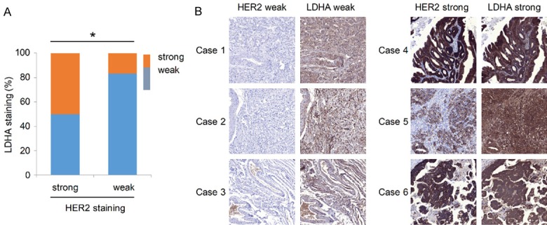
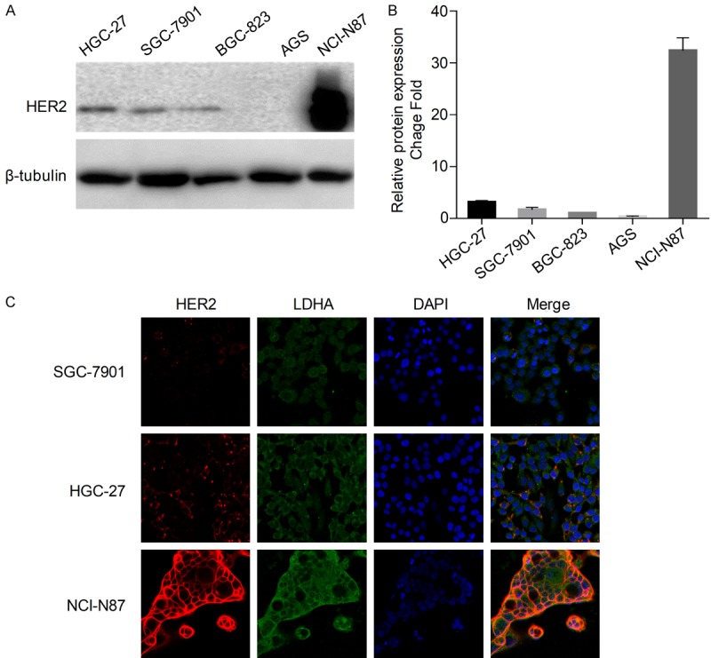
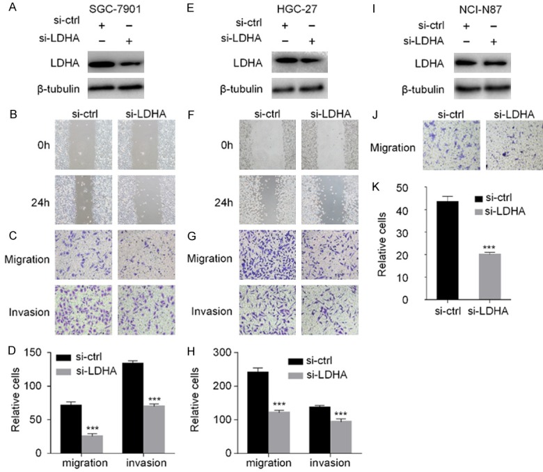
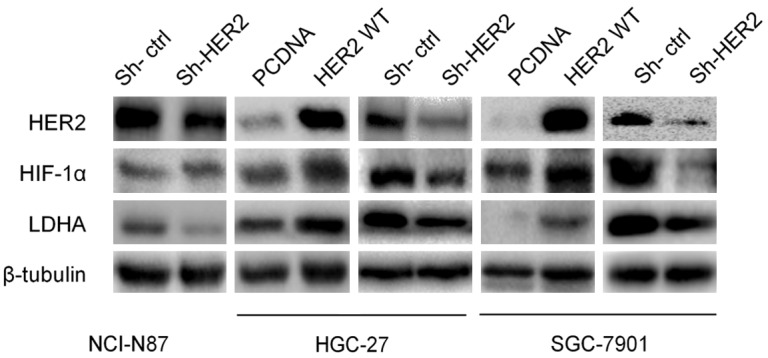
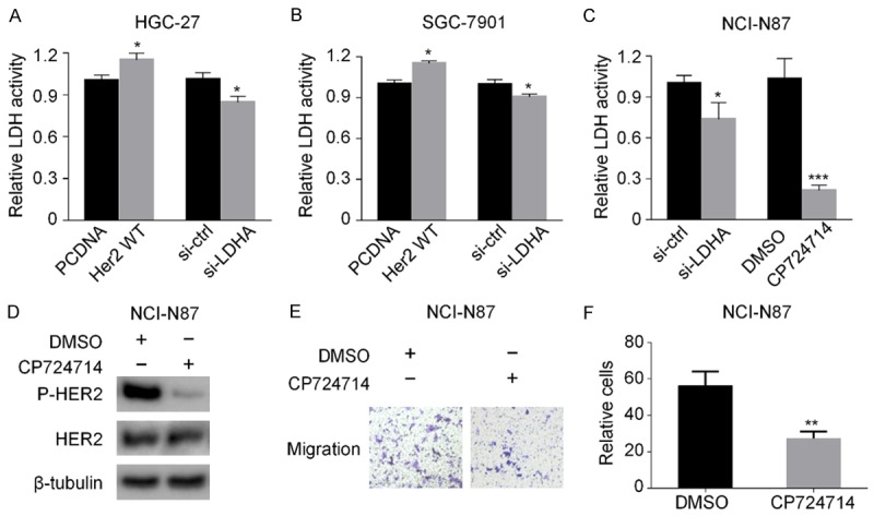
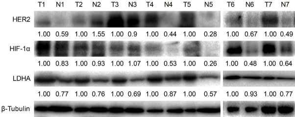
Results: HER2 expression and serum LDH levels were closely correlated (P = 0.027) in 179 GC patient cases. Immunohistochemical staining demonstrated a positive correlation between HER2 and LDHA in 12 pairs of GC tissues (P = 0.0308). Knocking down LDHA suppressed cell migration and invasion in GC cells. In addition, HER2 positively regulated hypoxia-inducible factor-1α (HIF-1α) and LDHA. Furthermore, the expressions of HER2, HIF-1α, and LDHA were consistent in 5/7 pairs of fresh GC tissues and adjacent normal tissues as well as in GC cell lines.

Conclusions: The HER2-HIF-1α-LDHA axis may serve as the basis for new methods and strategies for the treatment of GC.

Keywords: Gastric cancer; HER2; HIF-1α; LDHA; migration.


Conflict of interest statement

None.

Figures

Figure 1
Figure 1
HER2 and LDHA expressions in GC tissues. A. LDHA and HER2 expressions in 12 pairs of paraffin-embedded GC tissues. Key: (*) P < 0.05 (Spearman rank test). B. Typical immunohistochemical staining of HER2 and LDHA in six patients (cases 1-3: HER2 and LDHA both weak; cases 4-6: HER2 and LDHA both strong).
Figure 2
Figure 2
HER2 expression in GC cell lines. A. Detection of HER2 and β-tubulin by western blot analysis of cells from three metastatic GC cell lines (HGC-27, SGC-7901, NCI-N87) and two primary GC cell lines (BGC-823, AGS). B. HER2 bands were normalized to β-tubulin. The data are expressed as the means ± SD from three independent experiments. C. Immunofluorescence imaging of HER2 (red), LDHA (green), and nucleus labeled as DAPI (blue), and the co-localization of the three signals (merge) in SGC-7901, HGC-27, and NCI-N87.
Figure 3
Figure 3
Silencing of LDHA inhibits cell invasion and migration. SGC-7901, HGC-27, and NCI-N87 cells were transfected with LDHA siRNA for 48 h (A, E, I). The expression of LDHA in the three cell lines was detected by western blot analysis (B, F). The wound healing assay was carried out at 0 and 24 h to observe the migration ability of the HGC-27 and SGC-7901 after down-regulation of LDHA (C, G, J). Transwell migration and invasion experiments with SGC-7901, HGC-27, and NCI-N87 were performed and microscopic images (200× magnification) were acquired (D, H, K). Differences of the cell migration and invasion between the RNA interference group and the control group are shown. Each group of cells was counted by five fields of view. The data are expressed as the means ± SD from three independent experiments. The data were analyzed by a two-tailed Student’s t test. ***: P < 0.001.
Figure 4
Figure 4
HER2 regulates HIF-1α and LDHA expressions. NCI-N87 was transfected with HER2 shRNA plasmid and control for 48 h. HER2 WT, HER2 shRNA, and control plasmids were respectively transfected into HGC-27 and SGC-7901. Western blot analysis was used to detect HER2, HIF-1α, and LDHA expressions.
Figure 5
Figure 5
HER2 and LDHA regulate the activity of LDH in GC cells. A, B. HGC-27 and SGC-7901 cells were transfected with HER2 WT or LDHA siRNA, respectively, for 48 h. Then the activity of LDH was detected. C. NCI-N87 cells were transfected with LDHA siRNA or treated with HER2 tyrosine kinase inhibitor CP724714. Then the activity of LDH was detected. D. NCI-N87 cells were treated with CP724714. Then the expressions of HER2 and phosphorylated HER2 were determined by western blot analysis. E. NCI-N87 cells were treated with CP724714, the transwell migration experiments were performed, and microscopic images were acquired (200× magnification). F. The difference of the migrated NCI-N87 cells between the DMSO group and the CP724714 group. The data are expressed as the means ± SD from three independent experiments. The data were analyzed by a two-tailed Student’s t test. Key: (*) P < 0.05, (**) P < 0.01, (***) P < 0.001.
Figure 6
Figure 6
HER2, HIF-1α. and LDHA expressions in seven pairs of GC tissues and adjacent normal tissues. Western blot analysis was used to detect HER2, HIF-1α, and LDHA expressions in the seven pairs of GC tissues and adjacent normal tissues. Key: (N) paired non-cancerous gastric tissues, (T) gastric cancerous tissues.

References

    1. Torre LA, Bray F, Siegel RL, Ferlay J, Lortet-Tieulent J, Jemal A. Global cancer statistics, 2012. CA Cancer J Clin. 2015;65:87–108. - PubMed
    1. Siegel RL, Miller KD, Jemal A. Cancer statistics, 2016. CA Cancer J Clin. 2016;66:7–30. - PubMed
    1. Chen W, Zheng R, Baade PD, Zhang S, Zeng H, Bray F, Jemal A, Yu XQ, He J. Cancer statistics in China, 2015. CA Cancer J Clin. 2016;66:115–132. - PubMed
    1. Cunningham SC, Schulick RD. Palliative management of gastric cancer. Surg Oncol. 2007;16:267–275. - PubMed
    1. Huang D, Duan H, Huang H, Tong X, Han Y, Ru G, Qu L, Shou C, Zhao Z. Cisplatin resistance in GC cells is associated with HER2 upregulation-induced epithelial-mesenchymal transition. Sci Rep. 2016;6:20502. - PMC - PubMed

LinkOut - more resources