

Regional cortical thickness changes accompanying generalized tonic-clonic seizures
- PMID: 30094170
- PMCID: PMC6073085
- DOI: 10.1016/j.nicl.2018.07.015
Regional cortical thickness changes accompanying generalized tonic-clonic seizures
Abstract
Objective: Generalized tonic-clonic seizures are accompanied by cardiovascular and respiratory sequelae that threaten survival. The frequency of these seizures is a major risk factor for sudden unexpected death in epilepsy (SUDEP), a leading cause of untimely death in epilepsy. The circumstances accompanying such fatal events suggest a cardiovascular or respiratory failure induced by unknown neural processes rather than an inherent cardiac or lung deficiency. Certain cortical regions, especially the insular, cingulate, and orbitofrontal cortices, are key structures that integrate sensory input and influence diencephalic and brainstem regions regulating blood pressure, cardiac rhythm, and respiration; output from those cortical regions compromised by epilepsy-associated injury may lead to cardiorespiratory dysregulation. The aim here was to assess changes in cortical integrity, reflected as cortical thickness, relative to healthy controls. Cortical alterations in areas that influence cardiorespiratory action could contribute to SUDEP mechanisms.
Methods: High-resolution T1-weighted images were collected with a 3.0-Tesla MRI scanner from 53 patients with generalized tonic-clonic seizures (Mean age ± SD: 37.1 ± 12.6 years, 22 male) at Case Western Reserve University, University College London, and the University of California at Los Angeles. Control data included 530 healthy individuals (37.1 ± 12.6 years; 220 male) from UCLA and two open access databases (OASIS and IXI). Cortical thickness group differences were assessed at all non-cerebellar brain surface locations (P < 0.05 corrected).
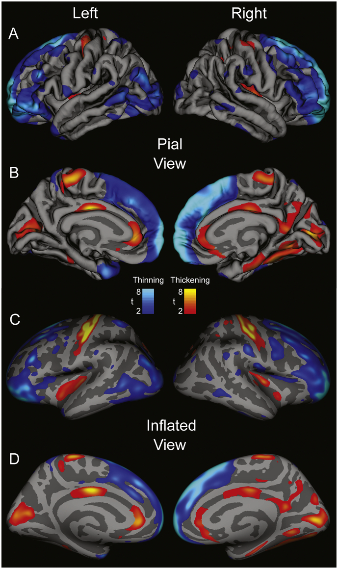
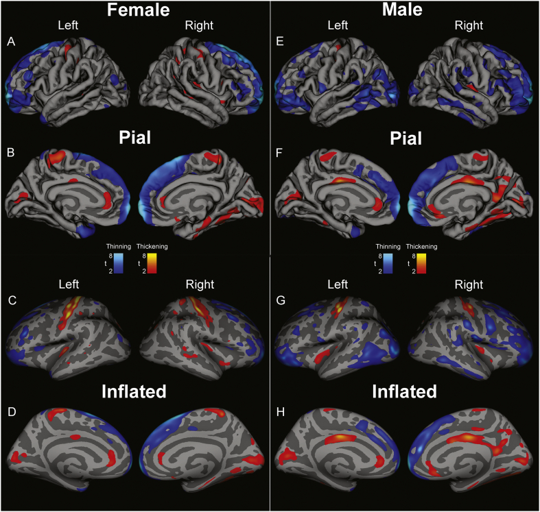
Results: Increased cortical thickness appeared in post-central gyri, insula, and subgenual, anterior, posterior, and isthmus cingulate cortices. Post-central gyri increases were greater in females, while males showed more extensive cingulate increases. Frontal and temporal cortex, lateral orbitofrontal, frontal pole, and lateral parietal and occipital cortices showed thinning. The extents of thickness changes were sex- and hemisphere-dependent, with only males exhibiting right-sided and posterior cingulate thickening, while females showed only left lateral orbitofrontal thinning. Regional cortical thickness showed modest correlations with seizure frequency, but not epilepsy duration.
Significance: Cortical thickening and thinning occur in patients with generalized tonic-clonic seizures, in cardiovascular and somatosensory areas, with extent of changes sex- and hemisphere-dependent. The data show injury in key autonomic and respiratory cortical areas, which may contribute to dysfunctional cardiorespiratory patterns during seizures, as well as to longer-term SUDEP risk.
Keywords: ACC, anterior cingulate cortex; Autonomic; CWRU, Case Western Reserve University; Cingulate; GTCS, generalized tonic-clonic seizures; Insula; OASIS, Open Access Series of Imaging Studies; PCC, posterior cingulate cortex; ROI, region of interest; Respiratory; SUDEP; SUDEP, sudden unexpected death in epilepsy; UCL, University College London; UCLA, University of California Los Angeles..
Figures
References
-
- Allen L.A., Harper R.M., Kumar R., Guye M., Ogren J.A., Lhatoo S.D., Lemieux L., Scott C.A., Vos S.B., Rani S., Diehl B. Dysfunctional brain networking among autonomic regulatory structures in temporal lobe epilepsy patients at high risk of sudden unexpected death in epilepsy. Front. Neurol. 2017;8(544) - PMC - PubMed
-
- Bernhardt B.C., Worsley K.J., Besson P., Concha L., Lerch J.P., Evans A.C., Bernasconi N. Mapping limbic network organization in temporal lobe epilepsy using morphometric correlations: insights on the relation between mesiotemporal connectivity and cortical atrophy. NeuroImage. 2008;42(2):515–524. - PubMed
-
- Bernhardt B.C., Rozen D.A., Worsley K.J., Evans A.C., Bernasconi N., Bernasconi A. Thalamo-cortical network pathology in idiopathic generalized epilepsy: insights from MRI-based morphometric correlation analysis. NeuroImage. 2009;46(2):373–381. - PubMed
Publication types
MeSH terms
Grants and funding
LinkOut - more resources
Full Text Sources
Other Literature Sources
Medical
